From 11:00 pm to 12:00 pm EST ( 8:00 pm to 9:00 pm PST ) on January 6th, the website will be under maintenance. We are sorry for the inconvenience. Please arrange your schedule properly.
Blue Tetrazolium (Tetrazolium blue), as a blue dye for microbial research, can be reduced into blue tetrazolium formazan (BTF). Blue Tetrazolium can be used to determine the activity of succinate dehydrogenase (SDH) in yeast strains, which has been reduced as a substrate. Use DMSO to extract BTF from cells, and test absorption spectrum of BTF. The BTF shows a wide wavelength range of 480-600 nm with maximal absorbance seen at 540 nm. Blue Tetrazolium, combines with succinate dehydrogenase (SDH) .
Blue Fluorescent Gelatin Methacryloyl (Blue Fluorescent GelMA) is methacryloyl gelatin (GelMA) with green fluorescence, which is obtained by "grafting" fluorescent molecules on GelMA. Blue Fluorescent Gelatin Methacryloyl acts as a scaffold and can be used to engineer tissue analogs from the vasculature to cartilage and bone, allowing cells to proliferate and spread. Blue Fluorescent Gelatin Methacryloyl is often used in cell culture, biological 3D printing, tissue engineering, etc .
Blue FPG-A trisodium is a selective antagonist of P2X1 receptor and P2Y1 receptor with IC50 values of 35.5 μM and 2.6 μM, respectively. Blue FPG-A trisodium is a structural isomer of the components of Reactive Blue 2 (RB2) .
Brilliant Blue FCF is an aromatic hydrocarbon, a synthetic dye produced from petroleum and used as a colorant for food and other substances. The solution has a maximum absorption at 628 nm.
Diphenyl Blue (Trypan Blue) is a cell active dye, the most commonly used dye for the identification of dead cells, of en used to test cell membrane integrity and cell viability. Diphenyl Blue staining is one of the methods for tissue and cell culture. When cells are deactivated or have incomplete cell membranes, Diphenyl Blue can stain them Blue. Normal living cells with intact cell membranes reject Diphenyl blue and do not stain them blue. However, macrophages are capable of phagocytosis of Diphenyl Blue, so it can be used as a living stain for macrophages .
Solvent Blue 35 (Sudan Blue II; Oil Blue 35) is a dye used for colouring alcoholic and hydrocarbon based solvents. It is used for staining triglycerides in animal tissues.
Patent Blue V (Acid blue 1) is a novel biological dye that can be used as an intraocular dye for retinectomy. Retinectomy refers to the removal of the translucent inner limiting membrane (ILM). The application of appropriate dyes in vitreoretinal surgery can achieve the purpose of complete removal. Patent Blue V can be used to stain retinal premembranous structures. Spectral analysis shows that Patent Blue V has strong absorption below 450 nm and above 600 nm, showing a blue-green color. Patent Blue V is also used as a marker in lymphangiography for resection of neoplastic lymph nodes .
Toluidine Blue (Toluidine Blue O) is an alkaline quinonimine dye (vivo dye) with high affinity for acidic tissue components, staining nuclei blue and polysaccharides purple. Toluidine Blue shows heterostaining properties for mast cells, mucins and chondrocytes. Toluidine Blue can stain different components of plant tissues and cells in different colours. Toluidine Blue is also used as a diagnostic aid to identify malignant lesions, such as cancer .
Methylene blue (Basic Blue 9) is a guanylyl cyclase (sGC), monoamine oxidase A (MAO-A) and NO synthase (NOS) inhibitor. Methylene blue is a vasopressor and is often used as a dye in several medical procedures. Methylene blue through the nitric oxide syntase/guanylate cyclase signalling pathway to reduce prepulse inhibition. Methylene blue is a REDOX cycling compound and able to cross the blood-brain barrier. Methylene blue is a Tau aggregation inhibitor. Methylene blue reduces cerebral edema, attenuated microglial activation and reduced neuroinflammation .
Methylene blue (Basic Blue 9) hydrate is a guanylyl cyclase (sGC), monoamine oxidase A (MAO-A) and NO synthase (NOS) inhibitor. Methylene blue is a vasopressor and is often used as a dye in several medical procedures. Methylene blue hydrate through the nitric oxide syntase/guanylate cyclase signalling pathway to reduce prepulse inhibition. Methylene blue hydrate is a REDOX cycling compound and able to cross the blood-brain barrier. Methylene blue hydrate is a Tau aggregation inhibitor. Methylene blue hydrate reduces cerebral edema, attenuated microglial activation and reduced neuroinflammation .
Direct Blue 86 (Solvent Blue 38) is a myelin-sheath stain, commonly utilized in microscopy to detect demyelination in the central nervous system. Direct Blue 86 also is a dye with various applications including as a commercial dye in the printing of cotton and mucilage glue fabrics .
Direct Blue 1 (Chicago Sky Blue 6B) is a complex dye for background autofluorescence in immunofluorescence histochemistry. Direct Blue 1 is a potent and competitive VGLUT inhibitor. Direct Blue 1 can inhibit the Aβ-binding small molecule PrP ligand. Direct Blue 1 has anti-inflammatory activity .
Reactive Blue 4 is an anthraquinone dye, as a single colorimetric chemosensor for sequential determination of multiple analytes with different optical responses in aqueous media. Reactive Blue 4 is phytotoxic, cytotoxic and genotoxic. Reactive Blue 4 .
Methylene blue trihydrate (C.I. Basic Blue 9 trihydrate) is a guanylyl cyclase (sGC), monoamine oxidase A (MAO-A) and NO synthase (NOS) inhibitor. Methylene blue trihydrate is a vasopressor and is often used as a dye in several medical procedures. Methylene blue trihydrate has antinociception, antimalarial, antidepressant and anxiolytic activity effects. Methylene Blue trihydrate has the potential for methemoglobinemias, neurodegenerative disorders and ifosfamide-induced encephalopathytreatment .
Basic Blue 20 is a very convenient red-emitting DNA stains. Basic Blue 20 has relatively narrow excitation and emission spectra, with peaks at 633 and 677 nm, respectively. Basic Blue 20 also has a very high resistance to photobleaching .
Basic blue 26 (Victoria blue B) is a synthetic cationic dye belonging to the class of triarylmethane dyes. It has a bright blue color and is commonly used as a colorant for a variety of applications, including textiles, paper and leather. Basic Blue 26 is also used as a biological stain for DNA and protein detection in laboratories. Due to its ability to bind negatively charged materials, it can be used as an indicator of the presence of specific molecules in biological samples. However, Basic blue 26 has been reported to have potentially harmful effects on human health and the environment and its use is regulated in some countries. Proper handling and disposal procedures are necessary to minimize its impact on the environment.
Leucomethylene blue (TRx0237) mesylate, an orally active second-generation tau protein aggregation inhibitor (Ki of 0.12 μM), could be used for the study of Alzheimer's Disease. Leucomethylene blue mesylate is a common reduced form of Methylene Blue, Methylene Blue is a member of the thiazine class of dyes .
Solvent blue 12 is a blue dye. Its series of products, such as Solvent orange 60 (HY-D1177), has been used in dyeing applications of plastic materials.
Solvent blue 97 is a blue dye. Its series of products, such as Solvent orange 60 (HY-D1177), has been used in dyeing applications of plastic materials.
Mordant Blue 13 is a synthetic dye used in the textile industry. It belongs to a class of metal complex dyes that are able to form strong bonds with fabrics and other materials. Mordant Blue 13 is commonly used for dyeing cotton, wool and silk fibres, it produces a fast blue color. It can be applied to textiles by a variety of methods including impregnation, padding and printing.
Toluidine Blue (Toluidine Blue O) purity 36% is an alkaline quinonimine dye (vivo dye) with high affinity for acidic tissue components, staining nuclei blue and polysaccharides purple. Toluidine Blue purity 36% shows heterostaining properties for mast cells, mucins and chondrocytes. Toluidine Blue purity 36% can stain different components of plant tissues and cells in different colours. Toluidine Blue purity 36% is also used as a diagnostic aid to identify malignant lesions, such as cancer .
Nile Blue A (Nile blue sulfate) is used to differentiate melanins and lipofuscins. It is also useful for staining fats and preparation of an amperometric glucose sensor .
Evans Blue (Direct Blue 53) is a potent inhibitor of L-glutamate uptake via the membrane bound excitatory amino acid transporter (EAAT). Evans Blue is a L-glutamate and kainate receptor-mediated currents inhibitor. Evans Blue has a strong affinity towards serum albumin, making it a high molecular weight protein tracer. Evans Blue is also used to study BBB (blood-brain barrier) permeability .
Thymol Blue sodium is an acid-base indicator used to indicate changes in pH. Thymol Blue sodium fades from red to yellow at pH 1.2 to 2.8 and from yellow to blue at pH 8.0 to 9.6 .
Celliton Fast Blue Green B (Disperse Blue 7), a blue-green dye used in textiles. The aqueous extract of Celliton Fast Blue Green B causes no signs of skin irritation and sensitization in laboratory animals. Celliton Fast Blue Green B colored textiles with no irritation in human .
Isosulfan blue is a blue dye for the identification of lymph vessels during lymphangiography. Isosulfan blueis is used during sentinel lymph node biopsies in breast cancer. Isosulfan blue is possible to have an allergic reaction during breast cancer operations .
Methyl blue belongs to the group of triaminotriphenylmethane dyes. Methyl blue is widely used as antiseptic dye in polychrome staining method and has applications in histological and microbiological staining solutions. Methyl blue has been used as a model to study the effect of various catalysts on photodegradation of dyes .
Calcein Blue, a membrane-impermeant fluorescent dye, is a coumarin derivative that contains an iminodiacetic acid structure. Calcein Blue is also a metallofluorochromic indicator .
Prussian blue insoluble (Iron(III) ferrocyanide) is a good adsorbent to be used as antidotes for poisoning with cesium or thallium ions. Prussian blue insoluble (Iron(III) ferrocyanide) has anticancerous and antibacterial properties. Prussian blue insoluble (Iron(III) ferrocyanide) can be used as a contrast agent in photoacoustic and magnetic resonance imaging (MRI). Prussian blue insoluble can be used for contrast agents, antidotes and cancer research .
Nile blue chloride is a highly fluorescent and photostable organic dye. Nile blue chloride and fluorescein isothiocyanate (FITC) can be used to construct a ratiometric pH sensitive probe for tracking the pH of the extracellular fluid between cancer cells in realtime. Nile Blue chloride has the potential for the research of nonlinear optics .
Celestine Blue is a electroactive indicator in DNA biosensors. Celestine Blue is strongly adsorbed on the spinel phases and CNT (carbon nanotubes), facilitates dispersion, acts as a capping agent and allows for the fabrication of spinel decorated CNT. Celestine Blue is an efficient charge transfer mediator, which allows for significant improvement of capacitive behavior. TiO2 nanoparticles doped with Celestine Blue can be used as a label in a sandwich immunoassay for the hepatitis C virus (HCV) core antigen .
Bromophenol blue is an acid phthalein dye, and it is used as a tracking dye for electrophoresis. Bromophenol blue is also used as a pH indicator, with a transition range of pH 3 to 4.6 .
Hydroxynaphthol Blue enables visual tube closure detection. Hydroxynaphthol Blue shows high specificity for the gene encoding Ara h 1 for visual field detection of peanut allergens .
Meldola blue is a biosensor for measuring lactate in serum based on screen-printed carbon electrodes modified with Meldola's Blue-Reinecke salt (MBRS-SPCE) coated with lactate dehydrogenase NAD+-dependent enzyme (from pig heart) and NAD+.
Patent Blue V has been widely used in sentinel lymph node mapping. Patent Blue V is also a food coloring agent and an alternative dye for trypan blue (TB) in descemet membrane endothelial keratoplasty (DMEK) .
Bromophenol blue sodium is a pH indicator. It changes from yellow at pH 3.0 to blue at pH 4.6. Bromophenol blue sodium is also used as a tracking dye to monitor the process of agarose gel electrophoresis and polyacrylamide gel electrophoresis .
Thiazolyl Blue (MTT) is a cell-permeable and positively charged tetrazolium dye that is used to detect reductive metabolism in cells. Thiazolyl Blue is taken up by cells through the plasma membrane and then reduced to formazan by intracellular NAD (P) H-oxidoreductases. Thiazolyl Blue is frequently used in colorimetric assays to measure cell proliferation, cytotoxicity, and apoptosis .
Msr-blue is a first turn-on fluorescent probe for methionine sulfoxide reductase with a more than 100-fold fluorescence increment. Msr-blue is used for monitoring the enzyme activity in live cells (λex=340 nm, λem=440 nm) .
CellTracker Blue CMAC is a fluorescent dye , whose chloromethyl group can form a covalent bond with cell proteins. CellTracker Blue CMAC providing a stable attachment permitting long-term cell tracking .
Aniline Blue sodium is a water-soluble dye commonly used as a biological stain for the detection of nucleic acids and proteins in various laboratory procedures such as electrophoresis and microscopy. Aniline Blue sodium has unique chemical properties that allow it to bind to specific cellular components, producing a color change that facilitates their visualization and analysis.
Fast Blue B Salt is a coloring agent that dissolves fats and phenolic compounds extracted from rye, as well as diazonium. Fast Blue B Salt can be used for the semiquantification of alkylresorcinols in rye and produces a color precipitate in the presence of acetone .
Reactive blue 5 is common textile dyes that can be adsorbed onto single-walled carbon nanotubes (SWCNTs) through electrostatic interactions, allowing the separation of residual dyes.
Reactive Blue 4 sodium is common textile dyes that can be adsorbed onto single-walled carbon nanotubes (SWCNTs) through electrostatic interactions, allowing the separation of residual dyes.
CellTracker Blue CMF2HC Dye is a blue dye, can be used in two-channel nuclei acid sequencing, with blue and purple excitation light (450-460 nm/400-405nm or 415-450 nm/480-525nm). CellTracker Blue CMF2HC Dye can be used to rapid determination of antibiotic sensitivity of microorganisms .
Victoria Blue R (Basic Blue 11) is a multifunctional dye. Dyes are important tools in biological experiments. They can help researchers observe and analyze cell structures, track biomolecules, evaluate cell functions, distinguish cell types, detect biomolecules, study tissue pathology and monitor microorganisms. Their applications range from basic scientific research to clinical A wide range of diagnostics. Dyes are also widely used in traditional fields such as textile dyeing, as well as in emerging fields such as functional textile processing, food pigments and dye-sensitized solar cells.
TMB (BM blue) is a chromogenic substrate used in staining procedures in immunohistochemistry as well as being a visualising reagent used in enzyme-linked immunosorbent assays (ELISA).
TMB dihydrochloride (BM blue dihydrochloride) is a chromogenic substrate used in staining procedures in immunohistochemistry as well as being a visualising reagent used in enzyme-linked immunosorbent assays (ELISA).
Acid blue 260 is an azo dye whose staining effect is effectively removed by multi-walled carbon nanotubes (MWCNTs). At 298 K, the adsorption capacity of MWCNT is 233.34 mg/g; and increases with the increase of dye concentration and temperature.
Brilliant Blue G-250 is a dye commonly used for the visualization of proteins separated by SDS-PAGE, offering a simple staining procedure and high quantitation. In the Bradford protein assay, protein concentrations are determined by the absorbance at 595 nm due to the binding of Brilliant Blue G-250 to proteins. Brilliant Blue G-250 is a safe highly selective P2×7R antagonist with promising consequent inactivation of NLRP3 inflammasome .
Hydroxy naphthol blue disodium, an azo dye, is a metal indicator for calcium and a colorimetric reagent for alkaline earth metal ions ( λmax = 650 nm). In the pH range between 12 and 13, the solution of the indicator is reddish pink in the presence of calcium ion and to deep blue in the presence of disodium EDTA .
True Blue (NCI 240899) is a fluorescent dye, as neuronal retrograde tracer (excitation wavelength 395-425 nm, barrier filter 450 nm). True Blue can label neuron and has no effects on neuronal survival .
C.I. Vat Blue 16 is the most important colorant used to add color or change the color of something and is chemically essentially unaffected by the carrier or medium into which it is incorporated. It can be widely used in textile, medicine, food, cosmetics, plastics, paint, ink, photography and paper industries.
Chrome Pure Blue BX (Sunchromine Pure Blue BX) is a multifunctional dye. Dyes are important tools in biological experiments. They can help researchers observe and analyze cell structures, track biomolecules, evaluate cell functions, distinguish cell types, detect biomolecules, study tissue pathology and monitor microorganisms. Their applications range from basic scientific research to clinical A wide range of diagnostics. Dyes are also widely used in traditional fields such as textile dyeing, as well as in emerging fields such as functional textile processing, food pigments and dye-sensitized solar cells.
Bromophenol blue indicator (3.0-4.6) is a synthetic dye commonly used as an acid-base indicator with a transition range of pH 3.0-4.6. Bromophenol blue indicator (3.0-4.6) is water soluble and changes color from yellow to blue as the pH of the solution changes from acidic to basic. Its unique chemical properties make it an important ingredient in a variety of scientific applications, especially in biochemistry and molecular biology. In addition, it can be used as a stain in microbiology and histology. However, Bromophenol blue indicator (3.0-4.6) has potential irritating and staining properties.
Toluidine blue (ZnCl2) is a basic thiazine dye commonly used as a biological stain for microscopy. It has a deep bluish-purple color and is commonly used to stain nucleic acids such as DNA and RNA, as well as to stain mast cells, cartilage, and other connective tissues. Toluidine blue (ZnCl2) stains the acidic components of these tissues, such as sulfated or carboxylated mucopolysaccharides. It is frequently used in histology, cytology, and pathology applications to aid in the diagnosis of various diseases and conditions. The dye is usually applied to tissue sections prior to microscopic examination and can be differentiated using an acidic alcohol solution. Toluidine blue (ZnCl2) is a relatively simple and inexpensive stain with good reproducibility, making it a popular choice for many laboratories.
(E)-5-O-Cinnamoylquinic acid is the isomer of 5-O-Cinnamoylquinic acid. 5-O-Cinnamoylquinic acid is a co-pigment. 5-O-Cinnamoylquinic acid could form the stable blue solution to clarify the mechanism of blue sepal-color development of hydrangea .
C.I. Solvent Blue 43 is the most important colorant used to add color or change the color of something and is chemically essentially unaffected by the carrier or medium into which it is incorporated. It can be widely used in textile, medicine, food, cosmetics, plastics, paint, ink, photography and paper industries.
C.I. Pigment blue 56 is the most important colorant used to add color or change the color of something and is chemically essentially unaffected by the carrier or medium into which it is incorporated. It can be widely used in textile, medicine, food, cosmetics, plastics, paint, ink, photography and paper industries.
C.I. Vat blue 22 is the most important colorant used to add color or change the color of something and is chemically essentially unaffected by the carrier or medium into which it is incorporated. It can be widely used in textile, medicine, food, cosmetics, plastics, paint, ink, photography and paper industries.
Nitro blue tetrazolium chloride (NBT) is a substrate for dehydrogenases; is used with the alkaline phosphatase substrate 5-Bromo-4-Chloro-3-Indolyl Phosphate (BCIP) in western blotting and immunohistological staining procedures .
Disperse blue 165 is a multifunctional dye. Dyes are important tools in biological experiments. They can help researchers observe and analyze cell structures, track biomolecules, evaluate cell functions, distinguish cell types, detect biomolecules, study tissue pathology and monitor microorganisms. Their applications range from basic scientific research to clinical A wide range of diagnostics. Dyes are also widely used in traditional fields such as textile dyeing, as well as in emerging fields such as functional textile processing, food pigments and dye-sensitized solar cells.
Disperse blue 291 is a multifunctional dye. Dyes are important tools in biological experiments. They can help researchers observe and analyze cell structures, track biomolecules, evaluate cell functions, distinguish cell types, detect biomolecules, study tissue pathology and monitor microorganisms. Their applications range from basic scientific research to clinical A wide range of diagnostics. Dyes are also widely used in traditional fields such as textile dyeing, as well as in emerging fields such as functional textile processing, food pigments and dye-sensitized solar cells.
7-Hydroxy-4-methylcoumarin-3-acetic acid, SE is a blue fluorophore that has pH-dependent and environment-sensitive fluorescence. It is widely used for preparing bioconjugates of blue fluorescence.
C.I. Acid blue 40 is a multifunctional dye. Dyes are important tools in biological experiments. They can help researchers observe and analyze cell structures, track biomolecules, evaluate cell functions, distinguish cell types, detect biomolecules, study tissue pathology and monitor microorganisms. Their applications range from basic scientific research to clinical A wide range of diagnostics. Dyes are also widely used in traditional fields such as textile dyeing, as well as in emerging fields such as functional textile processing, food pigments and dye-sensitized solar cells.
C.I. Acid blue 158 is a multifunctional dye. Dyes are important tools in biological experiments. They can help researchers observe and analyze cell structures, track biomolecules, evaluate cell functions, distinguish cell types, detect biomolecules, study tissue pathology and monitor microorganisms. Their applications range from basic scientific research to clinical A wide range of diagnostics. Dyes are also widely used in traditional fields such as textile dyeing, as well as in emerging fields such as functional textile processing, food pigments and dye-sensitized solar cells.
C.I. Disperse blue 284 is a multifunctional dye. Dyes are important tools in biological experiments. They can help researchers observe and analyze cell structures, track biomolecules, evaluate cell functions, distinguish cell types, detect biomolecules, study tissue pathology and monitor microorganisms. Their applications range from basic scientific research to clinical A wide range of diagnostics. Dyes are also widely used in traditional fields such as textile dyeing, as well as in emerging fields such as functional textile processing, food pigments and dye-sensitized solar cells.
Xylenol blue is a multifunctional dye. Dyes are important tools in biological experiments. They can help researchers observe and analyze cell structures, track biomolecules, evaluate cell functions, distinguish cell types, detect biomolecules, study tissue pathology and monitor microorganisms. Their applications range from basic scientific research to clinical A wide range of diagnostics. Dyes are also widely used in traditional fields such as textile dyeing, as well as in emerging fields such as functional textile processing, food pigments and dye-sensitized solar cells.
Oxazole blue is a multifunctional dye. Dyes are important tools in biological experiments. They can help researchers observe and analyze cell structures, track biomolecules, evaluate cell functions, distinguish cell types, detect biomolecules, study tissue pathology and monitor microorganisms. Their applications range from basic scientific research to clinical A wide range of diagnostics. Dyes are also widely used in traditional fields such as textile dyeing, as well as in emerging fields such as functional textile processing, food pigments and dye-sensitized solar cells.
Marina blue is a multifunctional dye. Dyes are important tools in biological experiments. They can help researchers observe and analyze cell structures, track biomolecules, evaluate cell functions, distinguish cell types, detect biomolecules, study tissue pathology and monitor microorganisms. Their applications range from basic scientific research to clinical A wide range of diagnostics. Dyes are also widely used in traditional fields such as textile dyeing, as well as in emerging fields such as functional textile processing, food pigments and dye-sensitized solar cells.
Amido Black 10B (Naphthol Blue Black) is a highly toxic azo dye for amino acid staining. Amido Black 10B can create several problems in the human respiratory system and may also cause skin and eye irritations .
Remazol marine blue is a multifunctional dye. Dyes are important tools in biological experiments. They can help researchers observe and analyze cell structures, track biomolecules, evaluate cell functions, distinguish cell types, detect biomolecules, study tissue pathology and monitor microorganisms. Their applications range from basic scientific research to clinical A wide range of diagnostics. Dyes are also widely used in traditional fields such as textile dyeing, as well as in emerging fields such as functional textile processing, food pigments and dye-sensitized solar cells.
3-Nitrotetrazolium blue (chloride) is a multifunctional dye. Dyes are important tools in biological experiments. They can help researchers observe and analyze cell structures, track biomolecules, evaluate cell functions, distinguish cell types, detect biomolecules, study tissue pathology and monitor microorganisms. Their applications range from basic scientific research to clinical A wide range of diagnostics. Dyes are also widely used in traditional fields such as textile dyeing, as well as in emerging fields such as functional textile processing, food pigments and dye-sensitized solar cells.
Nitro Blue Diformazan is a multifunctional dye. Dyes are important tools in biological experiments. They can help researchers observe and analyze cell structures, track biomolecules, evaluate cell functions, distinguish cell types, detect biomolecules, study tissue pathology and monitor microorganisms. Their applications range from basic scientific research to clinical A wide range of diagnostics. Dyes are also widely used in traditional fields such as textile dyeing, as well as in emerging fields such as functional textile processing, food pigments and dye-sensitized solar cells.
4-Methylumbelliferyl nonanoate is a fluorogenic substrate of esterases. 4-Methylumbelliferyl nonanoate can be hydrolyzed to 4-methylumbelliferone with bright blue fluorescence .
C.I. Food Blue 5:2 is a multifunctional dye. Dyes are important tools in biological experiments. They can help researchers observe and analyze cell structures, track biomolecules, evaluate cell functions, distinguish cell types, detect biomolecules, study tissue pathology and monitor microorganisms. Their applications range from basic scientific research to clinical A wide range of diagnostics. Dyes are also widely used in traditional fields such as textile dyeing, as well as in emerging fields such as functional textile processing, food pigments and dye-sensitized solar cells.
Viroxocin is a diterpenoid. Viroxocin can be isolated from the roots of Salvia viridis L. cvar. Blue Jeans. Viroxocin also has weak antibacterial activity .
4-Aminodiphenylamine sulfate (Variamine Blue RT sulfate) is a multifunctional dye. Dyes are important tools in biological experiments. They can help researchers observe and analyze cell structures, track biomolecules, evaluate cell functions, distinguish cell types, detect biomolecules, study tissue pathology and monitor microorganisms. Their applications range from basic scientific research to clinical A wide range of diagnostics. Dyes are also widely used in traditional fields such as textile dyeing, as well as in emerging fields such as functional textile processing, food pigments and dye-sensitized solar cells.
DL-Penicillamine [(±)-Penicillamine] is a copper chelating agent. DL-Penicillamine has antidotal effects in thallotoxicosis rats when co-treated with Prussian blue (HY-106594A). DL-Penicillamine can cause pyridoxine deficiency and then induce optic axial neuritis. DL-Penicillamine can also depress primary immune response .
Nile red (Nile blue oxazone) is a lipophilic stain. Nile red has environment-sensitive fluorescence. Nile red is intensely fluorescent in a lipid-rich environment while it has minimal fluorescence in aqueous media. Nile red is an excellent vital stain for the detection of intracellular lipid droplets by fluorescence microscopy and flow cytof uorometry. Nile red stains intracellular lipid droplets red. The fluorescence wavelength is 559/635 nm .
pyCTZ (Pyridyl CTZ) hydrochloride, a pyridyl Coelenterazine (CTZ) analog, and is an ATP-independent pyridyl substrate of LumiLuc luciferase. pyCTZ hydrochloride generates strong blue bioluminescence in the presence of luciferases. pyCTZ hydrochloride can be used for aequorin-based calcium sensing .
Coelenteramide is a oxidative product of Coelenterazine (HY-18743). Coelenteramide can form a complex with apoAequorin/Ca 2+, which is known as blue fluorescent protein (BFP) and shows continuous weak luminescence with Coelenterazine like a luciferase. Coelenteramide can be used as an imaging agent .
N-(9-Acridinyl)maleimide is a maleimide type fluorescent thiol reagent. N-(9-Acridinyl)maleimide shows no substantial fluorescence but its coupling products with thiol compounds exhibit strong blue fluorescence. N-(9-Acridinyl)maleimide is used for fluorometrical analysis of cysteine and glutathione .
pyCTZ (Pyridyl CTZ) TFA, a pyridyl Coelenterazine (CTZ) analog, and is an ATP-independent pyridyl substrate of LumiLuc luciferase. pyCTZ TFA generates strong blue bioluminescence in the presence of luciferases. pyCTZ TFA can be used for aequorin-based calcium sensing .
pyCTZ (Pyridyl CTZ), a pyridyl Coelenterazine (CTZ) analog, and is an ATP-independent pyridyl substrate of LumiLuc luciferase. pyCTZ generates strong blue bioluminescence in the presence of luciferases. pyCTZ can be used for aequorin-based calcium sensing .
7-Hydroxycoumarin-3-carboxylic acid N-succinimidyl ester is the amine-reactive succinimidyl ester of 7-Hydroxycoumarin-3-carboxylic acid. 7-Hydroxycoumarin-3-carboxylic acid N-succinimidyl ester is a blue fluorescent dye for labeling proteins and nucleic acids .
TMB monosulfate is a chromogenic substrate used in staining procedures in immunohistochemistry as well as being a visualizing reagent used in enzyme-linked immunosorbent assays (ELISA).
PDMPO, a lysosome pH indicator, is an excellent fluorescent acidotropic reagent for fluorescence imaging. PDMPO is a potent tool with which to study acidic organelles of live cells. PDMPO exhibits pH-dependent dual-excitation and dual-emission spectral peaks. PDMPO produces a blue fluorescence in weakly acidic organelles and shifts to yellow in more acidic lysosomes (Abs=329 nm; Em=440 nm) .
Dansyl chloride is a reagent that produces stable blue or blue-green fluorescent sulfonamide adducts in the reaction of aliphatic and aromatic amines with primary amino groups, and is widely used for modified amino acids, protein sequencing and amino acid analysis .
ER-Tracker dye is a derivative of BODIPY series dyes coupled with Glibenclamide (HY-15206), highly selective binding to the endoplasmic reticulum, non-toxic to cells at low concentrations, this type of dye is an environmentally sensitive probe, and formaldehyde treatment can still retain part of the fluorescence, with high fluorescence life, good extinction coefficient and other characteristics. Glibenclamide is an atp-dependent K + channel blocker (Kir6, KATP) and CFTR Cl-channel blocker that binds in the endoplasmic reticulum. ER-Tracker is not suitable for staining cells after fixation .
NOD1 antagonist-1 (compound 37) exhibits an antagonistic activity towards NOD1 and a weak NOD1/NOD2 selectivity, with IC50s of 9.18 μM and 20.8 μM, respectively .
X-GAL (BCIG) is a widely used chromogenic β-galactosidase substrate. X-GAL is a colorless compound until cleaved by β-galactosidase, at which point X-GAL turns to an insoluble and detectable blue compound, making X-GAL particularly useful in techniques such as blue-white screening for cloning in bacteria. X-GAL can also be used for detection of β-galactosidase activity .
Microcolin B is an extremely potent unusual acylpeptide, proline-containing potent immunosuppressant. Microcolin B is isolated from blue-green alga Lyngbya majuscule .
Pyrene phosphoramidite Du is a click chemistry reagent containing pyrene groups. The pyrene group in Pyrene phosphoramidite Du can be inserted into DNA with strong blue fluorescence.
Azure C is the product of sequential enzymatic oxidation of Methylene blue (MB) or Azure B (AB). Azure C serves as the substrate of horseradish peroxidase (HRP) .
Thymolphthalein is an electrochemically active dye due to the presence of thymol fragments. Thymolphthalein is a Phthalein dye used as an acid-base indicator. It is colourless in acid pH and is blue in basic pH .
Crystal Ponceau 6R is a red azo dye. Crystal Ponceau 6R used in histology, for staining fibrin with the martius, scarlet and blue (MSB) Trichrome stain .
Leishman's stain is an essential staining tool for for staining of the peripheral blood and bone marrow smears (displayed pale bluish-grey to deep blue under oil-immersion lens) .
Azure A (chloride) is a phenothiazine dye. Azure A (chlorine) is formed by oxidation of methylene blue and has strong metachromatic. Azure A (chlorine) can be used for the study of stains and redox media for electrochemical biosensing .
Dihydroethidium, also known as DHE, is a peroxide indicator. Dihydroethidium penetrates cell membranes to form a fluorescent protein complex with blue fluoresces. After entering the cells, Dihydroethidium is mainly localized in the cell membrane, cytoplasm and nucleus, and the staining effect is the strongest in the nucleus. Dihydroethidium produces inherent blue fluorescence with a maximum excitation wavelength of 370 nm and a maximum emission wavelength of 420 nm; after dehydrogenation, Dihydroethidium combines with RNA or DNA to produce red fluorescence with a maximum excitation wavelength of 300 nm and a maximum emission wavelength of 610 nm. 535 nm can also be used as the excitation wavelength for actual observation .
HMBR, an analog bearing an additional methyl group on the aromatic ring, is nonfluorescent by itself, but it fluoresces yellow light upon blue-light excitation when bound to Y-FAST. HMBR is nontoxic for zebrafish embryos. cell-permeant .
TSQ is a cytosolic zinc fluorescence probe that is membrane permeable and can be used for intracellular imaging of zinc proteins (λmax ~470 nm). TSQ can combine with Zn 2+ in the presence of Ca 2+ and Mg 2+ to produce blue fluorescence .
4-Methylumbelliferyl β-D-galactopyranoside is a fluorescent substrate for β-galactosidase which, when cleaved, produces a water-soluble blue fluorescent coumarin fluorophore that can be detected using a fluoroenzymeter or fluorometer .
Antitumor agent-150 (V10), an anti-breast cancer agent, is a PROTAC-based MDM2 protein degrader (Red: Ganoderic acid A; Black: 4O-PEG linker; Blue: VHL ligand) .
mCP-BP-SFAC is a luminogenic molecule. mCP-BP-SFAC exhibits strong sky-blue delayed fluorescence in neat films, with photoluminescence (PL) peaks at ~483 nm and delayed fluorescence lifetimes of 5.4 to 5.7 μs .
TCP-BP-SFAC is a luminogenic molecule. TCP-BP-SFAC exhibits strong sky-blue delayed fluorescence in neat films, with photoluminescence (PL) peaks at ~483 nm and delayed fluorescence lifetimes of 5.4 to 5.7 μs .
Bromocresol green is a pH-sensitive triphenylmethane dye commonly used for the determination of protein and albumin in serum. Bromocresol green is a bio-based dye with a yellow-green to blue-green color. Bromocresol green turns yellow (λmax=435 nm, protonated form) when placed in acidic solution (e.g. pH=4.15), and turns blue in basic solution (λmax=615 nm, deprotonated form). Bromocresol green is widely used as a pH indicator in the field of biochemical analysis. In addition, Bromocresol green is also used to detect the concentration of molecules such as creatinine, and to judge the viability of cells .
Hoechst 33258 is a marker dye in Hoechst series. Hoechst is A live nuclear marker dye. Hoechst binds to the grooves in the DNA double strand, which tends to be A/ T-rich DNA strand. Although it binds to all nucleic acids, the A/ T-rich double strand DNA significantly enhances fluorescence intensity Therefore,Hoechst dye can be used for living cell labeling. The fluorescence intensity of Hoechst dye increases with the increase of pH of solution .
Hoechst 33342 is a marker dye in Hoechst series. Hoechst is A live nuclear marker dye. Hoechst binds to the grooves in the DNA double strand, which tends to be A/ T-rich DNA strand. Although it binds to all nucleic acids, the A/ T-rich double strand DNA significantly enhances fluorescence intensity Therefore,Hoechst dye can be used for living cell labeling. The fluorescence intensity of Hoechst dye increases with the increase of pH of solution .
Hoechst 34580 is a marker dye in Hoechst series. Hoechst is A live nuclear marker dye. Hoechst binds to the grooves in the DNA double strand, which tends to be A/ T-rich DNA strand. Although it binds to all nucleic acids, the A/ T-rich double strand DNA significantly enhances fluorescence intensity Therefore,Hoechst dye can be used for living cell labeling. The fluorescence intensity of Hoechst dye increases with the increase of pH of solution .
Hoechst S 769121 is a marker dye in Hoechst series. Hoechst is A live nuclear marker dye. Hoechst binds to the grooves in the DNA double strand, which tends to be A/ T-rich DNA strand. Although it binds to all nucleic acids, the A/ T-rich double strand DNA significantly enhances fluorescence intensity Therefore,Hoechst dye can be used for living cell labeling. The fluorescence intensity of Hoechst dye increases with the increase of pH of solution .
HOE-S 785026 is a marker dye in Hoechst series. Hoechst is A live nuclear marker dye. Hoechst binds to the grooves in the DNA double strand, which tends to be A/ T-rich DNA strand. Although it binds to all nucleic acids, the A/ T-rich double strand DNA significantly enhances fluorescence intensity Therefore,Hoechst dye can be used for living cell labeling. The fluorescence intensity of Hoechst dye increases with the increase of pH of solution .
HOE 32021 is a marker dye in Hoechst series. Hoechst is A live nuclear marker dye. Hoechst binds to the grooves in the DNA double strand, which tends to be A/ T-rich DNA strand. Although it binds to all nucleic acids, the A/ T-rich double strand DNA significantly enhances fluorescence intensity Therefore,Hoechst dye can be used for living cell labeling. The fluorescence intensity of Hoechst dye increases with the increase of pH of solution .
meta-iodoHoechst 33258 is a marker dye in Hoechst series. Hoechst is A live nuclear marker dye. Hoechst binds to the grooves in the DNA double strand, which tends to be A/ T-rich DNA strand. Although it binds to all nucleic acids, the A/ T-rich double strand DNA significantly enhances fluorescence intensity Therefore,Hoechst dye can be used for living cell labeling. The fluorescence intensity of Hoechst dye increases with the increase of pH of solution .
Hoechst 33258 analog is a marker dye in Hoechst series. Hoechst is A live nuclear marker dye. Hoechst binds to the grooves in the DNA double strand, which tends to be A/ T-rich DNA strand. Although it binds to all nucleic acids, the A/ T-rich double strand DNA significantly enhances fluorescence intensity Therefore,Hoechst dye can be used for living cell labeling. The fluorescence intensity of Hoechst dye increases with the increase of pH of solution .
Hoechst 33258 analog 2 is a marker dye in Hoechst series. Hoechst is A live nuclear marker dye. Hoechst binds to the grooves in the DNA double strand, which tends to be A/ T-rich DNA strand. Although it binds to all nucleic acids, the A/ T-rich double strand DNA significantly enhances fluorescence intensity Therefore,Hoechst dye can be used for living cell labeling. The fluorescence intensity of Hoechst dye increases with the increase of pH of solution .
Hoechst 33258 analog 3 is a marker dye in Hoechst series. Hoechst is A live nuclear marker dye. Hoechst binds to the grooves in the DNA double strand, which tends to be A/ T-rich DNA strand. Although it binds to all nucleic acids, the A/ T-rich double strand DNA significantly enhances fluorescence intensity Therefore,Hoechst dye can be used for living cell labeling. The fluorescence intensity of Hoechst dye increases with the increase of pH of solution .
ortho-iodoHoechst 33258 is a marker dye in Hoechst series. Hoechst is A live nuclear marker dye. Hoechst binds to the grooves in the DNA double strand, which tends to be A/ T-rich DNA strand. Although it binds to all nucleic acids, the A/ T-rich double strand DNA significantly enhances fluorescence intensity Therefore,Hoechst dye can be used for living cell labeling. The fluorescence intensity of Hoechst dye increases with the increase of pH of solution .
Hoechst 33342 analog is a marker dye in Hoechst series. Hoechst is A live nuclear marker dye. Hoechst binds to the grooves in the DNA double strand, which tends to be A/ T-rich DNA strand. Although it binds to all nucleic acids, the A/ T-rich double strand DNA significantly enhances fluorescence intensity Therefore,Hoechst dye can be used for living cell labeling. The fluorescence intensity of Hoechst dye increases with the increase of pH of solution .
Hoechst 33258 analog 5 is a marker dye in Hoechst series. Hoechst is A live nuclear marker dye. Hoechst binds to the grooves in the DNA double strand, which tends to be A/ T-rich DNA strand. Although it binds to all nucleic acids, the A/ T-rich double strand DNA significantly enhances fluorescence intensity Therefore,Hoechst dye can be used for living cell labeling. The fluorescence intensity of Hoechst dye increases with the increase of pH of solution .
HOE 32020 is a marker dye in Hoechst series. Hoechst is A live nuclear marker dye. Hoechst binds to the grooves in the DNA double strand, which tends to be A/ T-rich DNA strand. Although it binds to all nucleic acids, the A/ T-rich double strand DNA significantly enhances fluorescence intensity Therefore,Hoechst dye can be used for living cell labeling. The fluorescence intensity of Hoechst dye increases with the increase of pH of solution .
Hoechst 33342 analog 2 is a marker dye in Hoechst series. Hoechst is A live nuclear marker dye. Hoechst binds to the grooves in the DNA double strand, which tends to be A/ T-rich DNA strand. Although it binds to all nucleic acids, the A/ T-rich double strand DNA significantly enhances fluorescence intensity Therefore,Hoechst dye can be used for living cell labeling. The fluorescence intensity of Hoechst dye increases with the increase of pH of solution .
Hoechst 33258 analog 6 is a marker dye in Hoechst series. Hoechst is A live nuclear marker dye. Hoechst binds to the grooves in the DNA double strand, which tends to be A/ T-rich DNA strand. Although it binds to all nucleic acids, the A/ T-rich double strand DNA significantly enhances fluorescence intensity Therefore,Hoechst dye can be used for living cell labeling. The fluorescence intensity of Hoechst dye increases with the increase of pH of solution .
para-iodoHoechst 33258 is a marker dye in Hoechst series. Hoechst is A live nuclear marker dye. Hoechst binds to the grooves in the DNA double strand, which tends to be A/ T-rich DNA strand. Although it binds to all nucleic acids, the A/ T-rich double strand DNA significantly enhances fluorescence intensity Therefore,Hoechst dye can be used for living cell labeling. The fluorescence intensity of Hoechst dye increases with the increase of pH of solution .
Hoechst 33342 trihydrochloride is a marker dye in Hoechst series. Hoechst is A live nuclear marker dye. Hoechst binds to the grooves in the DNA double strand, which tends to be A/ T-rich DNA strand. Although it binds to all nucleic acids, the A/ T-rich double strand DNA significantly enhances fluorescence intensity Therefore,Hoechst dye can be used for living cell labeling. The fluorescence intensity of Hoechst dye increases with the increase of pH of solution .
HOE-S 785026 trihydrochloride is a marker dye in Hoechst series. Hoechst is A live nuclear marker dye. Hoechst binds to the grooves in the DNA double strand, which tends to be A/ T-rich DNA strand. Although it binds to all nucleic acids, the A/ T-rich double strand DNA significantly enhances fluorescence intensity Therefore,Hoechst dye can be used for living cell labeling. The fluorescence intensity of Hoechst dye increases with the increase of pH of solution .
Hoechst 34580 tetrahydrochloride is a marker dye in Hoechst series. Hoechst is A live nuclear marker dye. Hoechst binds to the grooves in the DNA double strand, which tends to be A/ T-rich DNA strand. Although it binds to all nucleic acids, the A/ T-rich double strand DNA significantly enhances fluorescence intensity Therefore,Hoechst dye can be used for living cell labeling. The fluorescence intensity of Hoechst dye increases with the increase of pH of solution .
Hoechst 33342 analog 2 trihydrochloride is a marker dye in Hoechst series. Hoechst is A live nuclear marker dye. Hoechst binds to the grooves in the DNA double strand, which tends to be A/ T-rich DNA strand. Although it binds to all nucleic acids, the A/ T-rich double strand DNA significantly enhances fluorescence intensity Therefore,Hoechst dye can be used for living cell labeling. The fluorescence intensity of Hoechst dye increases with the increase of pH of solution .
Hoechst 33258 trihydrochloride is a marker dye in Hoechst series. Hoechst is A live nuclear marker dye. Hoechst binds to the grooves in the DNA double strand, which tends to be A/ T-rich DNA strand. Although it binds to all nucleic acids, the A/ T-rich double strand DNA significantly enhances fluorescence intensity Therefore,Hoechst dye can be used for living cell labeling. The fluorescence intensity of Hoechst dye increases with the increase of pH of solution .
HOE 33187 is a marker dye in Hoechst series. Hoechst is A live nuclear marker dye. Hoechst binds to the grooves in the DNA double strand, which tends to be A/ T-rich DNA strand. Although it binds to all nucleic acids, the A/ T-rich double strand DNA significantly enhances fluorescence intensity Therefore,Hoechst dye can be used for living cell labeling. The fluorescence intensity of Hoechst dye increases with the increase of pH of solution .
UNC8899 is a VHL-recruiting STINGPROTAC degrader (DC50: 0.0.924 μM). UNC8899 can be used for viral or bacterial infection research (Blue: VHL ligand, Black: linker; Pink: STING inhibitor) .
UNC8900 is a VHL recruiting STINGPROTAC degrader (DC50: 0.0.924 μM). UNC8900 can be used for research of viral or bacterial infection. (Blue: VHL ligand, black: linker; Pink: STING inhibitor) .
Tropaeolin O is an acidic monoazo dye that undergoes a coupling reaction under pH=10.5 conditions to form a blue disazo dye. Tropaeolin O can be used for the determination of palladium(II), osmium(IV), albumin, and casein .
Trx-red (NBL-SS perchlorate) is a red-emitting fluorescent probe derivatized from the nile blue fluorophore. Trx-red is used for selectively imaging thioredoxin (Trx) in live cells and in vivo (λex=615 nm, λem=661 nm) .
Zinquin ethyl ester is a fluorescent derivative of Zinquin and is a fluorescent probe of cytosolic zinc. Zinquin ethyl ester is able to penetrate cell membranes and is lipophilic and zinc-sensitive. Zinquin ethyl ester can combine with Zn 2+ in the presence of Ca 2+ and Mg 2+ to produce blue fluorescence .
HADA hydrochloride (HCC-Amino-D-alanine hydrochloride) is a blue (λem~450 nm) fluorescent D-amino acid (FDAA). FDAAs are efficiently incorporated into the peptidoglycans (PGs) of diverse bacterial species at the sites of PG biosynthesis, allowing specific and covalent probing of bacterial growth with minimal perturbation .
Acridine homodimer (NSC 219743), acridine dimer, is a fluorescence dye. Acridine homodimer emits a blue-green fluorescence when bound to DNA. Acridine homodimer has extremely high affinity for AT-rich regions of nucleic acids, can be used for chromosome banding .
AMCA-X-SE is a coumarin derivative that generates fixed blue fluorescence and an NHS-activated ester that forms stable amide bonds with primary amine groups. It is used as a reactive dye for labeling amino groups of peptides, proteins, and oligonucleotides. Maximum excitation/emission wavelength: 354/442 nm .
4-Methylumbelliferyl butyrate (4-MUB) is a coumarin-based fluorogenic substrate used for the identification of M. catarrhalis C4- esterase. 4-Methylumbelliferyl butyrate can converse to the blue-emissive 4-methylumbelliferone (4-MU; HY-N0187) .
7-Amino-4-methylcoumarin-3-acetic acid is a fluorescent protein labelling agent. 7-Amino-4-methylcoumarin-3-acetic acid emits in the blue region (440-460 nm) on activation with UV light (350 nm) .
2,3-Di(furan-2-yl)quinoxaline (Compound 5) is a quinoxaline exhibiting blue fluorescence. 2,3-Di(furan-2-yl)quinoxaline is cell permeable and sufficiently bright at a micromolar concentration (1.5 μM) .
3-Cyano-7-ethoxycoumarin is a fluorogenic cytochrome P-450 substrate that generates blue fluorescent product upon enzyme cleavage
Target: Cytochrome P450
3-Cyano-7-ethoxycoumarin is a fluorescent probe useful in microsomal dealkylase studies.
4-Methylumbelliferyl-α-D-galactopyranoside (4MU-α-Gal), a substrate for α-galactosidase A (GLA), is a blue pro-fluorogenic substrate. 4-Methylumbelliferyl-α-D-galactopyranoside forms two products, galactose and fluorescent 4MU, upon cleavage by GLA .
5-Bromo-1H-indol-3-yl octanoate is a chromogenic substrate for esterase with C8 activity. 5-Bromo-1H-indol-3-yl octanoate yields a blue precipitate when cleaved.
3-Indolyl-β-D-glucuronide, a chromogenic substrate for β-D-glucuronidase, employs in the detection and enumeration of E. coli, yielding a blue precipitate upon cleavage. 3-Indolyl-β-D-glucuronide in? patients' plasma act as a new indicator of renal failure .
DCIP sodium is a blue dye commonly used in various biochemical and biotechnological applications as an indicator of redox reactions. DCIP sodium has unique chemical properties that change color according to the oxidation state of the substance being tested. It is commonly used in enzyme assays, such as measuring the activity of succinate dehydrogenase, or in protein quantification methods, such as the Lowry assay.
L-Leucine-7-amido-4-methylcoumarin (Leu-AMC) hydrochloride is a bright blue fluorogenic peptidyl substrate for LAP3 (leucine aminopeptidase). L-Leucine-7-amido-4-methylcoumarin hydrochloride can be used for leucine aminopeptidase inhibition assays in vitro .
Urdamycin A (Compound 3b) is an angucycline antibiotic that can be isolated from Streptomyces fradiae. Urdamycin A is an orange indicator with a change of the color to ultramarine blue at pH 7.7. Urdamycin A has anticancer activity with IC50s of 2.4 and 0.55 μg/mL in proliferation and stem cell assays, respectively .
UNC9036 is a PROTAC-based STING degrader, with a DC50 of 227 nM. UNC9036-mediated STING degradation is proteasome and VHL dependent (Srtucture Note: Red, STING agonist diABZI (HY-112921A); Blue, VHL ligand VH032 (HY-120217); Black, linker) .
Wright's stain is a hematologic stain that facilitates the differentiation of blood cell types. Wright's stain is classically a mixture of eosin (red) and methylene blue dyes. It is used primarily to stain peripheral blood smears, urine samples, and bone marrow aspirates. Wright's stain provides a manual or automated stain for bone marrow and peripheral blood smears .
Bromocresol green sodium is an anionic dye. Bromocresol green sodium can be used for pH indication and DNA agarose gel electrophoresis. Bromocresol green sodium is also used in mammalian albumin measurement. Bromocresol green sodium deprotonates and produces the monoanionic form of yellow colour at lower pH (acidic condition), and produces dianionic blue colour at the basic condition .
alpha-Naphtholphthalein is an indicator that is light yellow at pH 6 and gradually shows a clear color change with increasing pH to light green-green-blue. alpha-Naphtholphthalein can be impregnated into cotton-blend fabrics and used to develop medical supplies for wound pH monitoring, such as medical gauze, hospital gowns and compression bandages .
3-Methoxybenzamide (3-MBA), an inhibitor of ADP-ribosyltransferase (ADPRTs) and PARP, inhibits cell division in Bacillus subtilis, leading to filamentation and eventually lysis of cells . 3-Methoxybenzamide (3-MBA) enhances in vitro plant growth, microtuberization, and transformation efficiency of blue potato (Solanum tuberosum L. subsp. andigenum) .
Gallocyanine chloride, a synthetic blue dyestuff, blocks DKK1 inhibitory activity by disrupting DKK1/LRP6 interaction. Its association with LRP6 is weak (IC50 of about 3 μM in the inhibition of DKK1 binding). Gallocyanine dye acts as a potential agent for the research of Alzheimer's disease and related neurodegenerative tauopathies .
DAPI (dilactate) is a blue fluorescent dye that preferentially binds dsDNA and binds to minor groove AT clusters. DAPI (dilactate) is combined with dsDNA, and the fluorescence was enhanced about 20-fold. DAPI (dilactate) can be used to identify the cell cycle and specifically stains the nucleus but not the cytoplasm. DAPI (dilactate) form is more soluble in water than DAPI (dihydrochloride) form.
KL001 is a first-in-class cryptochrome (CRY, a flavoproteins that are sensitive to blue light, and is involved in the circadian rhythms of plants and animals) stabilizer which specifically interacts with CRY1 and CRY2. KL001 prevents ubiquitin-dependent degradation of CRY, resulting in lengthening of the circadian period. KL001 has the potential to control fasting hormone-induced gluconeogenesis .
4,4'-Di-O-methylellagic acid (4,4'-DiOMEA; Nasutin C) can be isolated from the Australian termites. 4,4'-Di-O-methylellagic acid is blue-fluorescent under ultra-violet light . 4,4'-Di-O-methylellagic acid inhibits colon cancer cell proliferation via the wnt signal pathway .
D-Ala-Lys-AMCA is a known proton-coupled oligopeptide transporter 1 (PEPT1) substrate that emits blue fluorescence. D-Ala-Lys-AMCA may be transported into liver cancer cells and Caco-2 cells based on fluorescence analysis. D-Ala-Lys-AMCA can be used for characterizing PEPT1-specific substrates or inhibitors .
D-Ala-Lys-AMCA hydrochloride is a known proton-coupled oligopeptide transporter 1 (PEPT1) substrate that emits blue fluorescence. D-Ala-Lys-AMCA hydrochloride may be transported into liver cancer cells and Caco-2 cells based on fluorescence analysis. D-Ala-Lys-AMCA hydrochloride can be used for characterizing PEPT1-specific substrates or inhibitors .
Azure B is a cationic dye and the major metabolite of Methylene blue. Azure B is used in making Azure eosin stains for blood smear staining. Azure B is a high-potency, selective and reversible inhibitor of monoamine oxidases (MAO)-A, with IC50s of 11 and 968 nM for recombinant human MAO-A and MAO-B, respectively. Azure B possesses significant antidepressant-like effects .
X-Gluc sodium is a dye reagent for the detection of β-glucuronidase (GUS), an enzyme produced by E. coli. X-Gluc sodium can be used to detect E. coli contamination in food, water and the urinary tract (GUS as a specific detection indicator). X-Gluc sodium is also widely used in molecular biology experiments to label and detect the expression of target genes (reacts with the GUS gene, appears blue) .
STAT3-IN-17 is a moderate STAT3 inhibitor (IC50=0.7 μM; HEK-Blue IL-6), with antiproliferative activity in HeLa cells. STAT3-IN-17 has good pharmacokinetic characteristics. STAT3-IN-17 also inhibits pyruvate-ferredoxin oxidoreductase (PFOR), and inhibits Helicobacter pylori .
Coelenteramine (Coelenterazine) 400a hydrochloride, a derivative of Coelenterazine, is a Renilla luciferase (RLuc) substrate. In the presence of Coelenteramine 400a hydrochloride, RLuc can emit blue light at 395 nm . Coelenteramine 400a hydrochloride will causes color change in the bioluminescence reaction of Rluc by replacing the sulfur and oxygen heteroatoms of the methylene bridge. Coelenteramine 400a hydrochloride provides higher signal resolution and can be used in the research of bioluminescence resonance energy transfer (BRET) .
cis-MZ 1 hydrate is a negative control for BRD4-targeted PROTAC MZ 1 (HY-107425). cis-MZ 1 or MZ 1 is a combination of the von Hippel-Lindau ligand (red part in the structural formula) and the BRD4 ligand (blue part in the structural formula). The Kd of MZ 1 for BRD4 BD1/2 was 382 nM and 120 nM, respectively .
PROTAC NAMPT Degrader-1 is a potent PROTAC targeting NAMPT with aDC50 value of 217 nM. PROTAC NAMPT Degrader-1 has anti-proliferative activity with an IC50 value of 0.12μM against A2780 cells.
(Srtucture Note: PINK, NAMPT activator (HY-163445); Blue, VHL ligand (HY-163440); Black, linker) .
D-Ala-Lys-AMCA TFA is a known proton-coupled oligopeptide transporter 1 (PEPT1) substrate that emits blue fluorescence. D-Ala-Lys-AMCA TFA may be transported into liver cancer cells and Caco-2 cells based on fluorescence analysis. D-Ala-Lys-AMCA TFA can be used for characterizing PEPT1-specific substrates or inhibitors .
Coelenteramine 400a (Coelenterazine 400a), a derivative of Coelenterazine, is a Renilla luciferase (RLuc) substrate. In the presence of Coelenteramine 400a, RLuc can emit blue light at 395 nm . Coelenterazine 400a will causes color change in the bioluminescence reaction of Rluc by replacing the sulfur and oxygen heteroatoms of the methylene bridge. Coelenterazine 400a provides higher signal resolution and can be used in the research of bioluminescence resonance energy transfer (BRET) .
X-Gluc cyclohexanamine is a dye reagent for the detection of β-glucuronidase (GUS), an enzyme produced by E. coli. X-Gluc cyclohexanamine can be used to detect E. coli contamination in food, water and the urinary tract (GUS as a specific detection indicator). X-Gluc cyclohexanamine is also widely used in molecular biology experiments to label and detect the expression of target genes (reacts with the GUS gene, appears blue) .
Myricetin 3-O-α-L-arabinopyranoside is a quercetin derivative and plant flavonoid with antioxidant, antibacterial and antiurease effects. Myricetin 3-O-α-L-arabinopyranoside inhibits A2E photooxidation-induced RPE cell death. Myricetin 3-O-α-L-arabinopyranoside is protective against retinal degeneration and protects against blue light (BL)-induced damage in RPE cells and mouse models .
Pyridinium bisretinoid A2E-d4 TFA is the deuterium-labeled Pyridinium bisretinoid A2E (HY-134928). Pyridinium bisretinoid A2E (A2E)is an initiator of blue-light-induced apoptosis. Photoactivation of Pyridinium bisretinoid A2E mediates autophagy and the production of reactive oxygen species .
XY-52 (Compound 32) is a Stimulation-2 (ST2) inhibitor, with an IC50 value of 5.68 μM in AlphaLISA assay, and 4.59 μM in HEK-Blue assay. XY-52 increases proinflammatory T-cell proliferation. XY-52 reduces the plasma sST2 and IFNγ biomarkers in the graft versus host disease (GVHD) mice model .
MS8709 (10), a potential anticancer therapeutic, is a first-in-class G9a/GLPPROTAC degrader. MS8709 (10) is based on G9a/GLP inhibitor UNC0642 and recruits the von Hippel Lindau (VHL) E3 ligase (Red: G9a/GLP inhibitor UNC0642; Blue: VHL ligand; Black: linker) .
Resazurin (Diazoresorcinol) is a water-soluble, non-toxic, stable, membrane-permeable blue non-fluorescent dye (faintly fluorescent). Resazurin is used as a redox indicator, can be reduced to pink, highly fluorescent Resorufin (Ex=530-560 nm, Em=590 nm) in living cells. Resazurin can be used for the detection of cell viability, toxicity, proliferation, migration and invasion in cells (human, plant and animal, bacterial and fungal) .
Kaempferol-3-O-[2″,6″-di-O-E-p-coumaroyl]-β-D-glucopyranoside is a acylated keampferol glucoside. Kaempferol-3-O-[2″,6″-di-O-E-p-coumaroyl]-β-D-glucopyranoside can be isolated from the leaves of O. dentata, acts as a repellent against a fouling organism, the blue mussel M. edulis .
Pyridinium bisretinoid A2E (A2E) is a fluorophore that can be isolated from lipofuscin in the retinal pigment epithelium (RPE). Pyridinium bisretinoid A2E is an initiator of blue-light-induced apoptosis. Photoactivation of Pyridinium bisretinoid A2E mediates autophagy and the production of reactive oxygen species. Pyridinium bisretinoid A2E can be used in the study of retinal degenerative diseases .
3-Indolyl-β-D-glucuronide cyclohexanamine is the glucose component of X-Gluc staining buffer. 3-Indolyl-β-D-glucuronide cyclohexanamine can be used to detect gene expression. The active ingredient of the stain, β-Glucuronidase (GUS), reacts with the enzyme, causing the target gene to appear blue-purple in tissues or cells, so that the expression level and tissue distribution of the gene can be visually observed .
LHF418 is an effective SOS1 PROTAC degrader with a DC50 value of 209.4 nM in A549 cells. LHF418 can effectively inhibit RAS signaling and colony formation in KRAS-driven cancer cells. (Structural note: (Blue: Cereblon ligand (HY-A0003), Black: linker; Pink: SOS1 binder SOS1 Ligand intermediate-3 (HY-161452)) .
TMPD dihydrochloride, a readily oxidizable compound, is an enzymatically convert redox active substrate molecule. TMPD dihydrochloride is also an electron donor and serves as a reducing cosubstrate for heme peroxidases . TMPD dihydrochloride is also a complex IV substrate .
Both Ponceau S and Ponceau BS are synthetic dyes commonly used in biological research. They are commonly used as protein stains to visualize proteins in western blots and other protein detection analyses. Ponceau S is a red dye, while Ponceau BS is a blue shade of the same dye. Both dyes bind to positively charged amino acid residues in proteins for easy visualization. However, Ponceau S is more commonly used due to its higher sensitivity.
TOOS (TOOS sodium salt) is a highly water-soluble aniline derivative widely used in diagnostics and biological experiments. TOOS can be combined with 3-methyl-2-benzothiazolinone hydrazone hydrochloride (MBTH) to form a chromogenic system to measure oxidase activity. In the MBTH-TOOS chromogenic system, MBTH is catalytically oxidized to produce (-NH) free radicals, which react with TOOS to form colorless compounds. Furthermore, the colorless compound undergoes a disproportionation reaction to produce a blue-violet quinoid compound .
Cuprizone is a copper chelating agent that forms a deep blue copper ketone complex with copper (II). The copper ketone reaction can be used in colorimetric tests for the presence of trace copper. Cuprizone can be used to induce some schizophrenia-like behavior in mice. Cuprizone acts on copper enzymes, including SOD1, cytochrome oxidase, and DβH, thereby causing oxidative stress and increasing DA levels in certain brain regions such as the medial prefrontal cortex (PFC) .
Pyridinium bisretinoid A2E (A2E) TFA is a fluorophore that can be isolated from lipofuscin in the retinal pigment epithelium (RPE). Pyridinium bisretinoid A2E TFA is an initiator of blue-light-induced apoptosis. Photoactivation of Pyridinium bisretinoid A2E TFA mediates autophagy and the production of reactive oxygen species. Pyridinium bisretinoid A2E TFA can be used in the study of retinal degenerative diseases .
LC-1-40 is a PROTAC that selectively degrades NUDT1 (DC50=0.97 nM). LC-1-40 selectively inhibits MYCN-induced tumor growth in mouse models. LC-1-40 also induces nucleotide damage and apoptosis in MYCN-associated tumors. LC-1-40 can be used in cancer research . (Red: NUDT1 binder; Blue: CRBN ligand; Black: Linker).
4-(4-Nitrophenylazo)resorcinol is an azo purple dye used experimentally as a pH indicator, showing yellow when the pH value is lower than 11 and purple when the pH value is higher than 13. In slightly alkaline or alkaline environments, it also turns dark blue in the presence of magnesium salts. Azo Violet can also be used to test for the presence of ammonium ions. The color of the ammonium chloride or ammonium hydroxide solution will change depending on the concentration of azo violet used.
PROTAC AR/AR-V7 degrader-1 (27c) is a PROTAC-based and dual AR, AR-V7 degrader, with DC50 values of 2.67 and 2.64 μM for AR and AR-V7, respectively. PROTAC AR/AR-V7 degrader-1 (27c) induces apoptosis (Red: AR antagonist; Blue: E3 ligase ligand; Black: linker) .
PROTAC PARP1 degrader-1 (Compound CN0) is a PROTAC degrader of PARP1. PROTAC PARP1 degrader-1 activates the cGAS/STING immunity pathway and eventually enhances T cell killing of tumor cells. PROTAC PARP1 degrader-1 inhibits DNA damage repair, resulting in highly efficient accumulation of cytosolic DNA fragments (Blue: CRBN ligand, Black: linker; Pink: PARP1 inhibitor) .
7-Amino-4-methylcoumarin belongs to a group of coumarins. 7-Amino-4-methylcoumarin can be isolated from an endophytic fungus Xylaria sp., has broad antimicrobial activity. 7-Amino-4-methylcoumarin is additionally commonly used as an important laser dye emitting in the blue region, as well as a fluorescent probe enabling analyses of glycoproteins’ monosaccharides and N-linked oligosaccharides. The excitation wavelength and emission wavelength are 351 nm and 430 nm, respectively .
CU-T12-9 is a specific TLR1/2 agonist with EC50 of 52.9 nM in HEK-Blue hTLR2 SEAP assay. CU-T12-9 activates both the innate and the adaptive immune systems. CU-T12-9 selectively activates the TLR1/2 heterodimer, not TLR2/6. CU-T12-9 signals through NF-κB and invokes an elevation of the downstream effectors TNF-α, IL-10, and iNOS .
Pyrene is a polycyclic aromatic hydrocarbon (PAH) composed of four fused benzene rings. It has a distinct aromatic odor, produced by incomplete combustion of organic matter. Pyrene exhibits strong fluorescence, emitting in the blue region of the spectrum, making it useful as a probe for studying molecular interactions in solution and on surfaces. Pyrene is also used as a model compound for the study of PAHs in various environments and biological systems because of its ubiquity in these environments. However, long-term exposure to pyrene has been associated with potential health risks, including carcinogenicity and mutagenicity.
PRO-6E is an oral active PROTAC based on Cereblon ligand, and induces the degradation of MET with maximum degradation of 81.9% at 1 μM in MKN-45 cells. PRO-6E inhibits tumor growth in vivo and in vitro. PRO-6E induces cell apoptosis and induces cell arrest (Sturcture Note:(Blue: Cereblon ligand (HY-103596), Black: linker;Pink: ALK/c-Met inhibitor Crizotinib (HY-50878)) .
CBPD-409 is an orally active PROTAC degrader for CBP/p300, with DC50 of 0.2–0.4 nM. CBPD-409 exhibits antiproliferative effects in AR+ prostate cancer cell lines VCaP, LNCaP and 22Rv1, with IC50s of 1.2–2.0 nM. CBPD-409 exhibits antitumor efficacy (Red: CBP inhibitor GNE049 (HY-108435); Blue: CRBN/cullin 4A Thalidomide (HY-14658); Black: Linker) .
5-Bromo-4-chloro-3-indolyl β-D-glucopyranoside, a chromogenic substrate for the detection of β-galactosidase activity. It is commonly used in molecular biology techniques such as gene expression analysis and reporter gene analysis. When β-galactosidase cleaves X-Gluc, a blue precipitate is produced, which can be observed by microscopy or other detection methods. X-Gluc has high sensitivity and specificity for the detection of β-galactosidase activity, making it a widely used tool in molecular biology research.
CBPD-268 is a potent and orally active CBP/p300 PROTAC degrader with an DC50 value of ≤ 0.03 nM. CBPD-268 induces CBP/p300 degradation and inhibits cell growth. CBPD-268 shows antitumor activity. CBPD-268 has the potential for the research of AR-positive prostate cancer (Srtucture Note: Red, Androgen receptor degrader (HY-W248665A); Blue, CBP/p300 ligand (HY-161483); Black, Linker) .
DT2216 is a potent and selective BCL-XL (Bcl-2 family member) degrader based on PROTAC technology. DT2216 causes effective degradation of BCL-XL protein by recruiting Von Hippel-Lindau (VHL) E3 ubiquitin ligase. DT2216 inhibits various BCL-XL-dependent leukemia and cancer cells but considerably less toxic to platelets. DT2216 is composed of the Bcl-2 family protein inhibitor Navitoclax (HY-10087), a linker, and a VHL E3 ubiquitin ligase (Red: Navitoclax; Blue: VHL ligand; Black: linker) .
PROTAC Bcl2 degrader-1 (Compound C5) is a PROTAC based on Cereblon ligand, which potently and selectively induces the degradation of Bcl-2 (IC50, 4.94 μM; DC50, 3.0 μM) and Mcl-1 (IC50, 11.81 μM) by introducing the E3 ligase cereblon (CRBN)-binding ligand pomalidomide to Mcl-1/Bcl-2 dual inhibitor Nap-1 (Blue: CRBN ligand, Black: linker;Pink: Mcl-1/Bcl-2 inhibitor, Nap-1).
Solvent black 5 (Spirit nigrosine) is a synthetic dye belonging to the family of azo dyes. Also known as oil black or naphthol black, it is dark blue-black and has excellent solubility in organic solvents. Solvent black 5 is commonly used as a colorant in various industrial applications such as printing inks, coatings and plastics. It can also be used as an indicator dye to detect the presence of metals in solution. Furthermore, due to its high absorption and emission properties in the near-infrared region, it has been used in scientific research as a fluorescent biomarker for tissues and cells. However, Solvent black 5 has been reported to have potentially toxic effects on human health and the environment and its use is therefore regulated in some countries.
22-(4′-py)-JA is a semisynthetic derivative of junamycin A (JA) that can be isolated from the Thai blue sponge (Xestospongia sp.). 22-(4′-py)-JA has antimetastatic activity and can inhibit AKT/mTOR/p70S6K signaling. 22-(4′-py)-JA inhibits tumor cell invasion and tube formation in human umbilical vein endothelial cells (HUVEC), downregulates metalloproteinases (MMP-2 and MMP-9), hypoxia-inducible factor 1α (HIF-1α) and vascular endothelial growth factor (VEGF). 22-(4′-py)-JA has potent anticancer activity against non-small cell lung cancer (NSCLC) .
NX-5948 (BTK-IN-24) is an orally active chimeric targeting molecule (CTM) that induces specific BTK protein degradation by the cereblon E3 ligase (CRBN) complex without degradation of other cereblon neo-substrates. NX-5948 mediates potent anti-inflammatory activity via BTK degradation with resultant inhibition of B cell activation. NX-5948 exhibits potent tumor growth inhibition in TMD8 xenograft models that contain either wild-type BTK or BTKi-resistant mutations. NX-5948 is efficacious in a mouse collageninduced arthritis (CIA) model. NX-5948 can cross the blood brain barrier (BBB). NX-5948 is a PROTAC composed of the ligand for target protein, a linker, and a cereblon E3 ligase (CRBN) complex (Red: ligand for target protein; Blue: CRBN; Black: linker) .
β-Sheet Breaker Peptide iAβ5 is a potent degrader of cerebral amyloid-beta (Abeta). Abeta deposition is associatied with the Alzheimer disease (AD), due to its related toxicity linked to its beta-sheet conformation and/or aggregation. β-Sheet Breaker Peptide iAβ5 reproducibly induces in vivo disassembly of fibrillar amyloid deposits. Thus, β-Sheet Breaker Peptide iAβ5 prevents and/or reverses neuronal shrinkage caused by Abeta, and reduces the extent of interleukin-1beta positive microglia-like cells that surround the Abeta deposits. β-Sheet Breaker Peptide iAβ5 reduces the size and/or number of cerebral amyloid plaques in AD. β-Sheet Breaker Peptide iAβ5 labeled by hydrophobic benzyl alcohol (HBA) tag, can be used for quantitative assay by showing vivid blue color under acidic conditions .
MCE 5K Scaffold Library consists of 5,000 lead-like compounds. Each compound represents one unique scaffold. All compounds are compatible with Lipinski’s rule (Rule of 5) with multiple characteristics such as calculated good solubility (-3.2<logP<5), oral bioavailability (RotB<=10), drug transportability (PSA<120). Compounds contained within the library have been screened to remove any inappropriate chemical structures, avoiding “false hits”. The sufficient diverse of compound structure makes this library a powerful tool for drug screening.
MCE 50K Diversity Library consists of 50,000 lead-like compounds with multiple characteristics such as calculated good solubility (-3.2<logP<5), oral bioavailability (RotB<=10), drug transportability (PSA<120). These compounds were selected by dissimilarity search with an average Tanimoto Coefficient of 0.52. There are 36,857 unique scaffolds and each scaffold 1 to 7 compounds. What’s more, compounds with the same scaffold have as many functional groups as possible, which make abundant chemical spaces. This exceptionally diverse library is highly recommended for random screening against new as well as popular targets based its novel, diverse scaffolds, abundant chemical spaces and the convenience for subsequent modification.
Fragment-based drug discovery (FBDD) is well suited for discovering both drug leads and chemical probes of protein function. 3-dimensionality (3D) diversity is pivotal because the molecular shape is one of the most important factors in molecular recognition by a biomolecule. There is a developing appreciation that 3D fragments could offer opportunities that are not provided by 2D fragments.
MCE 3D Diverse Fragment Library consists of 5,196 non-flat fragment-like molecules (average Fsp3 value 0.58). More than 4,700 fragment compounds contain at least one chiral center in the structure. The key concepts that underlie the library design were 3D shape, structural diversity, reactive functionality and fragment-like. This 3D Diverse Fragment Library brings higher fragment hit optimization and increases the likelihood to find innovative hits in FBDD.
Brilliant Blue FCF is an aromatic hydrocarbon, a synthetic dye produced from petroleum and used as a colorant for food and other substances. The solution has a maximum absorption at 628 nm.
Diphenyl Blue (Trypan Blue) is a cell active dye, the most commonly used dye for the identification of dead cells, of en used to test cell membrane integrity and cell viability. Diphenyl Blue staining is one of the methods for tissue and cell culture. When cells are deactivated or have incomplete cell membranes, Diphenyl Blue can stain them Blue. Normal living cells with intact cell membranes reject Diphenyl blue and do not stain them blue. However, macrophages are capable of phagocytosis of Diphenyl Blue, so it can be used as a living stain for macrophages .
Solvent Blue 35 (Sudan Blue II; Oil Blue 35) is a dye used for colouring alcoholic and hydrocarbon based solvents. It is used for staining triglycerides in animal tissues.
Methylene blue (Basic Blue 9) is a guanylyl cyclase (sGC), monoamine oxidase A (MAO-A) and NO synthase (NOS) inhibitor. Methylene blue is a vasopressor and is often used as a dye in several medical procedures. Methylene blue through the nitric oxide syntase/guanylate cyclase signalling pathway to reduce prepulse inhibition. Methylene blue is a REDOX cycling compound and able to cross the blood-brain barrier. Methylene blue is a Tau aggregation inhibitor. Methylene blue reduces cerebral edema, attenuated microglial activation and reduced neuroinflammation .
Reactive Blue 4 is an anthraquinone dye, as a single colorimetric chemosensor for sequential determination of multiple analytes with different optical responses in aqueous media. Reactive Blue 4 is phytotoxic, cytotoxic and genotoxic. Reactive Blue 4 .
Basic blue 26 (Victoria blue B) is a synthetic cationic dye belonging to the class of triarylmethane dyes. It has a bright blue color and is commonly used as a colorant for a variety of applications, including textiles, paper and leather. Basic Blue 26 is also used as a biological stain for DNA and protein detection in laboratories. Due to its ability to bind negatively charged materials, it can be used as an indicator of the presence of specific molecules in biological samples. However, Basic blue 26 has been reported to have potentially harmful effects on human health and the environment and its use is regulated in some countries. Proper handling and disposal procedures are necessary to minimize its impact on the environment.
Leucomethylene blue (TRx0237) mesylate, an orally active second-generation tau protein aggregation inhibitor (Ki of 0.12 μM), could be used for the study of Alzheimer's Disease. Leucomethylene blue mesylate is a common reduced form of Methylene Blue, Methylene Blue is a member of the thiazine class of dyes .
Solvent blue 12 is a blue dye. Its series of products, such as Solvent orange 60 (HY-D1177), has been used in dyeing applications of plastic materials.
Solvent blue 97 is a blue dye. Its series of products, such as Solvent orange 60 (HY-D1177), has been used in dyeing applications of plastic materials.
Mordant Blue 13 is a synthetic dye used in the textile industry. It belongs to a class of metal complex dyes that are able to form strong bonds with fabrics and other materials. Mordant Blue 13 is commonly used for dyeing cotton, wool and silk fibres, it produces a fast blue color. It can be applied to textiles by a variety of methods including impregnation, padding and printing.
Toluidine Blue (Toluidine Blue O) purity 36% is an alkaline quinonimine dye (vivo dye) with high affinity for acidic tissue components, staining nuclei blue and polysaccharides purple. Toluidine Blue purity 36% shows heterostaining properties for mast cells, mucins and chondrocytes. Toluidine Blue purity 36% can stain different components of plant tissues and cells in different colours. Toluidine Blue purity 36% is also used as a diagnostic aid to identify malignant lesions, such as cancer .
Nile Blue A (Nile blue sulfate) is used to differentiate melanins and lipofuscins. It is also useful for staining fats and preparation of an amperometric glucose sensor .
Thymol Blue sodium is an acid-base indicator used to indicate changes in pH. Thymol Blue sodium fades from red to yellow at pH 1.2 to 2.8 and from yellow to blue at pH 8.0 to 9.6 .
Isosulfan blue is a blue dye for the identification of lymph vessels during lymphangiography. Isosulfan blueis is used during sentinel lymph node biopsies in breast cancer. Isosulfan blue is possible to have an allergic reaction during breast cancer operations .
Methyl blue belongs to the group of triaminotriphenylmethane dyes. Methyl blue is widely used as antiseptic dye in polychrome staining method and has applications in histological and microbiological staining solutions. Methyl blue has been used as a model to study the effect of various catalysts on photodegradation of dyes .
Calcein Blue, a membrane-impermeant fluorescent dye, is a coumarin derivative that contains an iminodiacetic acid structure. Calcein Blue is also a metallofluorochromic indicator .
Celestine Blue is a electroactive indicator in DNA biosensors. Celestine Blue is strongly adsorbed on the spinel phases and CNT (carbon nanotubes), facilitates dispersion, acts as a capping agent and allows for the fabrication of spinel decorated CNT. Celestine Blue is an efficient charge transfer mediator, which allows for significant improvement of capacitive behavior. TiO2 nanoparticles doped with Celestine Blue can be used as a label in a sandwich immunoassay for the hepatitis C virus (HCV) core antigen .
Thymol Blue sodium is an acid-base indicator used to indicate changes in pH. Thymol Blue sodium fades from red to yellow at pH 1.2 to 2.8 and from yellow to blue at pH 8.0 to 9.6 . Storage: protect from light.
Meldola blue is a biosensor for measuring lactate in serum based on screen-printed carbon electrodes modified with Meldola's Blue-Reinecke salt (MBRS-SPCE) coated with lactate dehydrogenase NAD+-dependent enzyme (from pig heart) and NAD+.
Thiazolyl Blue (MTT) is a cell-permeable and positively charged tetrazolium dye that is used to detect reductive metabolism in cells. Thiazolyl Blue is taken up by cells through the plasma membrane and then reduced to formazan by intracellular NAD (P) H-oxidoreductases. Thiazolyl Blue is frequently used in colorimetric assays to measure cell proliferation, cytotoxicity, and apoptosis .
Msr-blue is a first turn-on fluorescent probe for methionine sulfoxide reductase with a more than 100-fold fluorescence increment. Msr-blue is used for monitoring the enzyme activity in live cells (λex=340 nm, λem=440 nm) .
Aniline Blue sodium is a water-soluble dye commonly used as a biological stain for the detection of nucleic acids and proteins in various laboratory procedures such as electrophoresis and microscopy. Aniline Blue sodium has unique chemical properties that allow it to bind to specific cellular components, producing a color change that facilitates their visualization and analysis.
Reactive blue 5 is common textile dyes that can be adsorbed onto single-walled carbon nanotubes (SWCNTs) through electrostatic interactions, allowing the separation of residual dyes.
Reactive Blue 4 sodium is common textile dyes that can be adsorbed onto single-walled carbon nanotubes (SWCNTs) through electrostatic interactions, allowing the separation of residual dyes.
CellTracker Blue CMF2HC Dye is a blue dye, can be used in two-channel nuclei acid sequencing, with blue and purple excitation light (450-460 nm/400-405nm or 415-450 nm/480-525nm). CellTracker Blue CMF2HC Dye can be used to rapid determination of antibiotic sensitivity of microorganisms .
Victoria Blue R (Basic Blue 11) is a multifunctional dye. Dyes are important tools in biological experiments. They can help researchers observe and analyze cell structures, track biomolecules, evaluate cell functions, distinguish cell types, detect biomolecules, study tissue pathology and monitor microorganisms. Their applications range from basic scientific research to clinical A wide range of diagnostics. Dyes are also widely used in traditional fields such as textile dyeing, as well as in emerging fields such as functional textile processing, food pigments and dye-sensitized solar cells.
TMB (BM blue) is a chromogenic substrate used in staining procedures in immunohistochemistry as well as being a visualising reagent used in enzyme-linked immunosorbent assays (ELISA).
TMB dihydrochloride (BM blue dihydrochloride) is a chromogenic substrate used in staining procedures in immunohistochemistry as well as being a visualising reagent used in enzyme-linked immunosorbent assays (ELISA).
Acid blue 260 is an azo dye whose staining effect is effectively removed by multi-walled carbon nanotubes (MWCNTs). At 298 K, the adsorption capacity of MWCNT is 233.34 mg/g; and increases with the increase of dye concentration and temperature.
True Blue (NCI 240899) is a fluorescent dye, as neuronal retrograde tracer (excitation wavelength 395-425 nm, barrier filter 450 nm). True Blue can label neuron and has no effects on neuronal survival .
C.I. Vat Blue 16 is the most important colorant used to add color or change the color of something and is chemically essentially unaffected by the carrier or medium into which it is incorporated. It can be widely used in textile, medicine, food, cosmetics, plastics, paint, ink, photography and paper industries.
Chrome Pure Blue BX (Sunchromine Pure Blue BX) is a multifunctional dye. Dyes are important tools in biological experiments. They can help researchers observe and analyze cell structures, track biomolecules, evaluate cell functions, distinguish cell types, detect biomolecules, study tissue pathology and monitor microorganisms. Their applications range from basic scientific research to clinical A wide range of diagnostics. Dyes are also widely used in traditional fields such as textile dyeing, as well as in emerging fields such as functional textile processing, food pigments and dye-sensitized solar cells.
Toluidine blue (ZnCl2) is a basic thiazine dye commonly used as a biological stain for microscopy. It has a deep bluish-purple color and is commonly used to stain nucleic acids such as DNA and RNA, as well as to stain mast cells, cartilage, and other connective tissues. Toluidine blue (ZnCl2) stains the acidic components of these tissues, such as sulfated or carboxylated mucopolysaccharides. It is frequently used in histology, cytology, and pathology applications to aid in the diagnosis of various diseases and conditions. The dye is usually applied to tissue sections prior to microscopic examination and can be differentiated using an acidic alcohol solution. Toluidine blue (ZnCl2) is a relatively simple and inexpensive stain with good reproducibility, making it a popular choice for many laboratories.
C.I. Solvent Blue 43 is the most important colorant used to add color or change the color of something and is chemically essentially unaffected by the carrier or medium into which it is incorporated. It can be widely used in textile, medicine, food, cosmetics, plastics, paint, ink, photography and paper industries.
C.I. Pigment blue 56 is the most important colorant used to add color or change the color of something and is chemically essentially unaffected by the carrier or medium into which it is incorporated. It can be widely used in textile, medicine, food, cosmetics, plastics, paint, ink, photography and paper industries.
C.I. Vat blue 22 is the most important colorant used to add color or change the color of something and is chemically essentially unaffected by the carrier or medium into which it is incorporated. It can be widely used in textile, medicine, food, cosmetics, plastics, paint, ink, photography and paper industries.
Nitro blue tetrazolium chloride (NBT) is a substrate for dehydrogenases; is used with the alkaline phosphatase substrate 5-Bromo-4-Chloro-3-Indolyl Phosphate (BCIP) in western blotting and immunohistological staining procedures .
Disperse blue 165 is a multifunctional dye. Dyes are important tools in biological experiments. They can help researchers observe and analyze cell structures, track biomolecules, evaluate cell functions, distinguish cell types, detect biomolecules, study tissue pathology and monitor microorganisms. Their applications range from basic scientific research to clinical A wide range of diagnostics. Dyes are also widely used in traditional fields such as textile dyeing, as well as in emerging fields such as functional textile processing, food pigments and dye-sensitized solar cells.
Disperse blue 291 is a multifunctional dye. Dyes are important tools in biological experiments. They can help researchers observe and analyze cell structures, track biomolecules, evaluate cell functions, distinguish cell types, detect biomolecules, study tissue pathology and monitor microorganisms. Their applications range from basic scientific research to clinical A wide range of diagnostics. Dyes are also widely used in traditional fields such as textile dyeing, as well as in emerging fields such as functional textile processing, food pigments and dye-sensitized solar cells.
7-Hydroxy-4-methylcoumarin-3-acetic acid, SE is a blue fluorophore that has pH-dependent and environment-sensitive fluorescence. It is widely used for preparing bioconjugates of blue fluorescence.
C.I. Acid blue 40 is a multifunctional dye. Dyes are important tools in biological experiments. They can help researchers observe and analyze cell structures, track biomolecules, evaluate cell functions, distinguish cell types, detect biomolecules, study tissue pathology and monitor microorganisms. Their applications range from basic scientific research to clinical A wide range of diagnostics. Dyes are also widely used in traditional fields such as textile dyeing, as well as in emerging fields such as functional textile processing, food pigments and dye-sensitized solar cells.
C.I. Acid blue 158 is a multifunctional dye. Dyes are important tools in biological experiments. They can help researchers observe and analyze cell structures, track biomolecules, evaluate cell functions, distinguish cell types, detect biomolecules, study tissue pathology and monitor microorganisms. Their applications range from basic scientific research to clinical A wide range of diagnostics. Dyes are also widely used in traditional fields such as textile dyeing, as well as in emerging fields such as functional textile processing, food pigments and dye-sensitized solar cells.
C.I. Disperse blue 284 is a multifunctional dye. Dyes are important tools in biological experiments. They can help researchers observe and analyze cell structures, track biomolecules, evaluate cell functions, distinguish cell types, detect biomolecules, study tissue pathology and monitor microorganisms. Their applications range from basic scientific research to clinical A wide range of diagnostics. Dyes are also widely used in traditional fields such as textile dyeing, as well as in emerging fields such as functional textile processing, food pigments and dye-sensitized solar cells.
Xylenol blue is a multifunctional dye. Dyes are important tools in biological experiments. They can help researchers observe and analyze cell structures, track biomolecules, evaluate cell functions, distinguish cell types, detect biomolecules, study tissue pathology and monitor microorganisms. Their applications range from basic scientific research to clinical A wide range of diagnostics. Dyes are also widely used in traditional fields such as textile dyeing, as well as in emerging fields such as functional textile processing, food pigments and dye-sensitized solar cells.
Oxazole blue is a multifunctional dye. Dyes are important tools in biological experiments. They can help researchers observe and analyze cell structures, track biomolecules, evaluate cell functions, distinguish cell types, detect biomolecules, study tissue pathology and monitor microorganisms. Their applications range from basic scientific research to clinical A wide range of diagnostics. Dyes are also widely used in traditional fields such as textile dyeing, as well as in emerging fields such as functional textile processing, food pigments and dye-sensitized solar cells.
Marina blue is a multifunctional dye. Dyes are important tools in biological experiments. They can help researchers observe and analyze cell structures, track biomolecules, evaluate cell functions, distinguish cell types, detect biomolecules, study tissue pathology and monitor microorganisms. Their applications range from basic scientific research to clinical A wide range of diagnostics. Dyes are also widely used in traditional fields such as textile dyeing, as well as in emerging fields such as functional textile processing, food pigments and dye-sensitized solar cells.
Amido Black 10B (Naphthol Blue Black) is a highly toxic azo dye for amino acid staining. Amido Black 10B can create several problems in the human respiratory system and may also cause skin and eye irritations .
Remazol marine blue is a multifunctional dye. Dyes are important tools in biological experiments. They can help researchers observe and analyze cell structures, track biomolecules, evaluate cell functions, distinguish cell types, detect biomolecules, study tissue pathology and monitor microorganisms. Their applications range from basic scientific research to clinical A wide range of diagnostics. Dyes are also widely used in traditional fields such as textile dyeing, as well as in emerging fields such as functional textile processing, food pigments and dye-sensitized solar cells.
3-Nitrotetrazolium blue (chloride) is a multifunctional dye. Dyes are important tools in biological experiments. They can help researchers observe and analyze cell structures, track biomolecules, evaluate cell functions, distinguish cell types, detect biomolecules, study tissue pathology and monitor microorganisms. Their applications range from basic scientific research to clinical A wide range of diagnostics. Dyes are also widely used in traditional fields such as textile dyeing, as well as in emerging fields such as functional textile processing, food pigments and dye-sensitized solar cells.
Nitro Blue Diformazan is a multifunctional dye. Dyes are important tools in biological experiments. They can help researchers observe and analyze cell structures, track biomolecules, evaluate cell functions, distinguish cell types, detect biomolecules, study tissue pathology and monitor microorganisms. Their applications range from basic scientific research to clinical A wide range of diagnostics. Dyes are also widely used in traditional fields such as textile dyeing, as well as in emerging fields such as functional textile processing, food pigments and dye-sensitized solar cells.
C.I. Food Blue 5:2 is a multifunctional dye. Dyes are important tools in biological experiments. They can help researchers observe and analyze cell structures, track biomolecules, evaluate cell functions, distinguish cell types, detect biomolecules, study tissue pathology and monitor microorganisms. Their applications range from basic scientific research to clinical A wide range of diagnostics. Dyes are also widely used in traditional fields such as textile dyeing, as well as in emerging fields such as functional textile processing, food pigments and dye-sensitized solar cells.
4-Aminodiphenylamine sulfate (Variamine Blue RT sulfate) is a multifunctional dye. Dyes are important tools in biological experiments. They can help researchers observe and analyze cell structures, track biomolecules, evaluate cell functions, distinguish cell types, detect biomolecules, study tissue pathology and monitor microorganisms. Their applications range from basic scientific research to clinical A wide range of diagnostics. Dyes are also widely used in traditional fields such as textile dyeing, as well as in emerging fields such as functional textile processing, food pigments and dye-sensitized solar cells.
Nile red (Nile blue oxazone) is a lipophilic stain. Nile red has environment-sensitive fluorescence. Nile red is intensely fluorescent in a lipid-rich environment while it has minimal fluorescence in aqueous media. Nile red is an excellent vital stain for the detection of intracellular lipid droplets by fluorescence microscopy and flow cytof uorometry. Nile red stains intracellular lipid droplets red. The fluorescence wavelength is 559/635 nm .
Coelenteramide is a oxidative product of Coelenterazine (HY-18743). Coelenteramide can form a complex with apoAequorin/Ca 2+, which is known as blue fluorescent protein (BFP) and shows continuous weak luminescence with Coelenterazine like a luciferase. Coelenteramide can be used as an imaging agent .
N-(9-Acridinyl)maleimide is a maleimide type fluorescent thiol reagent. N-(9-Acridinyl)maleimide shows no substantial fluorescence but its coupling products with thiol compounds exhibit strong blue fluorescence. N-(9-Acridinyl)maleimide is used for fluorometrical analysis of cysteine and glutathione .
pyCTZ (Pyridyl CTZ), a pyridyl Coelenterazine (CTZ) analog, and is an ATP-independent pyridyl substrate of LumiLuc luciferase. pyCTZ generates strong blue bioluminescence in the presence of luciferases. pyCTZ can be used for aequorin-based calcium sensing .
7-Hydroxycoumarin-3-carboxylic acid N-succinimidyl ester is the amine-reactive succinimidyl ester of 7-Hydroxycoumarin-3-carboxylic acid. 7-Hydroxycoumarin-3-carboxylic acid N-succinimidyl ester is a blue fluorescent dye for labeling proteins and nucleic acids .
TMB monosulfate is a chromogenic substrate used in staining procedures in immunohistochemistry as well as being a visualizing reagent used in enzyme-linked immunosorbent assays (ELISA).
PDMPO, a lysosome pH indicator, is an excellent fluorescent acidotropic reagent for fluorescence imaging. PDMPO is a potent tool with which to study acidic organelles of live cells. PDMPO exhibits pH-dependent dual-excitation and dual-emission spectral peaks. PDMPO produces a blue fluorescence in weakly acidic organelles and shifts to yellow in more acidic lysosomes (Abs=329 nm; Em=440 nm) .
Dansyl chloride is a reagent that produces stable blue or blue-green fluorescent sulfonamide adducts in the reaction of aliphatic and aromatic amines with primary amino groups, and is widely used for modified amino acids, protein sequencing and amino acid analysis .
ER-Tracker dye is a derivative of BODIPY series dyes coupled with Glibenclamide (HY-15206), highly selective binding to the endoplasmic reticulum, non-toxic to cells at low concentrations, this type of dye is an environmentally sensitive probe, and formaldehyde treatment can still retain part of the fluorescence, with high fluorescence life, good extinction coefficient and other characteristics. Glibenclamide is an atp-dependent K + channel blocker (Kir6, KATP) and CFTR Cl-channel blocker that binds in the endoplasmic reticulum. ER-Tracker is not suitable for staining cells after fixation .
X-GAL (BCIG) is a widely used chromogenic β-galactosidase substrate. X-GAL is a colorless compound until cleaved by β-galactosidase, at which point X-GAL turns to an insoluble and detectable blue compound, making X-GAL particularly useful in techniques such as blue-white screening for cloning in bacteria. X-GAL can also be used for detection of β-galactosidase activity .
Pyrene phosphoramidite Du is a click chemistry reagent containing pyrene groups. The pyrene group in Pyrene phosphoramidite Du can be inserted into DNA with strong blue fluorescence.
Leishman's stain is an essential staining tool for for staining of the peripheral blood and bone marrow smears (displayed pale bluish-grey to deep blue under oil-immersion lens) .
Azure A (chloride) is a phenothiazine dye. Azure A (chlorine) is formed by oxidation of methylene blue and has strong metachromatic. Azure A (chlorine) can be used for the study of stains and redox media for electrochemical biosensing .
Dihydroethidium, also known as DHE, is a peroxide indicator. Dihydroethidium penetrates cell membranes to form a fluorescent protein complex with blue fluoresces. After entering the cells, Dihydroethidium is mainly localized in the cell membrane, cytoplasm and nucleus, and the staining effect is the strongest in the nucleus. Dihydroethidium produces inherent blue fluorescence with a maximum excitation wavelength of 370 nm and a maximum emission wavelength of 420 nm; after dehydrogenation, Dihydroethidium combines with RNA or DNA to produce red fluorescence with a maximum excitation wavelength of 300 nm and a maximum emission wavelength of 610 nm. 535 nm can also be used as the excitation wavelength for actual observation .
HMBR, an analog bearing an additional methyl group on the aromatic ring, is nonfluorescent by itself, but it fluoresces yellow light upon blue-light excitation when bound to Y-FAST. HMBR is nontoxic for zebrafish embryos. cell-permeant .
TSQ is a cytosolic zinc fluorescence probe that is membrane permeable and can be used for intracellular imaging of zinc proteins (λmax ~470 nm). TSQ can combine with Zn 2+ in the presence of Ca 2+ and Mg 2+ to produce blue fluorescence .
4-Methylumbelliferyl β-D-galactopyranoside is a fluorescent substrate for β-galactosidase which, when cleaved, produces a water-soluble blue fluorescent coumarin fluorophore that can be detected using a fluoroenzymeter or fluorometer .
Hoechst 33258 is a marker dye in Hoechst series. Hoechst is A live nuclear marker dye. Hoechst binds to the grooves in the DNA double strand, which tends to be A/ T-rich DNA strand. Although it binds to all nucleic acids, the A/ T-rich double strand DNA significantly enhances fluorescence intensity Therefore,Hoechst dye can be used for living cell labeling. The fluorescence intensity of Hoechst dye increases with the increase of pH of solution .
Hoechst 33342 is a marker dye in Hoechst series. Hoechst is A live nuclear marker dye. Hoechst binds to the grooves in the DNA double strand, which tends to be A/ T-rich DNA strand. Although it binds to all nucleic acids, the A/ T-rich double strand DNA significantly enhances fluorescence intensity Therefore,Hoechst dye can be used for living cell labeling. The fluorescence intensity of Hoechst dye increases with the increase of pH of solution .
Hoechst 34580 is a marker dye in Hoechst series. Hoechst is A live nuclear marker dye. Hoechst binds to the grooves in the DNA double strand, which tends to be A/ T-rich DNA strand. Although it binds to all nucleic acids, the A/ T-rich double strand DNA significantly enhances fluorescence intensity Therefore,Hoechst dye can be used for living cell labeling. The fluorescence intensity of Hoechst dye increases with the increase of pH of solution .
Hoechst S 769121 is a marker dye in Hoechst series. Hoechst is A live nuclear marker dye. Hoechst binds to the grooves in the DNA double strand, which tends to be A/ T-rich DNA strand. Although it binds to all nucleic acids, the A/ T-rich double strand DNA significantly enhances fluorescence intensity Therefore,Hoechst dye can be used for living cell labeling. The fluorescence intensity of Hoechst dye increases with the increase of pH of solution .
HOE-S 785026 is a marker dye in Hoechst series. Hoechst is A live nuclear marker dye. Hoechst binds to the grooves in the DNA double strand, which tends to be A/ T-rich DNA strand. Although it binds to all nucleic acids, the A/ T-rich double strand DNA significantly enhances fluorescence intensity Therefore,Hoechst dye can be used for living cell labeling. The fluorescence intensity of Hoechst dye increases with the increase of pH of solution .
HOE 32021 is a marker dye in Hoechst series. Hoechst is A live nuclear marker dye. Hoechst binds to the grooves in the DNA double strand, which tends to be A/ T-rich DNA strand. Although it binds to all nucleic acids, the A/ T-rich double strand DNA significantly enhances fluorescence intensity Therefore,Hoechst dye can be used for living cell labeling. The fluorescence intensity of Hoechst dye increases with the increase of pH of solution .
meta-iodoHoechst 33258 is a marker dye in Hoechst series. Hoechst is A live nuclear marker dye. Hoechst binds to the grooves in the DNA double strand, which tends to be A/ T-rich DNA strand. Although it binds to all nucleic acids, the A/ T-rich double strand DNA significantly enhances fluorescence intensity Therefore,Hoechst dye can be used for living cell labeling. The fluorescence intensity of Hoechst dye increases with the increase of pH of solution .
Hoechst 33258 analog is a marker dye in Hoechst series. Hoechst is A live nuclear marker dye. Hoechst binds to the grooves in the DNA double strand, which tends to be A/ T-rich DNA strand. Although it binds to all nucleic acids, the A/ T-rich double strand DNA significantly enhances fluorescence intensity Therefore,Hoechst dye can be used for living cell labeling. The fluorescence intensity of Hoechst dye increases with the increase of pH of solution .
Hoechst 33258 analog 2 is a marker dye in Hoechst series. Hoechst is A live nuclear marker dye. Hoechst binds to the grooves in the DNA double strand, which tends to be A/ T-rich DNA strand. Although it binds to all nucleic acids, the A/ T-rich double strand DNA significantly enhances fluorescence intensity Therefore,Hoechst dye can be used for living cell labeling. The fluorescence intensity of Hoechst dye increases with the increase of pH of solution .
Hoechst 33258 analog 3 is a marker dye in Hoechst series. Hoechst is A live nuclear marker dye. Hoechst binds to the grooves in the DNA double strand, which tends to be A/ T-rich DNA strand. Although it binds to all nucleic acids, the A/ T-rich double strand DNA significantly enhances fluorescence intensity Therefore,Hoechst dye can be used for living cell labeling. The fluorescence intensity of Hoechst dye increases with the increase of pH of solution .
ortho-iodoHoechst 33258 is a marker dye in Hoechst series. Hoechst is A live nuclear marker dye. Hoechst binds to the grooves in the DNA double strand, which tends to be A/ T-rich DNA strand. Although it binds to all nucleic acids, the A/ T-rich double strand DNA significantly enhances fluorescence intensity Therefore,Hoechst dye can be used for living cell labeling. The fluorescence intensity of Hoechst dye increases with the increase of pH of solution .
Hoechst 33342 analog is a marker dye in Hoechst series. Hoechst is A live nuclear marker dye. Hoechst binds to the grooves in the DNA double strand, which tends to be A/ T-rich DNA strand. Although it binds to all nucleic acids, the A/ T-rich double strand DNA significantly enhances fluorescence intensity Therefore,Hoechst dye can be used for living cell labeling. The fluorescence intensity of Hoechst dye increases with the increase of pH of solution .
Hoechst 33258 analog 5 is a marker dye in Hoechst series. Hoechst is A live nuclear marker dye. Hoechst binds to the grooves in the DNA double strand, which tends to be A/ T-rich DNA strand. Although it binds to all nucleic acids, the A/ T-rich double strand DNA significantly enhances fluorescence intensity Therefore,Hoechst dye can be used for living cell labeling. The fluorescence intensity of Hoechst dye increases with the increase of pH of solution .
HOE 32020 is a marker dye in Hoechst series. Hoechst is A live nuclear marker dye. Hoechst binds to the grooves in the DNA double strand, which tends to be A/ T-rich DNA strand. Although it binds to all nucleic acids, the A/ T-rich double strand DNA significantly enhances fluorescence intensity Therefore,Hoechst dye can be used for living cell labeling. The fluorescence intensity of Hoechst dye increases with the increase of pH of solution .
Hoechst 33342 analog 2 is a marker dye in Hoechst series. Hoechst is A live nuclear marker dye. Hoechst binds to the grooves in the DNA double strand, which tends to be A/ T-rich DNA strand. Although it binds to all nucleic acids, the A/ T-rich double strand DNA significantly enhances fluorescence intensity Therefore,Hoechst dye can be used for living cell labeling. The fluorescence intensity of Hoechst dye increases with the increase of pH of solution .
Hoechst 33258 analog 6 is a marker dye in Hoechst series. Hoechst is A live nuclear marker dye. Hoechst binds to the grooves in the DNA double strand, which tends to be A/ T-rich DNA strand. Although it binds to all nucleic acids, the A/ T-rich double strand DNA significantly enhances fluorescence intensity Therefore,Hoechst dye can be used for living cell labeling. The fluorescence intensity of Hoechst dye increases with the increase of pH of solution .
para-iodoHoechst 33258 is a marker dye in Hoechst series. Hoechst is A live nuclear marker dye. Hoechst binds to the grooves in the DNA double strand, which tends to be A/ T-rich DNA strand. Although it binds to all nucleic acids, the A/ T-rich double strand DNA significantly enhances fluorescence intensity Therefore,Hoechst dye can be used for living cell labeling. The fluorescence intensity of Hoechst dye increases with the increase of pH of solution .
Hoechst 33342 trihydrochloride is a marker dye in Hoechst series. Hoechst is A live nuclear marker dye. Hoechst binds to the grooves in the DNA double strand, which tends to be A/ T-rich DNA strand. Although it binds to all nucleic acids, the A/ T-rich double strand DNA significantly enhances fluorescence intensity Therefore,Hoechst dye can be used for living cell labeling. The fluorescence intensity of Hoechst dye increases with the increase of pH of solution .
HOE-S 785026 trihydrochloride is a marker dye in Hoechst series. Hoechst is A live nuclear marker dye. Hoechst binds to the grooves in the DNA double strand, which tends to be A/ T-rich DNA strand. Although it binds to all nucleic acids, the A/ T-rich double strand DNA significantly enhances fluorescence intensity Therefore,Hoechst dye can be used for living cell labeling. The fluorescence intensity of Hoechst dye increases with the increase of pH of solution .
Hoechst 34580 tetrahydrochloride is a marker dye in Hoechst series. Hoechst is A live nuclear marker dye. Hoechst binds to the grooves in the DNA double strand, which tends to be A/ T-rich DNA strand. Although it binds to all nucleic acids, the A/ T-rich double strand DNA significantly enhances fluorescence intensity Therefore,Hoechst dye can be used for living cell labeling. The fluorescence intensity of Hoechst dye increases with the increase of pH of solution .
Hoechst 33342 analog 2 trihydrochloride is a marker dye in Hoechst series. Hoechst is A live nuclear marker dye. Hoechst binds to the grooves in the DNA double strand, which tends to be A/ T-rich DNA strand. Although it binds to all nucleic acids, the A/ T-rich double strand DNA significantly enhances fluorescence intensity Therefore,Hoechst dye can be used for living cell labeling. The fluorescence intensity of Hoechst dye increases with the increase of pH of solution .
Hoechst 33258 trihydrochloride is a marker dye in Hoechst series. Hoechst is A live nuclear marker dye. Hoechst binds to the grooves in the DNA double strand, which tends to be A/ T-rich DNA strand. Although it binds to all nucleic acids, the A/ T-rich double strand DNA significantly enhances fluorescence intensity Therefore,Hoechst dye can be used for living cell labeling. The fluorescence intensity of Hoechst dye increases with the increase of pH of solution .
HOE 33187 is a marker dye in Hoechst series. Hoechst is A live nuclear marker dye. Hoechst binds to the grooves in the DNA double strand, which tends to be A/ T-rich DNA strand. Although it binds to all nucleic acids, the A/ T-rich double strand DNA significantly enhances fluorescence intensity Therefore,Hoechst dye can be used for living cell labeling. The fluorescence intensity of Hoechst dye increases with the increase of pH of solution .
Trx-red (NBL-SS perchlorate) is a red-emitting fluorescent probe derivatized from the nile blue fluorophore. Trx-red is used for selectively imaging thioredoxin (Trx) in live cells and in vivo (λex=615 nm, λem=661 nm) .
Zinquin ethyl ester is a fluorescent derivative of Zinquin and is a fluorescent probe of cytosolic zinc. Zinquin ethyl ester is able to penetrate cell membranes and is lipophilic and zinc-sensitive. Zinquin ethyl ester can combine with Zn 2+ in the presence of Ca 2+ and Mg 2+ to produce blue fluorescence .
HADA hydrochloride (HCC-Amino-D-alanine hydrochloride) is a blue (λem~450 nm) fluorescent D-amino acid (FDAA). FDAAs are efficiently incorporated into the peptidoglycans (PGs) of diverse bacterial species at the sites of PG biosynthesis, allowing specific and covalent probing of bacterial growth with minimal perturbation .
AMCA-X-SE is a coumarin derivative that generates fixed blue fluorescence and an NHS-activated ester that forms stable amide bonds with primary amine groups. It is used as a reactive dye for labeling amino groups of peptides, proteins, and oligonucleotides. Maximum excitation/emission wavelength: 354/442 nm .
7-Amino-4-methylcoumarin-3-acetic acid is a fluorescent protein labelling agent. 7-Amino-4-methylcoumarin-3-acetic acid emits in the blue region (440-460 nm) on activation with UV light (350 nm) .
3-Cyano-7-ethoxycoumarin is a fluorogenic cytochrome P-450 substrate that generates blue fluorescent product upon enzyme cleavage
Target: Cytochrome P450
3-Cyano-7-ethoxycoumarin is a fluorescent probe useful in microsomal dealkylase studies.
5-Bromo-1H-indol-3-yl octanoate is a chromogenic substrate for esterase with C8 activity. 5-Bromo-1H-indol-3-yl octanoate yields a blue precipitate when cleaved.
DCIP sodium is a blue dye commonly used in various biochemical and biotechnological applications as an indicator of redox reactions. DCIP sodium has unique chemical properties that change color according to the oxidation state of the substance being tested. It is commonly used in enzyme assays, such as measuring the activity of succinate dehydrogenase, or in protein quantification methods, such as the Lowry assay.
L-Leucine-7-amido-4-methylcoumarin (Leu-AMC) hydrochloride is a bright blue fluorogenic peptidyl substrate for LAP3 (leucine aminopeptidase). L-Leucine-7-amido-4-methylcoumarin hydrochloride can be used for leucine aminopeptidase inhibition assays in vitro .
Wright's stain is a hematologic stain that facilitates the differentiation of blood cell types. Wright's stain is classically a mixture of eosin (red) and methylene blue dyes. It is used primarily to stain peripheral blood smears, urine samples, and bone marrow aspirates. Wright's stain provides a manual or automated stain for bone marrow and peripheral blood smears .
Bromocresol green sodium is an anionic dye. Bromocresol green sodium can be used for pH indication and DNA agarose gel electrophoresis. Bromocresol green sodium is also used in mammalian albumin measurement. Bromocresol green sodium deprotonates and produces the monoanionic form of yellow colour at lower pH (acidic condition), and produces dianionic blue colour at the basic condition .
DAPI (dilactate) is a blue fluorescent dye that preferentially binds dsDNA and binds to minor groove AT clusters. DAPI (dilactate) is combined with dsDNA, and the fluorescence was enhanced about 20-fold. DAPI (dilactate) can be used to identify the cell cycle and specifically stains the nucleus but not the cytoplasm. DAPI (dilactate) form is more soluble in water than DAPI (dihydrochloride) form.
D-Ala-Lys-AMCA is a known proton-coupled oligopeptide transporter 1 (PEPT1) substrate that emits blue fluorescence. D-Ala-Lys-AMCA may be transported into liver cancer cells and Caco-2 cells based on fluorescence analysis. D-Ala-Lys-AMCA can be used for characterizing PEPT1-specific substrates or inhibitors .
D-Ala-Lys-AMCA hydrochloride is a known proton-coupled oligopeptide transporter 1 (PEPT1) substrate that emits blue fluorescence. D-Ala-Lys-AMCA hydrochloride may be transported into liver cancer cells and Caco-2 cells based on fluorescence analysis. D-Ala-Lys-AMCA hydrochloride can be used for characterizing PEPT1-specific substrates or inhibitors .
Azure B is a cationic dye and the major metabolite of Methylene blue. Azure B is used in making Azure eosin stains for blood smear staining. Azure B is a high-potency, selective and reversible inhibitor of monoamine oxidases (MAO)-A, with IC50s of 11 and 968 nM for recombinant human MAO-A and MAO-B, respectively. Azure B possesses significant antidepressant-like effects .
X-Gluc sodium is a dye reagent for the detection of β-glucuronidase (GUS), an enzyme produced by E. coli. X-Gluc sodium can be used to detect E. coli contamination in food, water and the urinary tract (GUS as a specific detection indicator). X-Gluc sodium is also widely used in molecular biology experiments to label and detect the expression of target genes (reacts with the GUS gene, appears blue) .
D-Ala-Lys-AMCA TFA is a known proton-coupled oligopeptide transporter 1 (PEPT1) substrate that emits blue fluorescence. D-Ala-Lys-AMCA TFA may be transported into liver cancer cells and Caco-2 cells based on fluorescence analysis. D-Ala-Lys-AMCA TFA can be used for characterizing PEPT1-specific substrates or inhibitors .
Coelenteramine 400a (Coelenterazine 400a), a derivative of Coelenterazine, is a Renilla luciferase (RLuc) substrate. In the presence of Coelenteramine 400a, RLuc can emit blue light at 395 nm . Coelenterazine 400a will causes color change in the bioluminescence reaction of Rluc by replacing the sulfur and oxygen heteroatoms of the methylene bridge. Coelenterazine 400a provides higher signal resolution and can be used in the research of bioluminescence resonance energy transfer (BRET) .
Resazurin (Diazoresorcinol) is a water-soluble, non-toxic, stable, membrane-permeable blue non-fluorescent dye (faintly fluorescent). Resazurin is used as a redox indicator, can be reduced to pink, highly fluorescent Resorufin (Ex=530-560 nm, Em=590 nm) in living cells. Resazurin can be used for the detection of cell viability, toxicity, proliferation, migration and invasion in cells (human, plant and animal, bacterial and fungal) .
Both Ponceau S and Ponceau BS are synthetic dyes commonly used in biological research. They are commonly used as protein stains to visualize proteins in western blots and other protein detection analyses. Ponceau S is a red dye, while Ponceau BS is a blue shade of the same dye. Both dyes bind to positively charged amino acid residues in proteins for easy visualization. However, Ponceau S is more commonly used due to its higher sensitivity.
4-(4-Nitrophenylazo)resorcinol is an azo purple dye used experimentally as a pH indicator, showing yellow when the pH value is lower than 11 and purple when the pH value is higher than 13. In slightly alkaline or alkaline environments, it also turns dark blue in the presence of magnesium salts. Azo Violet can also be used to test for the presence of ammonium ions. The color of the ammonium chloride or ammonium hydroxide solution will change depending on the concentration of azo violet used.
7-Amino-4-methylcoumarin belongs to a group of coumarins. 7-Amino-4-methylcoumarin can be isolated from an endophytic fungus Xylaria sp., has broad antimicrobial activity. 7-Amino-4-methylcoumarin is additionally commonly used as an important laser dye emitting in the blue region, as well as a fluorescent probe enabling analyses of glycoproteins’ monosaccharides and N-linked oligosaccharides. The excitation wavelength and emission wavelength are 351 nm and 430 nm, respectively .
Pyrene is a polycyclic aromatic hydrocarbon (PAH) composed of four fused benzene rings. It has a distinct aromatic odor, produced by incomplete combustion of organic matter. Pyrene exhibits strong fluorescence, emitting in the blue region of the spectrum, making it useful as a probe for studying molecular interactions in solution and on surfaces. Pyrene is also used as a model compound for the study of PAHs in various environments and biological systems because of its ubiquity in these environments. However, long-term exposure to pyrene has been associated with potential health risks, including carcinogenicity and mutagenicity.
Solvent black 5 (Spirit nigrosine) is a synthetic dye belonging to the family of azo dyes. Also known as oil black or naphthol black, it is dark blue-black and has excellent solubility in organic solvents. Solvent black 5 is commonly used as a colorant in various industrial applications such as printing inks, coatings and plastics. It can also be used as an indicator dye to detect the presence of metals in solution. Furthermore, due to its high absorption and emission properties in the near-infrared region, it has been used in scientific research as a fluorescent biomarker for tissues and cells. However, Solvent black 5 has been reported to have potentially toxic effects on human health and the environment and its use is therefore regulated in some countries.
Patent Blue V (Acid blue 1) is a novel biological dye that can be used as an intraocular dye for retinectomy. Retinectomy refers to the removal of the translucent inner limiting membrane (ILM). The application of appropriate dyes in vitreoretinal surgery can achieve the purpose of complete removal. Patent Blue V can be used to stain retinal premembranous structures. Spectral analysis shows that Patent Blue V has strong absorption below 450 nm and above 600 nm, showing a blue-green color. Patent Blue V is also used as a marker in lymphangiography for resection of neoplastic lymph nodes .
Evans Blue (Direct Blue 53) is a potent inhibitor of L-glutamate uptake via the membrane bound excitatory amino acid transporter (EAAT). Evans Blue is a L-glutamate and kainate receptor-mediated currents inhibitor. Evans Blue has a strong affinity towards serum albumin, making it a high molecular weight protein tracer. Evans Blue is also used to study BBB (blood-brain barrier) permeability .
Blue Tetrazolium (Tetrazolium blue), as a blue dye for microbial research, can be reduced into blue tetrazolium formazan (BTF). Blue Tetrazolium can be used to determine the activity of succinate dehydrogenase (SDH) in yeast strains, which has been reduced as a substrate. Use DMSO to extract BTF from cells, and test absorption spectrum of BTF. The BTF shows a wide wavelength range of 480-600 nm with maximal absorbance seen at 540 nm. Blue Tetrazolium, combines with succinate dehydrogenase (SDH) .
Blue Fluorescent Gelatin Methacryloyl (Blue Fluorescent GelMA) is methacryloyl gelatin (GelMA) with green fluorescence, which is obtained by "grafting" fluorescent molecules on GelMA. Blue Fluorescent Gelatin Methacryloyl acts as a scaffold and can be used to engineer tissue analogs from the vasculature to cartilage and bone, allowing cells to proliferate and spread. Blue Fluorescent Gelatin Methacryloyl is often used in cell culture, biological 3D printing, tissue engineering, etc .
Hydroxynaphthol Blue enables visual tube closure detection. Hydroxynaphthol Blue shows high specificity for the gene encoding Ara h 1 for visual field detection of peanut allergens .
Bromophenol blue sodium is a pH indicator. It changes from yellow at pH 3.0 to blue at pH 4.6. Bromophenol blue sodium is also used as a tracking dye to monitor the process of agarose gel electrophoresis and polyacrylamide gel electrophoresis .
Fast Blue B Salt is a coloring agent that dissolves fats and phenolic compounds extracted from rye, as well as diazonium. Fast Blue B Salt can be used for the semiquantification of alkylresorcinols in rye and produces a color precipitate in the presence of acetone .
Brilliant Blue G-250 is a dye commonly used for the visualization of proteins separated by SDS-PAGE, offering a simple staining procedure and high quantitation. In the Bradford protein assay, protein concentrations are determined by the absorbance at 595 nm due to the binding of Brilliant Blue G-250 to proteins. Brilliant Blue G-250 is a safe highly selective P2×7R antagonist with promising consequent inactivation of NLRP3 inflammasome .
Bromophenol blue indicator (3.0-4.6) is a synthetic dye commonly used as an acid-base indicator with a transition range of pH 3.0-4.6. Bromophenol blue indicator (3.0-4.6) is water soluble and changes color from yellow to blue as the pH of the solution changes from acidic to basic. Its unique chemical properties make it an important ingredient in a variety of scientific applications, especially in biochemistry and molecular biology. In addition, it can be used as a stain in microbiology and histology. However, Bromophenol blue indicator (3.0-4.6) has potential irritating and staining properties.
Levine Eosin-Methylene Blue Agar Medium (Levine EMB Agar) is a weakly selective medium for the isolation of intestinal pathogens, especially E. coli. The composition of 1 liter of Levine Eosin-Methylene Blue Agar Medium: 10.0 g Peptone, 10.0 g lactose, 2.0 g dipotassium hydrogen phosphate, 15.0 g agar, 0.4 g eosin, 0.065 g methylene blue .
m-Cresol purple sodium (Metacresol purple sodium) is a pH indicator. m-Cresol purple sodium solution from yellow to blue in the pH range from 7.5 to 9.0 .
Thymolphthalein is an electrochemically active dye due to the presence of thymol fragments. Thymolphthalein is a Phthalein dye used as an acid-base indicator. It is colourless in acid pH and is blue in basic pH .
Bromocresol green is a pH-sensitive triphenylmethane dye commonly used for the determination of protein and albumin in serum. Bromocresol green is a bio-based dye with a yellow-green to blue-green color. Bromocresol green turns yellow (λmax=435 nm, protonated form) when placed in acidic solution (e.g. pH=4.15), and turns blue in basic solution (λmax=615 nm, deprotonated form). Bromocresol green is widely used as a pH indicator in the field of biochemical analysis. In addition, Bromocresol green is also used to detect the concentration of molecules such as creatinine, and to judge the viability of cells .
Tropaeolin O is an acidic monoazo dye that undergoes a coupling reaction under pH=10.5 conditions to form a blue disazo dye. Tropaeolin O can be used for the determination of palladium(II), osmium(IV), albumin, and casein .
alpha-Naphtholphthalein is an indicator that is light yellow at pH 6 and gradually shows a clear color change with increasing pH to light green-green-blue. alpha-Naphtholphthalein can be impregnated into cotton-blend fabrics and used to develop medical supplies for wound pH monitoring, such as medical gauze, hospital gowns and compression bandages .
TOOS (TOOS sodium salt) is a highly water-soluble aniline derivative widely used in diagnostics and biological experiments. TOOS can be combined with 3-methyl-2-benzothiazolinone hydrazone hydrochloride (MBTH) to form a chromogenic system to measure oxidase activity. In the MBTH-TOOS chromogenic system, MBTH is catalytically oxidized to produce (-NH) free radicals, which react with TOOS to form colorless compounds. Furthermore, the colorless compound undergoes a disproportionation reaction to produce a blue-violet quinoid compound .
5-Bromo-4-chloro-3-indolyl β-D-glucopyranoside, a chromogenic substrate for the detection of β-galactosidase activity. It is commonly used in molecular biology techniques such as gene expression analysis and reporter gene analysis. When β-galactosidase cleaves X-Gluc, a blue precipitate is produced, which can be observed by microscopy or other detection methods. X-Gluc has high sensitivity and specificity for the detection of β-galactosidase activity, making it a widely used tool in molecular biology research.
D-Ala-Lys-AMCA hydrochloride is a known proton-coupled oligopeptide transporter 1 (PEPT1) substrate that emits blue fluorescence. D-Ala-Lys-AMCA hydrochloride may be transported into liver cancer cells and Caco-2 cells based on fluorescence analysis. D-Ala-Lys-AMCA hydrochloride can be used for characterizing PEPT1-specific substrates or inhibitors .
β-Sheet Breaker Peptide iAβ5 is a potent degrader of cerebral amyloid-beta (Abeta). Abeta deposition is associatied with the Alzheimer disease (AD), due to its related toxicity linked to its beta-sheet conformation and/or aggregation. β-Sheet Breaker Peptide iAβ5 reproducibly induces in vivo disassembly of fibrillar amyloid deposits. Thus, β-Sheet Breaker Peptide iAβ5 prevents and/or reverses neuronal shrinkage caused by Abeta, and reduces the extent of interleukin-1beta positive microglia-like cells that surround the Abeta deposits. β-Sheet Breaker Peptide iAβ5 reduces the size and/or number of cerebral amyloid plaques in AD. β-Sheet Breaker Peptide iAβ5 labeled by hydrophobic benzyl alcohol (HBA) tag, can be used for quantitative assay by showing vivid blue color under acidic conditions .
D-Ala-Lys-AMCA is a known proton-coupled oligopeptide transporter 1 (PEPT1) substrate that emits blue fluorescence. D-Ala-Lys-AMCA may be transported into liver cancer cells and Caco-2 cells based on fluorescence analysis. D-Ala-Lys-AMCA can be used for characterizing PEPT1-specific substrates or inhibitors .
D-Ala-Lys-AMCA TFA is a known proton-coupled oligopeptide transporter 1 (PEPT1) substrate that emits blue fluorescence. D-Ala-Lys-AMCA TFA may be transported into liver cancer cells and Caco-2 cells based on fluorescence analysis. D-Ala-Lys-AMCA TFA can be used for characterizing PEPT1-specific substrates or inhibitors .
AMCA Phalloidin is Phalloidin conjugated to the fluorescent dye AMCA. Phalloidin binds F-actins with high selectivity while AMCA provides stable and bright blue fluorescence.
2× PCR Master Mix (with Dye) is provided as a simple-to-use, stabilized 2× formulation that includes all components for PCR except sample DNA, primers and water, in which bromophenol blue dye is included.
2× Fast PCR Master Mix (with Dye) is provided as a simple-to-use, stabilized 2× formulation that includes all components for fast PCR except sample DNA, primers and water, in which bromophenol blue dye is included.
(E)-5-O-Cinnamoylquinic acid is the isomer of 5-O-Cinnamoylquinic acid. 5-O-Cinnamoylquinic acid is a co-pigment. 5-O-Cinnamoylquinic acid could form the stable blue solution to clarify the mechanism of blue sepal-color development of hydrangea .
Viroxocin is a diterpenoid. Viroxocin can be isolated from the roots of Salvia viridis L. cvar. Blue Jeans. Viroxocin also has weak antibacterial activity .
Microcolin B is an extremely potent unusual acylpeptide, proline-containing potent immunosuppressant. Microcolin B is isolated from blue-green alga Lyngbya majuscule .
3-Indolyl-β-D-glucuronide, a chromogenic substrate for β-D-glucuronidase, employs in the detection and enumeration of E. coli, yielding a blue precipitate upon cleavage. 3-Indolyl-β-D-glucuronide in? patients' plasma act as a new indicator of renal failure .
Urdamycin A (Compound 3b) is an angucycline antibiotic that can be isolated from Streptomyces fradiae. Urdamycin A is an orange indicator with a change of the color to ultramarine blue at pH 7.7. Urdamycin A has anticancer activity with IC50s of 2.4 and 0.55 μg/mL in proliferation and stem cell assays, respectively .
4,4'-Di-O-methylellagic acid (4,4'-DiOMEA; Nasutin C) can be isolated from the Australian termites. 4,4'-Di-O-methylellagic acid is blue-fluorescent under ultra-violet light . 4,4'-Di-O-methylellagic acid inhibits colon cancer cell proliferation via the wnt signal pathway .
Myricetin 3-O-α-L-arabinopyranoside is a quercetin derivative and plant flavonoid with antioxidant, antibacterial and antiurease effects. Myricetin 3-O-α-L-arabinopyranoside inhibits A2E photooxidation-induced RPE cell death. Myricetin 3-O-α-L-arabinopyranoside is protective against retinal degeneration and protects against blue light (BL)-induced damage in RPE cells and mouse models .
Kaempferol-3-O-[2″,6″-di-O-E-p-coumaroyl]-β-D-glucopyranoside is a acylated keampferol glucoside. Kaempferol-3-O-[2″,6″-di-O-E-p-coumaroyl]-β-D-glucopyranoside can be isolated from the leaves of O. dentata, acts as a repellent against a fouling organism, the blue mussel M. edulis .
The hemagglutinin HA1 protein is essential for attachment of virions to host cells by binding to sialic acid-containing receptors, inducing virion internalization via clathrin-dependent or clathrin- and caveolin-independent pathways. As a class I viral fusion protein, HA1 determines host range restriction and virulence, mediating fusion between the virion membrane and the endosomal membrane. HA/Hemagglutinin Protein, H10N7 (ADH96304, sf9, His) is the recombinant Virus-derived HA/Hemagglutinin protein, expressed by Sf9 insect cells , with C-His labeled tag. The total length of HA/Hemagglutinin Protein, H10N7 (ADH96304, sf9, His) is 525 a.a., with molecular weight of ~58 kDa.
The hemagglutinin HA1 protein is essential for attachment of virions to host cells by binding to sialic acid-containing receptors, inducing virion internalization via clathrin-dependent or clathrin- and caveolin-independent pathways. As a class I viral fusion protein, HA1 determines host range restriction and virulence, mediating fusion between the virion membrane and the endosomal membrane. HA1/Hemagglutinin Protein, H10N7 (ADH96304, HEK293, His) is the recombinant Virus-derived HA1/Hemagglutinin protein, expressed by HEK293 , with C-His labeled tag. The total length of HA1/Hemagglutinin Protein, H10N7 (ADH96304, HEK293, His) is 340 a.a., with molecular weight of ~36.7 kDa.
Pyridinium bisretinoid A2E-d4 TFA is the deuterium-labeled Pyridinium bisretinoid A2E (HY-134928). Pyridinium bisretinoid A2E (A2E)is an initiator of blue-light-induced apoptosis. Photoactivation of Pyridinium bisretinoid A2E mediates autophagy and the production of reactive oxygen species .
Pyrene phosphoramidite Du is a click chemistry reagent containing pyrene groups. The pyrene group in Pyrene phosphoramidite Du can be inserted into DNA with strong blue fluorescence.
Inquiry Online
Your information is safe with us. * Required Fields.


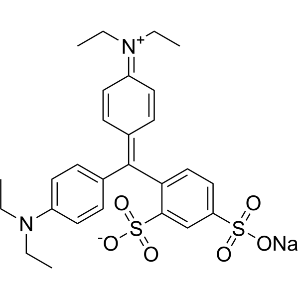
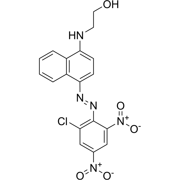
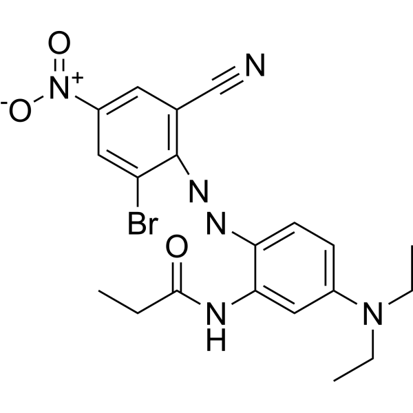
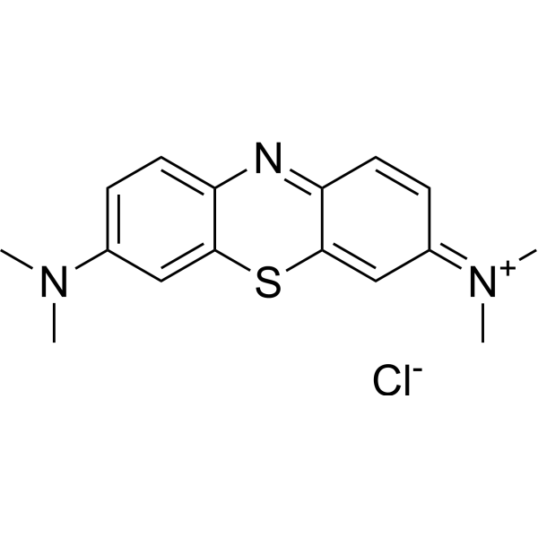
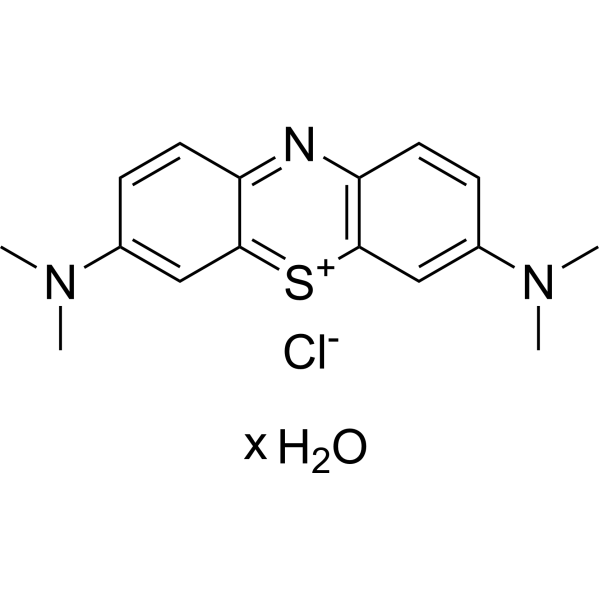

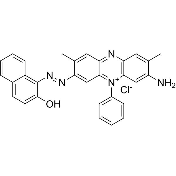
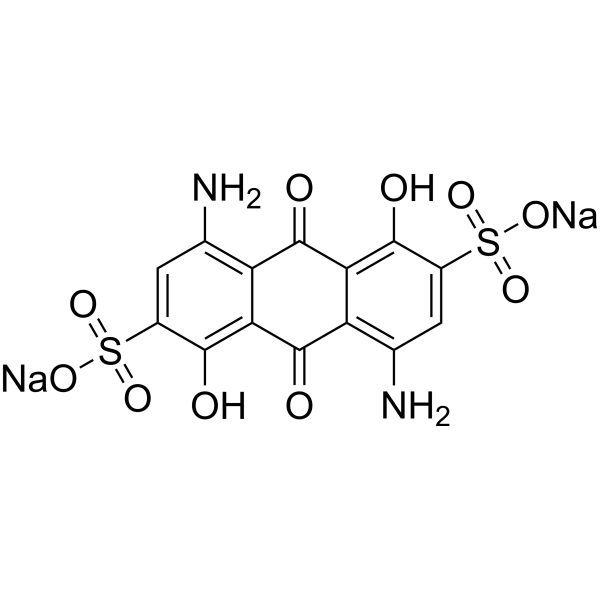
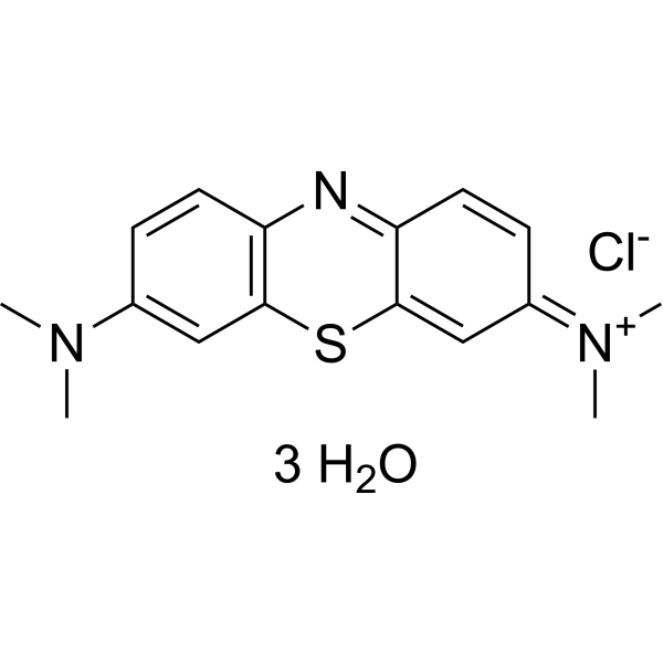
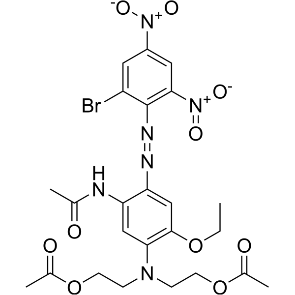
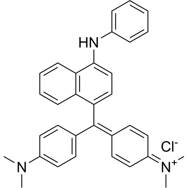
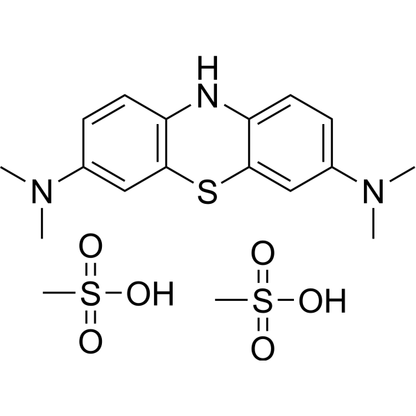
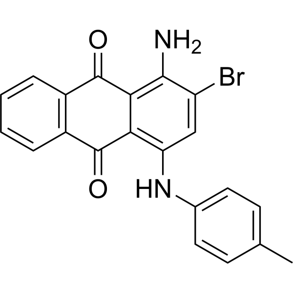

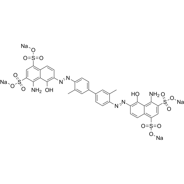
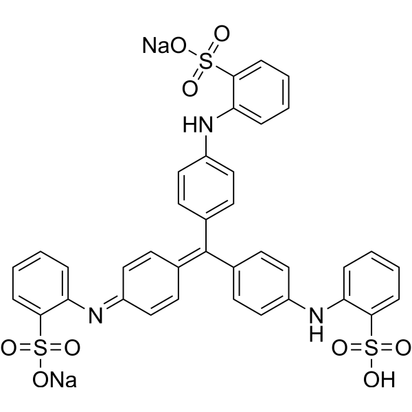
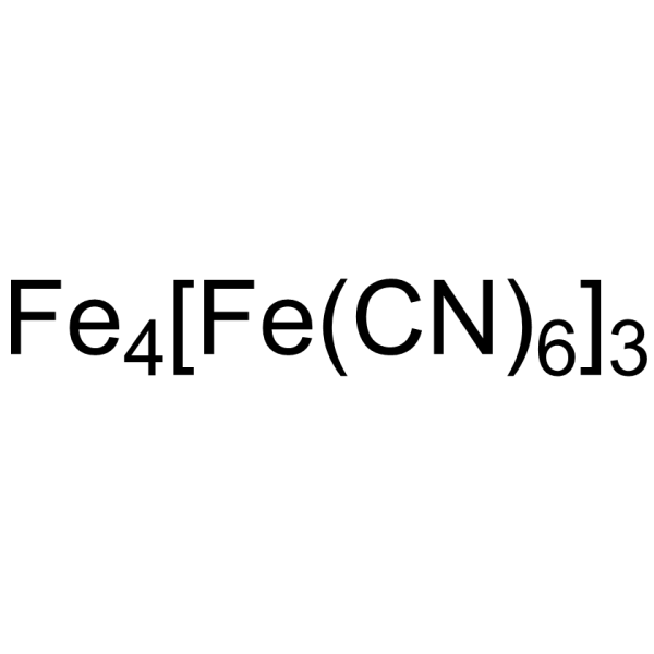
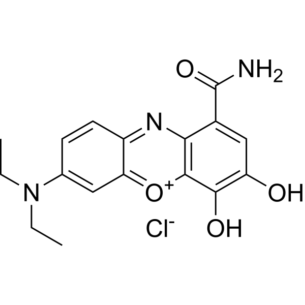


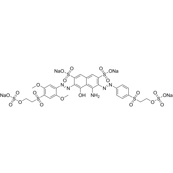
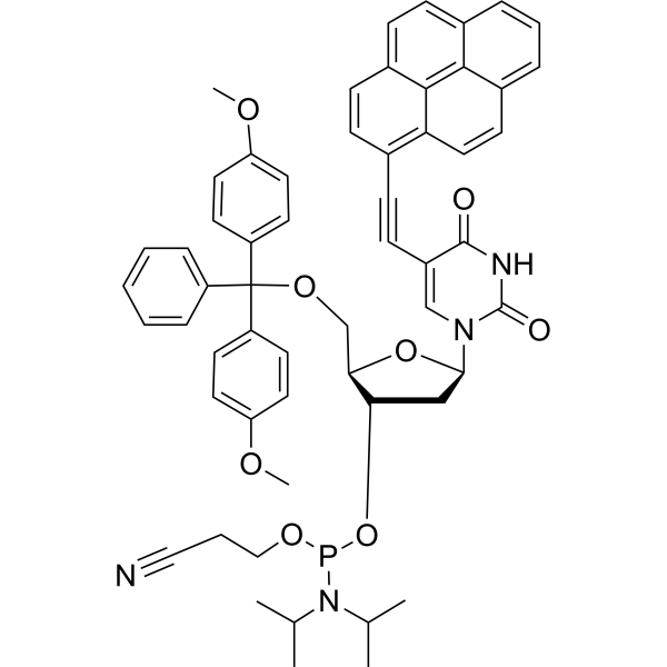
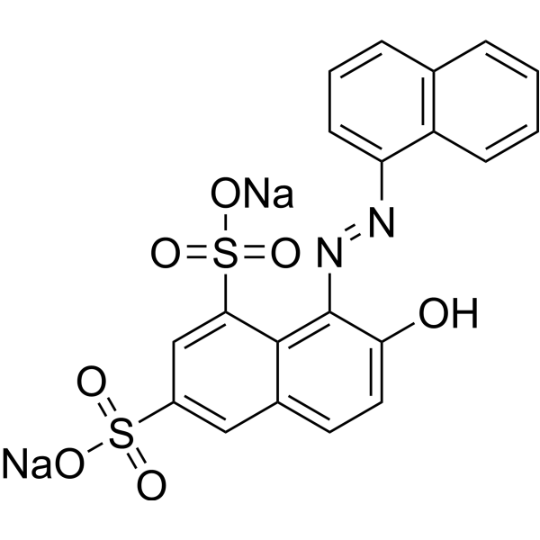

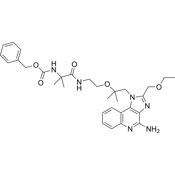
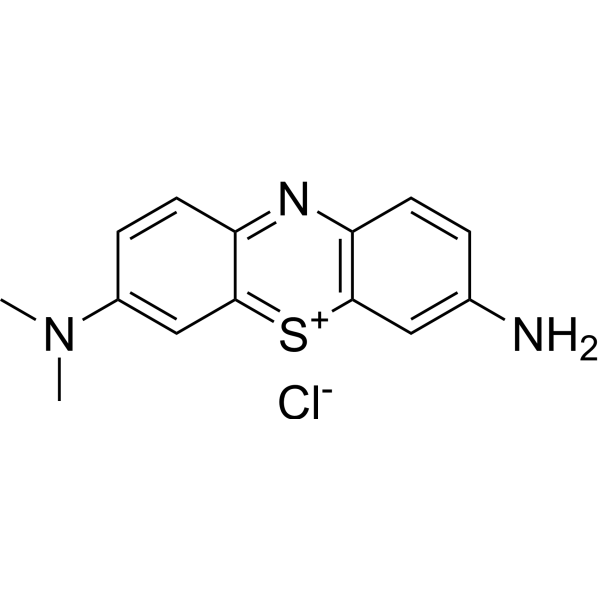
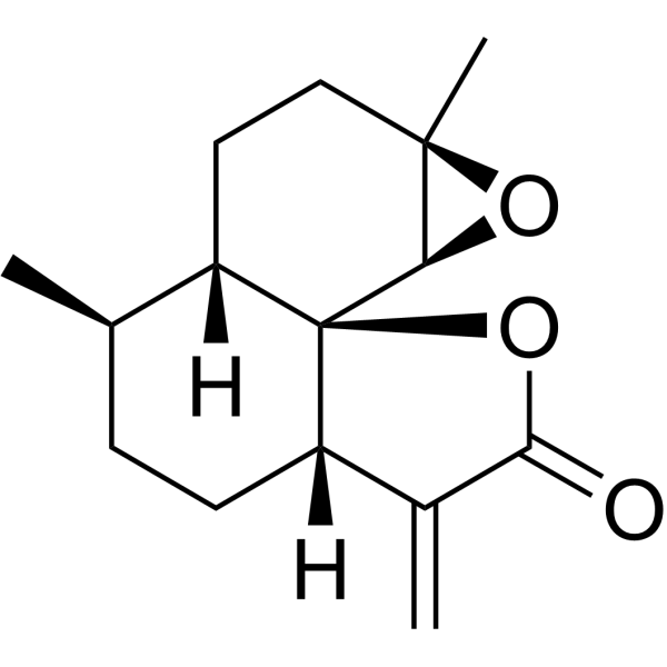


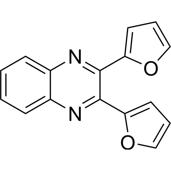

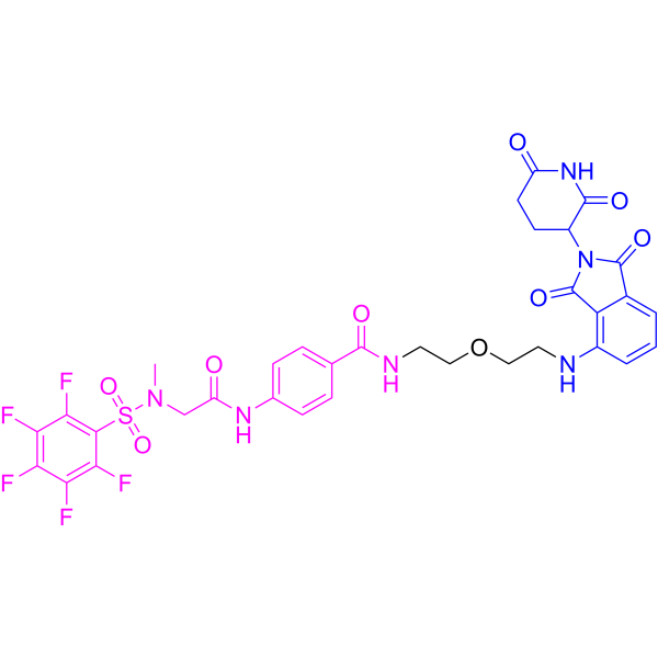

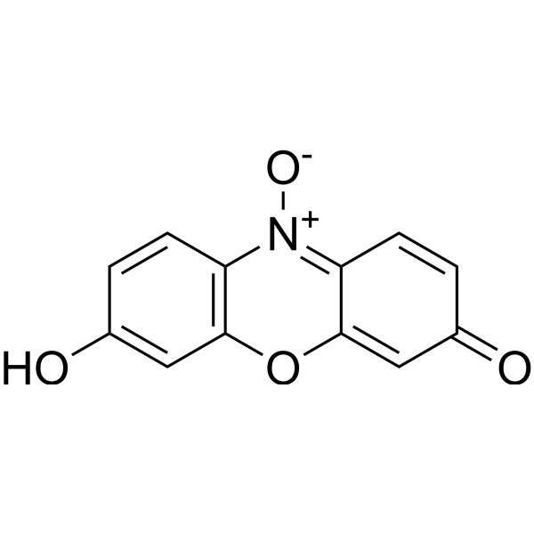
![Kaempferol-3-O-[2″,6″-di-O-E-p-coumaroyl]-β-D-glucopyranoside](http://file.medchemexpress.com/product_pic/hy-n10784.gif)