From 11:00 pm to 12:00 pm EST ( 8:00 pm to 9:00 pm PST ) on January 6th, the website will be under maintenance. We are sorry for the inconvenience. Please arrange your schedule properly.
Aging Without Dying -- The secret of longevity interpreted by high score literature
Aging, the inexorable destination of the passage of time, is a spontaneous process in many living organisms characterized by structural degeneration and decline of physiological functions.
Aging increases the risk of malignant or chronic diseases such as cancer, diabetes, and cardiovascular disease and reduces the ability to defend against external harm. For example, due to the weakened function of the immune system, the transmission of the initial warning signal of SARS-CoV-2 virus after infection is slow, and the virus is prone to a rapid proliferation in the body (Fig. 1). Therefore, the symptoms of COVID-19 infection are more severe in the elderly[1]. Understanding the mechanism of aging not only has guiding significance for prolonging human life but also has important clinical significance for the prevention and treatment of diseases in the elderly population, thus, improving their life quality and well-being.
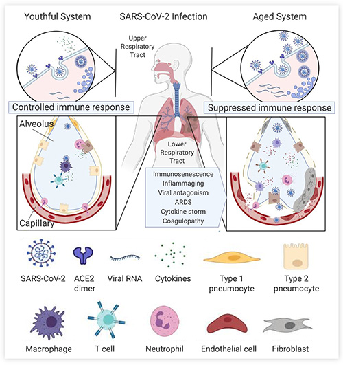
Fig. 1 Immune system clearance of SARS-CoV-2 in young and old adults[1].
After the SARS-CoV-2 virus is endocytosed in the airway epithelial cells of the upper respiratory tract, the virus spreads to the alveoli and can be recognized by alveolar macrophages or dendritic cells, resulting in the immune response. However, in the aged immune system, the viral alarm signals are initially slow, hereby leading to massive virus replication. Defective macrophages and T cells with limited receptors are less effective at killing infected cells (lower right), and more cells were infected, triggering a storm of high levels of inflammatory cytokines.
Over the past 30 years, scientists in the field of aging studies have shifted the focus from identifying the aging phenotypes to the genetic pathways that regulate the phenotypes. Researchers have identified multiple genes and signal pathways related to aging, including the nutritional sense-related mTOR pathway, insulin-like signaling pathway, mitochondria, and oxidative stress pathways etc.[2]. Carlos Lopez-Otin et al. summarized nine common features of aging (Fig. 2): genomic instability, telomere depletion, epigenetic changes, loss of protein homeostasis, nutrient dysregulation, mitochondrial dysfunction, cellular senescence, stem cell failure, and altered intercellular communication[3]. These studies on aging characteristics and related pathways are of great significance for developing anti-aging drugs. For example, Metformin , which is known to regulate mitochondrial function, can play an anti-aging role by inhibiting mitochondrial respiratory chain complex I, reactive oxygen species (ROS), and pro-inflammatory cytokines.
Fig. 2 The hallmarks of aging: genomic instability, telomere attrition, epigenetic alterations, loss of proteostasis, deregulated nutrient sensing, mitochondrial dysfunction, cellular senescence, stem cell exhaustion, and altered intercellular communication[3].
Aging is inevitable for everyone. With time, the definition of "old" keeps updating constantly, and aging research keeps moving. Let's see what discoveries have had recently.
Longevity and Anti-aging
· Longevity and low activity ribosome pathways
Ribosome and protein biosynthesis are highly energy-demanding cellular processes. Under-expression of some related genes could reduce the synthesis and save energy, suggesting that some disease-inducing genes are not overexpressed. Ribosomal pathway inhibition could extend the lifespan in some model organisms as demonstrated in other species models. For example, inhibition of mitochondrial ribosomal genes MRPs (Mitochondrial Ribosomal Proteins) by RNAi extended the lifespan of C. elegans[7].
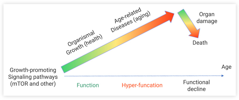
Fig. 3 Schematic diagram of cellular hyperactivity theory[5]
Aging is also considered to be caused by cellular hyperfunction. Excessive activation of some pathways, such as mTOR pathway, is more likely to cause loss of function in certain cells and organs, leading to diseases or even death (Fig. 3)[5]. Lifespan is correlated with health status. The occurrence of the age-related diseases (e.g., cardiovascular and neurodegenerative diseases) is often delayed in healthy older adults.
To find the genetic root of longevity, the team led by Dr. Qingpeng Kong from the Kunming Institute of Zoology, Chinese Academy of Sciences, analyzed the transcriptome of peripheral leukocytes of 185 long-lived elderly females (98.9 ± 3.8 years old) and their 86 offsprings (57.4 ± 9.0 years old) from Lingshui and Linggao county, Hainan Province, China (their offsprings were chosen because of their similar living conditions which would minimize variations on the living environment). The results showed that the ETS1 (ETS proto-oncogene 1) gene and ribosome pathway regulation-related genes were significantly down-regulated in the long-lived elderly, which may be one of the reasons for the prolonged life of the long-lived elderly. The work was published in Science Advances[6].
Kong team found that the ribosome-pathway-related genes, particularly the ribosomal protein genes (RPGs) were significantly down-regulated in these long-lived elderly females. Among the down-regulated RPG promoters, the TF motifs with the highest score involved the ETS family, ETS1 was the one that showed the most significant difference in expression. The ETS1 gene is positively co-expressed with RPGs (positively co-express) therefore its low expression can down-regulate RPGs. The ETS1 gene has been identified as a potential key transcription factor for RPG transcription in long-lived elders.
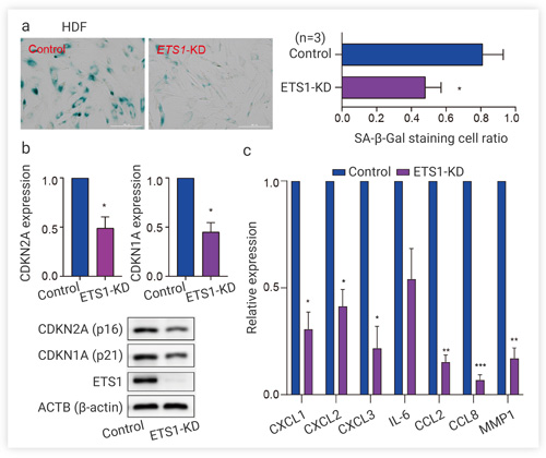
Through siRNA knockdown of ETS1 in human dermal fibroblasts (HDF) and human embryonic lung fibroblasts (IMR-90), the researchers verified whether ETS1 downregulation could slow down aging. The results showed that low expression of ETS1 significantly decreased the proportion of the senescent cells stained with β-galactosidase (SA-β-Gal) (Fig. 4a) and reduced the expression of CDKN2A (p16) and CDKN1A (p21) (Fig. 4b) (SA-β-Gal is one of the most widely used markers for cell aging; CDKN2A and CDKN1A are two important regulators of senescence). In addition, the expression of senescence-associated secretory phenotype (SASP) genes (including CXCL1, CXCL1, IL-6, etc.) was decreased (Fig. 4c). RNA-seq were performed on ETS1-knockdown HDFs and controls, and the results showed that 60 of the 61 differentially expressed RPG were significantly down-regulated in ETS1-knockdown HDF cells.
Fig. 4 Functional study of down-regulated ETS1 at cellular level[6]
a. Senescent cells (β-galactosidase staining) were significantly reduced in ETS1 knockdown cells; b. The expression of CDKN2A (p16) and CDKN1A (p21) were significantly decreased in ETS1 knockdown cells; c. The expression of the SASP gene was decreased considerably in ETS1 knockdown cells
These results suggest that there is a decrease in ribosome biosynthesis caused by the downregulation of ETS1 in or at least in, some of the long-lived elderly females. Energy saving through protein synthesis reduction can be a conserved longevity mechanism in various species, including human[6].
· Anti-aging and BDNF-TrkB pathway
Cell senescence is a state in which cells are in a stable cell cycle arresting. The uncontrolled senescent cell accumulation in organs may lead to tissue dysfunction and disease. Therefore, removing harmful senescent cells from tissues and organs has become a new strategy for treating aging-related diseases.
Carlos Anerillas et al. found that the BDNF-TrkB pathway was associated with anti-aging. They proved that autocrine/paracrine activating of TrkB by BDNF maintained senescent cell viability. and that TrkB inhibitors induced senescent cell apoptosis in mice, thereby reducing the level of liver aging markers. These research results were published in Nature Communications on October 20, 2022[8].
Tips: the Trk family, encoded by the NTRK gene, plays a vital role in the development of the nervous system. Trk receptors are activated by a family of secreted ligands called neurotrophins, including NGF, NTF3, NTF4, and BDNF 15, which are essential for neuronal function.
The researchers first screened a library of 43 inhibitors of the MAPK signaling cascade mediators, using β-galactosidase as an indicator, to identify compounds that killed senescent cells but did not affect the viability of proliferating cells (non-senescent cells). The results showed that the TrkB receptor inhibitors GNF 5837 and ANA 12 had the best performance. Then they investigated the role of the Trk pathway in aging. By immunofluorescence detection, TrkB was found to be very abundant in the aging cell membrane. Increasing the level of TrkB can enhance the resistance of senescent cells to apoptosis. The BDNF mRNA, the most important ligand of TrkB, also sustained to increase. TrkB and BDNF levels increased significantly after two days of induced senescence and silencing TrkB or BDNF increased senescent cell death.
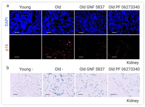
The researchers injected TrkB inhibitors GNF 5837, ANA 12, and PF 06273340 into naturally aged mice (21 months of age). The results showed that GNF 5837 and PF 06273340 significantly reduced the elevated levels of aging markers p16 (Fig. 5a), p21, and β-galactosidase (Fig. 5b) in the kidney, lung, and liver of the aged mice. These results suggest that the TrkB inhibitors GNF 5837 and PF 06273340 may alleviate the functional decline in senescent organs by reducing the accumulation of senescent cells.
Fig. 5 The expression levels of p16 (a) β-galactosidase (b) in the kidney of naturally aged mice were significantly decreased after injection of GNF 5837 and PF 06273340[8].
Study of Aging Mechanisms
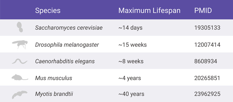
Whether studying aging phenotypes or the mechanisms behind the agingthem, researchers usually investigated it in vivo and in vitro, using model animals (Table 1) or cells to simulate human aging processes and phenotypes.
Table 1. Common model animals in aging studies[9].
For the aging study, the first step is to induce aging in cells or animal models. Various antibiotics (such as Etoposide[8]), irradiation (causing DNA damage), hydrogen peroxide (H2O2)[8], etc., can be used in cells for that purpose. Irradiation[10], antibiotics (e.g., Trametinib[10]), D-galactose[11], etc., can also be used in animals. Besides, genes knockdown or knock-out is a more powerful tool for verifying a gene’s function on aging. For example, the Kong team used siRNA to knockdown EST1 to verify the gene’s effect on aging[6].
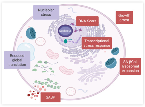
Detection senescence indicator is a straight way to prove senescence in cells or animals. During cell aging, DNA, proteins, secreted factors, and morphology all changes (Fig. 6), including cell size enlargement, increased expression of cyclin-dependent kinase (CDK) inhibitors (such as p16 and p21), lysosome expansion, and a large number of pro-inflammatory factors secretion[12]. These substances can be used as markers for the detection. The commonly used markers include β-galactosidase, p53, p21, p16, SASP, Rb, etc.. Other detection methods include phenotypic observation[10], and transcriptome or metabolome sequencing[8].
Fig. 6 Intracellular changes during cell senescence[12].
DNA damage, cell proliferation arrest, expression of stress response proteins (p16, Rb, p53, and p21), lysosome expansion, and β-galactosidase staining were positive at pH 6. The production and secretion of SASP increased
Including 190+ natural products with validated anti-aging activity, which is a useful tool for the study of aging-related diseases drugs and pharmacology.
Including 1100+ compounds that act as antioxidants for high throughput screening (HTS) and high content screening (HCS). This library is a useful tool for discovery new antioxidants and oxidative stress research.
Metformin inhibits the mitochondrial respiratory chain in the liver, leading to activation of AMPK, enhancing insulin sensitivity for type 2 diabetes research. Metformin can cross the blood-brain barrier and triggers autophagy.
Rapamycin is a potent and specific mTOR inhibitor with an IC50 of 0.1 nM in HEK293 cells. Rapamycin binds to FKBP12 and specifically acts as an allosteric inhibitor of mTORC1. Rapamycin is an autophagy activator, an immunosuppressant.
As the situation with COVID-19 continues to unfold in every community, MedChemExpress (MCE) is responding to the uncertainty caused by this outbreak thoughtfully and cautiously.
Your need for high quality reagent doesn't stop during difficult times, and neither do we.
We're doing our best to keep everyone healthy and safe in the workplace while also avoiding the interruptions to our day-to-day operations.
If you need to change the delivery plan for items ordered, please contact us via email [email protected].
Thank you for being a loyal MedChemExpress (MCE) customer, we are here to assist you as needed.