1. PI3K/Akt/mTOR
  2. MELK
  3. OTSSP167 hydrochloride

OTSSP167 hydrochloride  (Synonyms: OTS167 hydrochloride)

Cat. No.: HY-15512A Purity: 99.84%
COA Handling Instructions

OTSSP167 (OTS167) hydrochloride is a highly potent and ATP-competitive MELK inhibitor with IC50 value of 0.41 nM.

For research use only. We do not sell to patients.

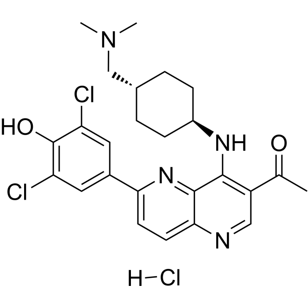
OTSSP167 hydrochloride Chemical Structure

OTSSP167 hydrochloride Chemical Structure

CAS No. : 1431698-10-0

Size Price Stock Quantity
Free Sample (0.1 - 0.5 mg)   Apply Now  
Solution
10 mM * 1 mL in DMSO USD 115 In-stock
Estimated Time of Arrival: December 31
Solid + Solvent
10 mM * 1 mL
ready for reconstitution
USD 115 In-stock
Estimated Time of Arrival: December 31
Solid
5 mg USD 100 In-stock
Estimated Time of Arrival: December 31
10 mg USD 150 In-stock
Estimated Time of Arrival: December 31
25 mg USD 250 In-stock
Estimated Time of Arrival: December 31
50 mg USD 450 In-stock
Estimated Time of Arrival: December 31
100 mg USD 750 In-stock
Estimated Time of Arrival: December 31
200 mg   Get quote  
500 mg   Get quote  

* Please select Quantity before adding items.

Customer Review

Based on 26 publication(s) in Google Scholar

Other Forms of OTSSP167 hydrochloride:

Top Publications Citing Use of Products

    OTSSP167 hydrochloride purchased from MCE. Usage Cited in: Blood Cancer J. 2019 Nov 18;9(12):87.  [Abstract]

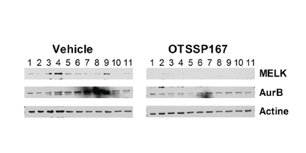
    Expression of MELK and Aurora B kinase (AurB) is determined in samples from vehicle and OTSSP167-treated mice. β-actin is used as loading control.

    OTSSP167 hydrochloride purchased from MCE. Usage Cited in: Clin Cancer Res. 2018 Nov 15;24(22):5645-5657.  [Abstract]

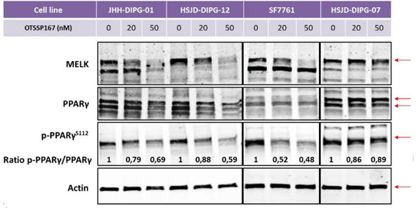
    Western blot showing protein levels of MELK, PPARγ and phospho-PPARγS112 in JHH-DIPG-01, HSJD-DIPG-12, SF7761 and HSJD-DIPG-07 cells after 48 hours treatment with either 20 nM or 50 nM OTSSP167.

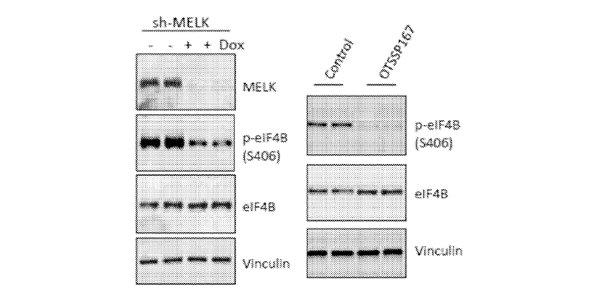
    OTSSP167 hydrochloride purchased from MCE. Usage Cited in: EMBO Mol Med. 2018 Mar;10(3). pii: e8274.  [Abstract]

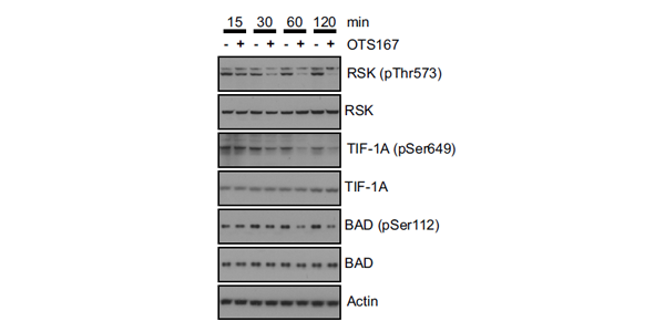
    Validation of effects of OTS167 on phosphorylation of p90RSK and its targets. C4-2b cells are treated with vehicle or 30 nM OTS167 for the indicated times, and levels of total and phosphorylated proteins are determined by Western blot analysis. β-Actin is used as a loading control.

    OTSSP167 hydrochloride purchased from MCE. Usage Cited in: Gynecol Oncol. 2017 Apr;145(1):159-166.  [Abstract]

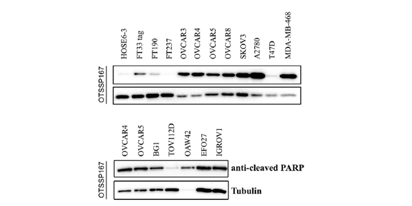
    Western blotting demonstrates that 100 nM OTSSP167 for 48 h produces marked PARP-1 cleavage in HGSOC cell lines whereas no or weak PARP-1 cleavage is observed in normal ovarian surface epithelial cells (HOSE6-3) and in the fallopian tubal cells (FT33-tag, FT190, FT237).

    OTSSP167 hydrochloride purchased from MCE. Usage Cited in: Sci Rep. 2017 Feb 14;7:42502.  [Abstract]

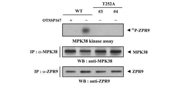
    In vitro kinase assays are performed using MPK38 immunoprecipitates, which are obtained from cell lysates of wild-type and clonal CRISPR/Cas9 ZPR9 (T252A) KI isolates, treated with (+) or without (−) OTSSP167 (1 μM, 2 h), a MPK38-specific inhibitor, in the presence of ZPR9 immunoprecipitates as the substrate. Recombinant MPK38 proteins (lower panels), which were expressed in bacterial cells, treated with (+) or without (−) OTSSP167 are also used instead of the MPK38 immunoprecipitates (upper pan

    OTSSP167 hydrochloride purchased from MCE. Usage Cited in: PLoS One. 2017 Feb 24;12(2):e0172832.  [Abstract]

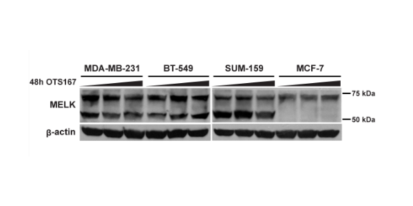
    OTS167 reduces proliferation and regulates MELK transcript but not protein levels in TNBC cells. The MELK 52 kDa variant is highly expressed in the TNBC cell lines but not in luminal A MCF-7.

    OTSSP167 hydrochloride purchased from MCE. Usage Cited in: Oncotarget. 2017 Dec 20;9(2):2591-2602.  [Abstract]

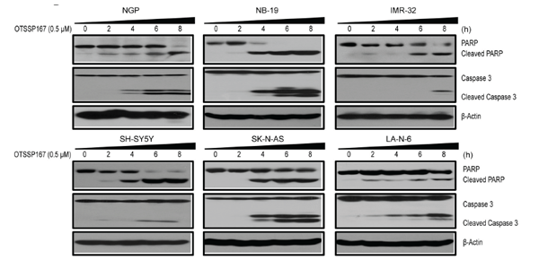
    Effects of OTSSP167 on PARP and Caspase-3 cleavages in NB cells are shown by immunoblotting assay.

    OTSSP167 hydrochloride purchased from MCE. Usage Cited in: Patent. US20160291017A1.

    Mitotic MDA-MB-468 cells are treated for 30 min with vehicle or 200 nM OTSSP167, a MELK inhibitor. Lysates are used for immunoblotting.

    OTSSP167 hydrochloride purchased from MCE. Usage Cited in: Mol Cancer. 2014 May 4;13:100.  [Abstract]

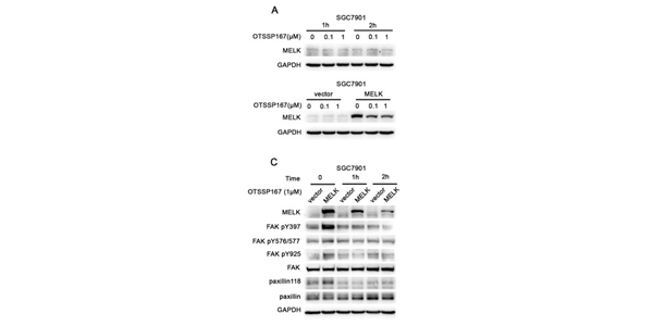
    MELK specific inhibitor OTSSP167 suppresses cell migration and invasion. A, Immunoblotting representing MELK expression in SGC7901 cell following OTSSP167 treatment for indicated period. In the upper panel, SGC7901 cell are treated with different concentration (0, 0.1, 1 μM) of OTSSP167 for 1 h and 2 h. In the lower panel, SGC7901/vector and SGC7901/MELK cell are treated with 1 μM OTSSP167 for 2 h. C, SGC7901/vector and SGC7901/MELK cells are treated with 1 μM OTSSP167 for indicated period and
    • Biological Activity

    • Protocol

    • Purity & Documentation

    • References

    • Customer Review

    Description

    OTSSP167 (OTS167) hydrochloride is a highly potent and ATP-competitive MELK inhibitor with IC50 value of 0.41 nM.

    IC50 & Target

    IC50: 0.41 nM (MELK)

    In Vitro

    OTSSP167 inhibits the growth of A549 (lung), T47D (breast), DU4475 (breast), 22Rv1 (prostate) and HT1197 (bladder) cancer cells with IC50 values of 6.7, 4.3, 2.3, 6.0 and 97 nM, respectively[1]. OTSSP167 can abrogate the mitotic checkpoint, disrupt MCC and MCC-APC/C interaction in MCF7 cells. OTSSP167 causes GFP-MELK localization to cell cortex in prometaphase cells[2]. OTSSP167 is a MELK selective inhibitor, exhibits a strong in vitro activity, conferring an IC50 of 0.41 nM[3].

    MCE has not independently confirmed the accuracy of these methods. They are for reference only.

    In Vivo

    OTSSP167 (20 mg/kg, i.v.) results in tumor growth inhibition (TGI) of 73% in xenograft mouse model; OTSSP167 (1, 5, and 10 mg/kg, p.o.) reveals TGI of 51, 91, and 108%, respectively. OTSSP167 (20 mg/kg, p.o.) shows no tumor growth suppressive effect on PC-14 xenografts[1].

    MCE has not independently confirmed the accuracy of these methods. They are for reference only.

    Molecular Weight

    523.88

    Appearance

    Solid

    Formula

    C25H29Cl3N4O2

    CAS No.
    SMILES

    ClC1=CC(C2=CC=C(N=CC(C(C)=O)=C3N[[email protected]]4CC[[email protected]](CN(C)C)CC4)C3=N2)=CC(Cl)=C1O.[H]Cl

    Shipping

    Room temperature in continental US; may vary elsewhere.

    Storage

    4°C, sealed storage, away from moisture

    *In solvent : -80°C, 6 months; -20°C, 1 month (sealed storage, away from moisture)

    Solvent & Solubility
    In Vitro: 

    DMSO : 33.33 mg/mL (63.62 mM; Need ultrasonic)

    H2O : 7.14 mg/mL (13.63 mM; Need ultrasonic)

    Preparing
    Stock Solutions
    Concentration Solvent Mass 1 mg 5 mg 10 mg
    1 mM 1.9088 mL 9.5442 mL 19.0883 mL
    5 mM 0.3818 mL 1.9088 mL 3.8177 mL
    10 mM 0.1909 mL 0.9544 mL 1.9088 mL
    *Please refer to the solubility information to select the appropriate solvent.
    In Vivo:
    • 1.

      Add each solvent one by one:  10% DMSO    40% PEG300    5% Tween-80    45% saline

      Solubility: ≥ 3 mg/mL (5.73 mM); Clear solution

    • 2.

      Add each solvent one by one:  10% DMSO    90% (20% SBE-β-CD in saline)

      Solubility: ≥ 3 mg/mL (5.73 mM); Clear solution

    • 3.

      Add each solvent one by one:  10% DMSO    90% corn oil

      Solubility: 3 mg/mL (5.73 mM); Suspended solution; Need ultrasonic

    • 4.

      Add each solvent one by one:  PBS

      Solubility: 1 mg/mL (1.91 mM); Clear solution; Need ultrasonic

    *All of the co-solvents are available by MCE.
    Purity & Documentation

    Purity: 99.84%

    References
    Kinase Assay
    [1]

    For in vitro kinase assay, MELK recombinant protein (0.4 μg) is mixed with 5 μg of each substrate in 20 μL of kinase buffer containing 30 mM Tris-HCl (pH), 10 mM DTT, 40 mM NaF, 10 mM MgCl2, 0.1 mM EGTA with 50 μM cold-ATP and 10 Ci of [γ-32P]ATP for 30 min at 30°C. The reaction Is terminated by addition of SDS sample buffer and boiled for 5 min prior to SDS-PAGE. The gel is dried and autoradiographed with intensifying screens at room temperature. OTSSP167 (final concentration of 10 nM) is dissolved in DMSO and added to kinase buffer before the incubation.

    MCE has not independently confirmed the accuracy of these methods. They are for reference only.

    Cell Assay
    [1]

    In vitro cell viability is measured by the colorimetric assay using Cell Counting Kit-8. Cells are plated in 100 μL in 96-well plates at a density that generates continual linear growth (A549, 1×10³ cells; T47D, 3×10³ cells; DU4475, 4×10³ cells; 22Rv1, 6×10³ cells; and HT1197, 2×10³ cells, in 100 μL per well). The cells are allowed to adhere overnight before exposure to OTSSP167 for 72 hours at 37°C. Plates are read using a spectrophotometer at a wavelength of 450 nm. All assays are carried out in triplicate.

    MCE has not independently confirmed the accuracy of these methods. They are for reference only.

    Animal Administration
    [1]

    MDA-MB-231 cells are injected into the mammary fat pads of NOD.CB17-Prkdcscid/J mice. A549, MIAPaCa-2 and PC-14 cells (1×10⁵ cells) are injected subcutaneously in the left flank of female BALB/cSLC-nu/nu mice. DU145 cells are injected subcutaneously in the left flank of male BALB/cSLC-nu/nu mice. When MDA-MB-231, A549, DU145, MIAPaCa-2, and PC-14 xenografts has reached an average volume of 100, 210, 110, 250, and 250 mm3, respectively, animals are randomized into groups of 6 mice (except for PC-14, for which groups of 3 mice are used). For oral administration, OTSSP167 and other compounds are prepared in a vehicle of 0.5% methylcellulose and given by oral garbage at the indicated dose and schedule. For intravenous administration, compounds are formulated in 5% glucose and injected into the tail vein. An administration volume of 10 mL per kg of body weight is used for both administration routes. Tumor volumes are determined every other day using a caliper.

    MCE has not independently confirmed the accuracy of these methods. They are for reference only.

    References
    • No file chosen (Maximum size is: 1024 Kb)
    • If you have published this work, please enter the PubMed ID.
    • Your name will appear on the site.
    Help & FAQs
    • Do most proteins show cross-species activity?

      Species cross-reactivity must be investigated individually for each product. Many human cytokines will produce a nice response in mouse cell lines, and many mouse proteins will show activity on human cells. Other proteins may have a lower specific activity when used in the opposite species.

    • Molarity Calculator

    • Dilution Calculator
    The molarity calculator equation

    Mass (g) = Concentration (mol/L) × Volume (L) × Molecular Weight (g/mol)

    Mass   Concentration   Volume   Molecular Weight *
    = × ×

    The dilution calculator equation

    Concentration (start) × Volume (start) = Concentration (final) × Volume (final)

    This equation is commonly abbreviated as: C1V1 = C2V2

    Concentration (start) × Volume (start) = Concentration (final) × Volume (final)
    × = ×
    C1   V1   C2   V2

    Your Recently Viewed Products:

    Inquiry Online

    Your information is safe with us. * Required Fields.

    Product Name


    Salutation

    Applicant Name *


    Email address *

    Phone number *


    Organization name *

    Department *


    Requested quantity *

    Country or Region *


    Remarks

    Bulk Inquiry

    Inquiry Information

    Product Name:
    OTSSP167 hydrochloride
    Cat. No.:
    HY-15512A
    Quantity:
    MCE Japan Authorized Agent: