



Cathepsin B Protein, Human (HEK293, His)
Based on 2 publication(s) in Google Scholar
Cathepsin B Protein, Human (HEK293, His) functions in intracellular protein catabolism and in certain situations may also be involved in other physiological processes, such as processing of antigens in the immune response, hormone activation and bone turnover.
- Species: Human
- Source: HEK293
-
Storage:Stored at -20°C for 2 years from date of receipt. After reconstitution, it is stable at 4°C for 1 week or -20°C for longer (with carrier protein). It is recommended to freeze aliquots at -20°C or -80°C for extended storage.


To place orders, for customer services and technical support, please contact: MedChemExpress USA
Tel: 609-228-6898 E-mail: [email protected] [email protected]


-
 Biological Activity
-
Technical Parameters
-
Product Properties
- Documentation
- Help & FAQs


-

HY-P7754A
Cathepsin L1 Protein, Human (HEK293, His) -
HY-P7748A
Cathepsin D Protein, Human (HEK293, His) -
HY-P7756
Cathepsin S Protein, Human (HEK293, His) -

HY-15141
Staurosporine -
HY-15917
DL-Dithiothreitol -

HY-100558
Bafilomycin A1 -
HY-148905
p-Aminophenylmercuric acetate -
HY-D0858
MES -
HY-Y0873
PEG300 -

HY-P1645
Papain -
HY-P73296
MMP-2 Protein, Human (HEK293) -
HY-100410
FCCP


Biological Activity
Description
Cathepsin B Protein, Human (HEK293, His) functions in intracellular protein catabolism and in certain situations may also be involved in other physiological processes, such as processing of antigens in the immune response, hormone activation and bone turnover[1].
Background
Cathepsin B is a lysosomal cysteine protease of the papain family. It functions in intracellular protein catabolism and in certain situations may also be involved in other physiological processes, such as processing of antigens in the immune response, hormone activation and bone turnover. There is also evidence that cathepsin B is implicated in the pathology of chronic inflammatory diseases of airways and joints, and in cancer and pancreatitis[1].
Verified Bioactivity
Measured by its ability to cleave the fluorogenic peptide substrate Z-LR-AMC. Read at excitation and emission wavelengths of 380 nm and 460 nm. The specific activity is 6500-20000 pmol/min/µg, as measured under the described conditions. (Activation description: The proenzyme needs to be activated in acid reducing buffer for an activated form.)
MCE Validation Data
-
Purity - SDS-PAGE
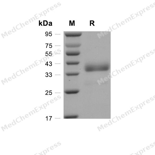
Purity - SDS-PAGE
-
Bioactivity - Enzymatic Activity Assay
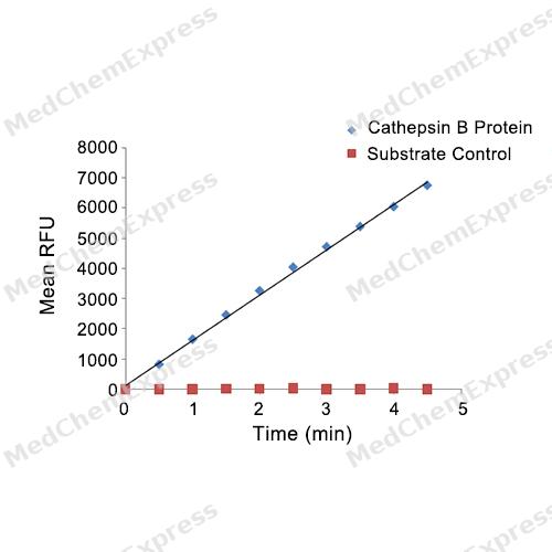
Bioactivity - Enzymatic Activity Assay

Assay Procedure
Materials
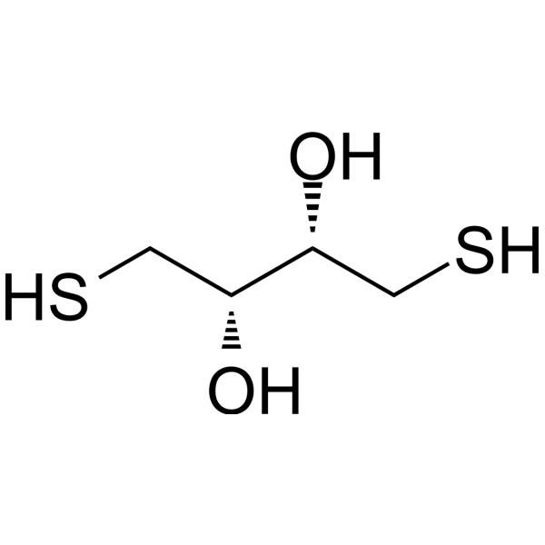
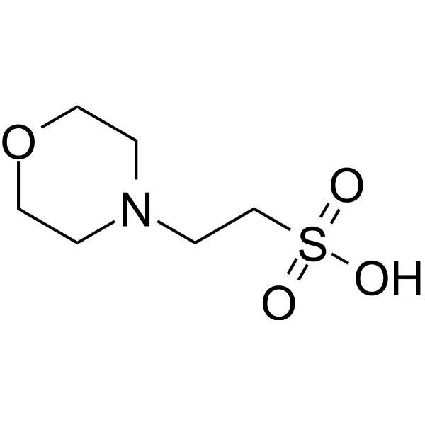
Activation buffer: 25 mM MES, 5 mM DTT, pH 5.0
Assay buffer: 25 mM MES, 0.02% Brij-35, pH 5.0
Test protein: Cathepsin B Protein, Human (HEK293, C-His) (HY-P7993A)
Substrate: Z-Leu-Arg-AMC (HY-142021)
Standard: 7-amino, 4-Methyl Coumarin (AMC, HY-D0027)
Procedure
1. Standard Curve: Dilute AMC in assay buffer to concentrations of 0, 0.4, 0.8, 1.2, 1.6, 2, 6.25, 12.5, 25, 50, and 100 μmol/L, respectively. Add 100 μL of each dilution into black microplate wells. Set the excitation and emission wavelengths to 380 nm and 460 nm, respectively. Plot the standard curve with the measured RFU (Relative Fluorescence Units) on the x-axis and the amount of standard substance on the y-axis to obtain the standard curve equation.
2. Dilute the protein sample to 10 μg/mL in activation buffer.
3. Incubate at room temperature for 15 minutes.
4. Dilute Human Cathepsin B to 0.2 μg/mL in assay buffer.
5. Dilute the substrate to 20 μM in assay buffer.
6. Add 50 μL of each protein concentration into the assay wells and add 50 μL of 20 μM substrate to initiate the reaction. For the blank wells, add 50 μL of assay buffer and 50 μL of 20 μM substrate without protein.
7. Set the excitation and emission wavelengths to 380 nm and 460 nm, respectively. Read the fluorescence in kinetic mode for 5 minutes.
8. Calculate Specific Activity:
Specific Activity (pmol/min/μg) = | Adjusted Vmax* (RFU/min) x Conversion Factor (pmol/RFU) ** |
| amount of enzyme (μg) |
**Derived using calibration standard AMC.


Publications (2)
-
Journal Impact Factor
-
Most Recent
-
Adv Sci (Weinh)
Lysosomal Cathepsin S Escape Facilitates Near Infrared Light-Triggered Pyroptosis Via an Antibody-Indocyanine Green Conjugate. [Abstract]2025 Sep;12(34):e04851. PMID: 40539828 -


Technical Parameters
-
Species Human
-
Source HEK293
-
Tag C-6*His
-
Accession
P07858 (R18-I339)
-
Gene ID1508
-
Molecular Construction
-
N-term
-
Cathepsin B (R18-I339)
Accession # P07858 -
6*His
-
C-term
-
-
Protein Length
Full Length of Mature Protein
-
Synonyms
CTSB; Keratolytic Winter Erythema (Oudtshoorn Skin Disease); Cathepsin B; Epididymis Secretory Sperm Binding Protein; APP Secretase; Amyloid Precursor Protein Secretase; Cathepsin B1; Cysteine Protease; APPS; RECEUP; CPSB; KWE
-
AA Sequence
RSRPSFHPLSDELVNYVNKRNTTWQAGHNFYNVDMSYLKRLCGTFLGGPKPPQRVMFTEDLKLPASFDAREQWPQCPTIKEIRDQGSCGSCWAFGAVEAISDRICIHTNAHVSVEVSAEDLLTCCGSMCGDGCNGGYPAEAWNFWTRKGLVSGGLYESHVGCRPYSIPPCEHHVNGSRPPCTGEGDTPKCSKICEPGYSPTYKQDKHYGYNSYSVSNSEKDIMAEIYKNGPVEGAFSVYSDFLLYKSGVYQHVTGEMMGGHAIRILGWGVENGTPYWLVANSWNTDWGDNGFFKILRGQDHCGIESEVVAGIPRTDQYWEKI
-
Predicted Molecular Mass
36.7 kDa
-
Molecular Weight
Approximately 37-45 kDa, based on SDS-PAGE under reducing conditions, due to the glycosylation.
-
Glycosylation
Yes
-
Purity
≥ 95%, as determined by reducing SDS-PAGE.


Product Properties
Lyophilized powder
1.Lyophilized from a 0.22 μm filtered solution of PBS, pH 7.4.
2.Lyophilized from a 0.22 μm filtered solution of PBS, pH 7.4, 8% trehalose.
Please refer to the lot-specific COA for specific buffer information.
<1 EU/μg, determined by LAL method.
It is not recommended to reconstitute to a concentration less than 100 μg/mL in ddH2O. For long term storage it is recommended to add a carrier protein (0.1% BSA, 5% HSA, 10% FBS or 5% Trehalose).
Stored at -20°C for 2 years from date of receipt. After reconstitution, it is stable at 4°C for 1 week or -20°C for longer (with carrier protein). It is recommended to freeze aliquots at -20°C or -80°C for extended storage.
Room temperature in continental US; may vary elsewhere.


Documentation
-
Data Sheet (237 KB)
-
SDS (251 KB)
- English - EN (251 KB)
- Français - FR (251 KB)
- Deutsch - DE (251 KB)
- Norwegian - NO (251 KB)
- Español - ES (251 KB)
- Swedish - SV (251 KB)
- Italian - IT (251 KB)
- Korean - KR (251 KB)
- Portuguese - PT (251 KB)
-
Handling Instructions (2659 KB)

Calculators
Concentration (start) × Volume (start) = Concentration (final) × Volume (final)


 Powered by Bioz

