1. Signaling Pathways
  2. Others
  3. Fluorescent Dye

Fluorescent Dye

Fluorescence is a phenomenon of photoluminescence in luminescence. When an atom is irradiated by light, some electrons around the nucleus jump from their original orbitals to higher energy orbitals under the influence of light energy, i.e., from the ground state to the first or second excited single-line state, etc. Due to the instability of the first or second excited singlet state, the electron returns from the excited singlet state to the ground state accompanied by released energy, resulting in fluorescence.

Fluorescent dyes can be covalently bound or physically adsorbed in the structure of the substance. The fluorescence properties allow for the characterization, localization and quantitative analysis of the labeled object. Fluorescent dyes have shown great potential for the detection of proteins, nucleic acids, cells, and immunoassays, attributed to the advantages in non-radioactive contamination, simple experimental operation and easy observation.

Fluorescent dyes have penetrated many fields such as pharmacology, physiology, environmental science, and information science, as well as functional studies of proteins and drug screening.

Fluorescent Dye Related Products (1861):

Cat. No. Product Name Effect Purity Chemical Structure
  • HY-D1055
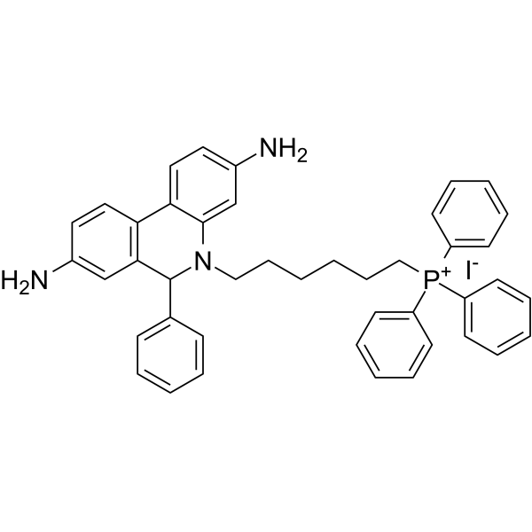
    MitoSOX Red
    MitoSOX Red is a live cell fluorescent probe that specifically targets mitochondria and is cell membrane permeable. MitoSOX Red enters mitochondria and is oxidized by superoxide but not by other ROS or RNS generating systems. The oxidized MitoSOX Red then binds to nucleic acids in mitochondria/nucleus, producing strong red fluorescence. MitoSOX Red can be used as a fluorescent indicator to specifically detect superoxide. In addition, superoxide dismutase (SOD) can prevent the oxidation of MitoSOX Red.
    Excitation/emission wavelength: 510/580 nm.
    MitoSOX Red
  • HY-W090090
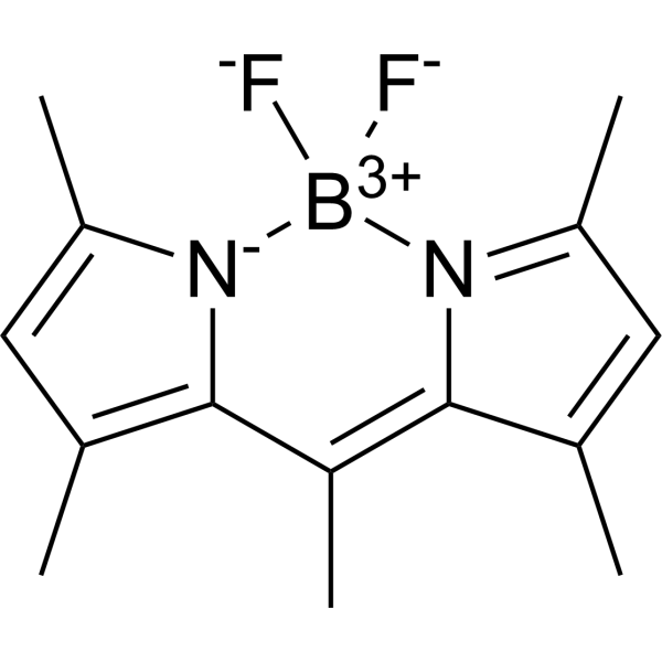
    BODIPY 493/503
    99.48%
    BODIPY493/503 is a BODIPY dye. BODIPY dye is a small molecule dye with strong ultraviolet absorption ability, its fluorescence peak is relatively sharp, and the quantum yield is high. They are relatively insensitive to the polarity and pH of the environment and are relatively stable under different physiological conditions. Due to its structural asymmetry, BODIPY derives a variety of structural products. BODIPY lipid droplet dyes can well pass through the cell membrane into the cell, and localize the polar lipids in the cell to specifically stain the lipid droplets, which can be used for labeling of live cells and fixed cells. Maximum excitation/emission wavelength: 493/503 nm.
    BODIPY 493/503
  • HY-12591B
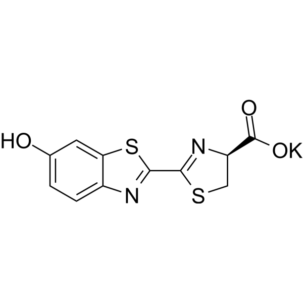
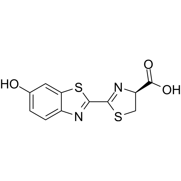
    D-Luciferin potassium
    99.97%
    D-luciferin is the natural substrate of the enzyme luciferase (Luc) that catalyzes the production of the typical yellowgreen light of fireflies. The 560 nm chemiluminescence from this reaction peaks within seconds, with light output that is proportional to luciferase concentration when the substrate luciferin is present in excess. The luciferase (luc) gene is a popular reporter gene for research and agent screening. Chemiluminescent techniques are virtually background-free, making the luc reporter gene ideal for detecting low-level gene expression. As little as 0.02 pg of luciferase can be reliably measured in a standard scintillation counter. In addition to its role as a reporter of gene expression, luciferase is commonly used in an extremely sensitive assay for ATP. We offer the firefly luciferase (HY-P1004), luciferin free acid (HY-12591A), as well as its water-soluble sodium salts (HY-12591) and potassium salts (HY-12591B) .
    D-Luciferin potassium
  • HY-D0718
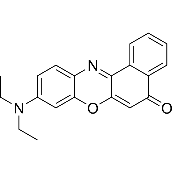
    Nile Red
    98.15%
    Nile red (Nile blue oxazone) is a lipophilic stain. Nile red has environment-sensitive fluorescence. Nile red is intensely fluorescent in a lipid-rich environment while it has minimal fluorescence in aqueous media. Nile red is an excellent vital stain for the detection of intracellular lipid droplets by fluorescence microscopy and flow cytof uorometry. Nile red stains intracellular lipid droplets red. The fluorescence wavelength is 559/635 nm.
    Nile Red
  • HY-66019
    FITC
    98.60%
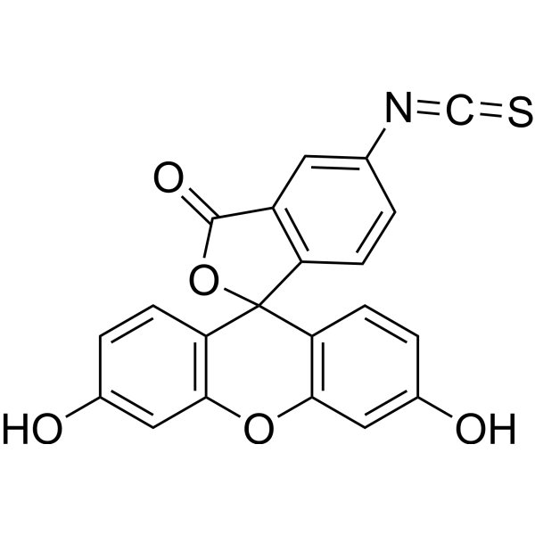
    FITC (Fluorescein Isothiocyanate), is one of the green fluorescein derivatives widely used in biology. FITC has the characteristics of high absorptivity, excellent fluorescence quantum yield and good water solubility. The isothiocyanate group of FITC can be combined with amino, sulfhydryl, imidazole, tyrosyl, carbonyl and other groups on the protein, so as to achieve protein labeling including antibodies and lectins. In addition to its use as a protein marker, FITC can also be used as a fluorescent protein tracer to rapidly identify pathogens by labeling antibodies, or for microsequencing of proteins and peptides (HPLC). The maximum excitation wavelength of FITC is 494 nm. Once excited, it fluoresces yellow-green at a maximum emission wavelength of 520 nm.
    FITC
  • HY-D1730A
    AF488 NHS ester TEA
    AF488 NHS ester TEA is an analog of AF488 NHS ester. AF488 NHS ester TEA is used in DNA sequencing research.
    AF488 NHS ester TEA
  • HY-D2119
    CQ-Lyso
    98.95%
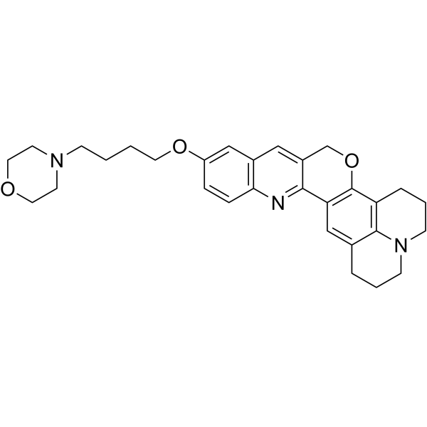
    CQ-Lyso is a lysosome-targeting chromenoquinoline. CQ-Lyso is a fluorescent probe for measuring the pH of lysosomes in living cells using only single wavelength excitation. CQ-Lyso can visualize the dynamic pH changes in lysosomes.
    CQ-Lyso
  • HY-D1063
    IR-780
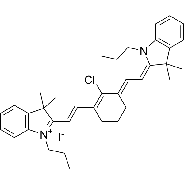
    IR780 is a prototypic NIR heptamethine cyanine agent, with a high molar extinction coefficient. The maximum excitation wavelength of IR780 was 777-780 nm, and the maximum emission wavelength was 798-823 nm. IR780 can be used in cancer PTT/PDT and imaging.
    IR-780
  • HY-15534
    JC-1
    99.0%
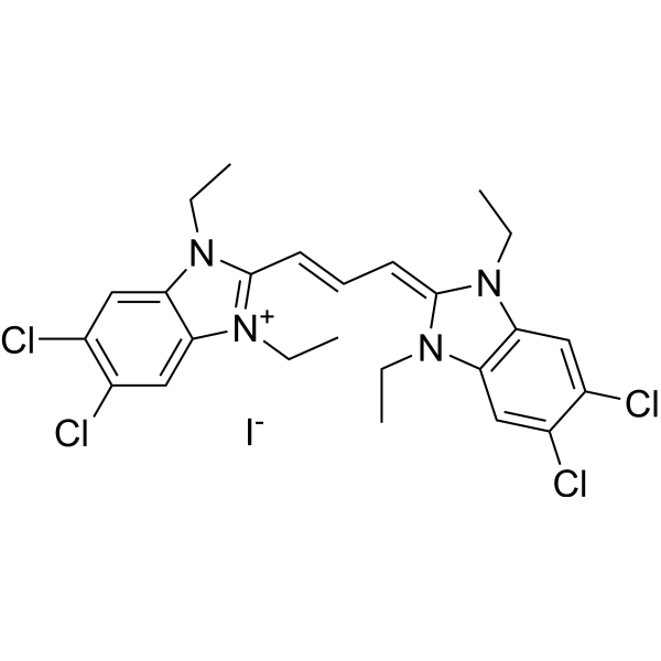
    JC-1 (CBIC2) is an ideal fluorescent probe widely used to detect mitochondrial membrane potential. JC-1 accumulates in mitochondria in a potential dependent manner and can be used to detect the membrane potential of cells, tissues or purified mitochondria. In normal mitochondria, JC-1 aggregates in the mitochondrial matrix to form a polymer, which emits strong red fluorescence (Ex=585nm, Em=590nm); When the mitochondrial membrane potential is low, JC-1 cannot aggregate in the matrix of mitochondria and produce green fluorescence (ex=514nm, em=529nm).
    JC-1
  • HY-D0815
    Propidium Iodide
    99.68%
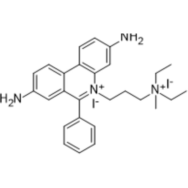
    Propidium Iodide (PI) is a nuclear staining agent that stains DNA. Propidium Iodide is an analogue of ethidine bromide that emits red fluorescence upon embedding in double-stranded DNA. Propidium Iodide cannot pass through living cell membranes, but it can pass through damaged cell membranes to stain the nucleus. Propidium Iodide has a fluorescence wavelength of 493/617 nm and a wavelength of 536/635 nm after Mosaic with DNA. Propidium Iodide is commonly used in the detection of apoptosis (apoptosis) or necrosis (necrosis), and is often used in flow cytometry analysis.
    Propidium Iodide
  • HY-D0938
    CFDA-SE
    99.01%
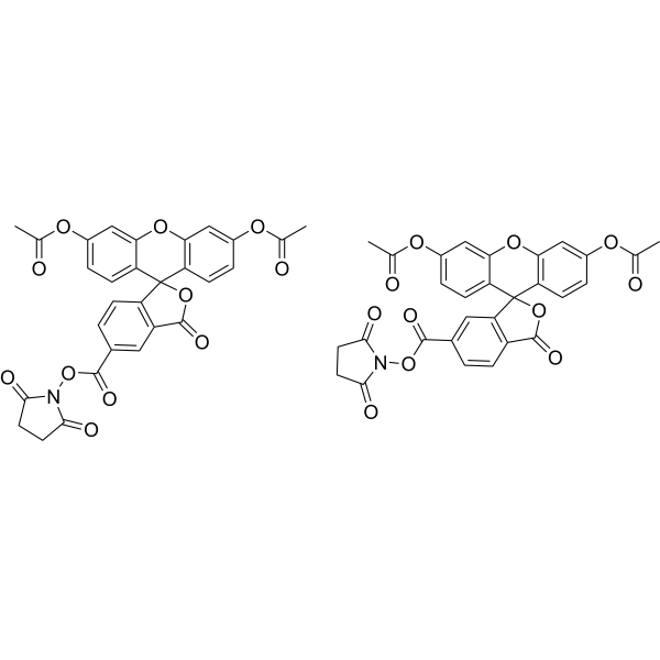
    CFDA-SE is a fluorescent dye that can penetrate the cell membrane. It can react with the free amine group in the cytoskeleton protein inside the cell, and finally form a protein complex with fluorescence. After entering the cell, CFDA-SE locates in the cell membrane, cytoplasm and nucleus, and the fluorescence staining is strongest in the nucleus. CFDA-SE dye can be uniformly inherited by the cells with cell division and proliferation, and its attenuation is proportional to the number of cell divisions. This phenomenon can be detected and analyzed by flow cytometry under the excitation light of 488 nm, and can be used to detect the proliferation of cells.
    CFDA-SE
  • HY-128868A
    FITC-Dextran (MW 4000)
    FITC-Dextran (MW 4000) is a fluorescent probe for fluorescein isothiocyanate (FITC) dextran (Ex=495 nm; Em=525 nm). FITC-Dextran (MW 4000) can be used as a marker to reveal heat shock-induced cell damage and to study the early and late stages of apoptosis. FITC-Dextran (MW 4000) can also be used for cell permeability studies, such as blood-brain barrier permeability and determination of the extent of blood-brain barrier disruption.
    FITC-Dextran (MW 4000)
  • HY-D1048
    DiR
    98.20%
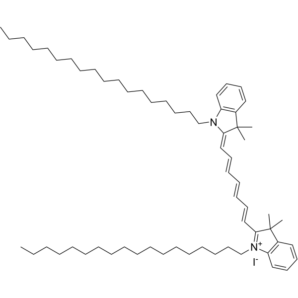
    DiR is a long-chain carbocyanine dye. Carbocyanine dyes are widely used as Di to label cells, organelles, liposomes, viruses and lipoproteins.
    DiR
  • HY-D0079
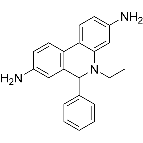
    Dihydroethidium
    Dihydroethidium, also known as DHE, is a peroxide indicator. Dihydroethidium penetrates cell membranes to form a fluorescent protein complex with blue fluoresces. After entering the cells, Dihydroethidium is mainly localized in the cell membrane, cytoplasm and nucleus, and the staining effect is the strongest in the nucleus. Dihydroethidium produces inherent blue fluorescence with a maximum excitation wavelength of 370 nm and a maximum emission wavelength of 420 nm; after dehydrogenation, Dihydroethidium combines with RNA or DNA to produce red fluorescence with a maximum excitation wavelength of 300 nm and a maximum emission wavelength of 610 nm. 535 nm can also be used as the excitation wavelength for actual observation.
    Dihydroethidium
  • HY-116215
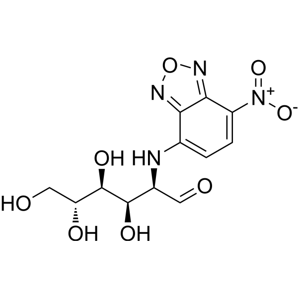
    2-NBDG
    98.92%
    2-NBDG is a fluorescently-labeled deoxyglucose analog that is used primarily to directly monitor glucose uptake by living cells and tissues. It is also used as a topical contrast reagent for the detection of neoplasia. 2-NBDG can be used in real-time confocal, high-resolution, or wide-field fluorescence microscopy as well as in flow cytometry. The probe can be excited by the Argon laser at 488 nm to give the environment-sensitive fluorescence. It has lower photostability than the rhodamine-based fluorescent probes.
    2-NBDG
  • HY-128868
    FITC-Dextran (MW 10000)
    FITC-Dextran (MW 10000) is a fluorescent probe for fluorescein isothiocyanate (FITC) dextran (Ex=495 nm; Em=525 nm). FITC-Dextran (MW 10000) can be used as a marker to reveal heat shock-induced cell damage and to study the early and late stages of apoptosis. FITC-Dextran (MW 10000) can also be used for cell permeability studies, such as blood-brain barrier permeability and determination of the extent of blood-brain barrier disruption.
    FITC-Dextran (MW 10000)
  • HY-D0041
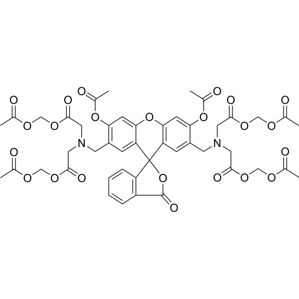
    Calcein-AM
    ≥98.0%
    Calcein AM, has cell membrane permeability and can easily enter the cell. Calcein AM has no fluorescence and is hydrolyzed by endogenous esterase in the cell to produce polar molecule Calcein (Calcein), which has strong negative charge and cannot permeate the cell membrane. Calcein can emit strong green fluorescence, so it is often used with Propidium Iodide for cell viability/virulence detection, excitation/emission wavelength: 494/515 nm.
    Calcein-AM
  • HY-12591A
    D-Luciferin
    99.87%
    D-luciferin is the natural substrate of the enzyme luciferase (Luc) that catalyzes the production of the typical yellowgreen light of fireflies. The 560 nm chemiluminescence from this reaction peaks within seconds, with light output that is proportional to luciferase concentration when the substrate luciferin is present in excess. The luciferase (luc) gene is a popular reporter gene for research and agent screening. Chemiluminescent techniques are virtually background-free, making the luc reporter gene ideal for detecting low-level gene expression. As little as 0.02 pg of luciferase can be reliably measured in a standard scintillation counter. In addition to its role as a reporter of gene expression, luciferase is commonly used in an extremely sensitive assay for ATP. We of er the firefly luciferase (HY-P1004), luciferin free acid (HY-12591A), as well as its water-soluble sodium salts (HY-12591) and potassium salts (HY-12591B) .
    D-Luciferin
  • HY-15924
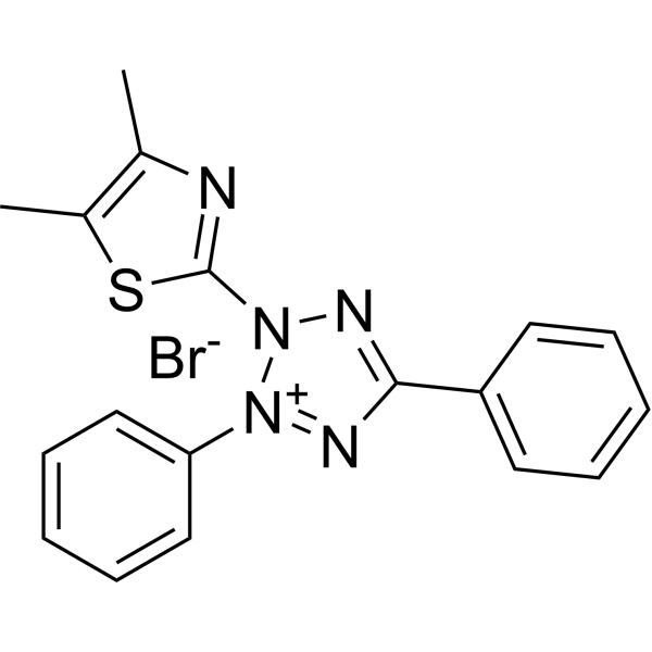
    Thiazolyl Blue
    99.84%
    Thiazolyl Blue (MTT) is a cell-permeable and positively charged tetrazolium dye that is used to detect reductive metabolism in cells. Thiazolyl Blue is taken up by cells through the plasma membrane and then reduced to formazan by intracellular NAD (P) H-oxidoreductases. Thiazolyl Blue is frequently used in colorimetric assays to measure cell proliferation, cytotoxicity, and apoptosis.
    Thiazolyl Blue
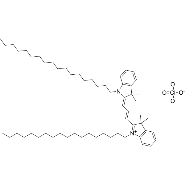
  • HY-D0083
    DiI
    99.92%
    DiI is a long-chain carbocyanine dye. Carbocyanine dyes are widely used as Di to label cells, organelles, liposomes, viruses and lipoproteins.
    DiI