


JC-1
Based on 217 publication(s) in Google Scholar
JC-1 (CBIC2) is an ideal fluorescent probe widely used to detect mitochondrial membrane potential. JC-1 accumulates in mitochondria in a potential dependent manner and can be used to detect the membrane potential of cells or purified mitochondria. In normal mitochondria, JC-1 aggregates in the mitochondrial matrix to form a polymer, which emits strong red fluorescence (Ex=585 nm, Em=590 nm); When the mitochondrial membrane potential is low, JC-1 cannot aggregate in the matrix of mitochondria and produce green fluorescence (ex=510 nm, em= 527 nm).
For research use only. We do not sell to patients.
- Purity: 98.05%
- CAS No.: 3520-43-2
- Formula: C25H27Cl4IN4
- Molecular Weight:652.23
-
Storage:
4°C, sealed storage, away from moisture and light
* In solvent : -80°C, 6 months; -20°C, 1 month (sealed storage, away from moisture and light)


-
 Biological Activity
-
Chemical Information
-
Solvent & Solubility
- Purity & Documentation
- References
-
 Help & FAQs


Publications Citing Use of MedChemExpress (MCE) JC-1
More- Mol Cancer. 2025 Jan 28;24(1):34. [Abstract]
- Mol Cancer. 2019 Apr 10;18(1):85. [Abstract]
- Adv Mater. 2025 Apr 28:e2500156. [Abstract]
- Adv Mater. 2025 Jan 10:e2411595. [Abstract]
- Bioact Mater. 2026 Jan 7:59:642-661. [Abstract]
- Bioact Mater. 2024 Apr 23:37:393-406. [Abstract]
- Bioact Mater. 2022 Aug 11;21:20-31. [Abstract]
- Adv Funct Mater. 2025 Oct 21.
- Cancer Res. 2023 Jul 5;83(13):2187-2207. [Abstract]
- ACS Nano. 2025 Aug 12;19(31):28353-28371. [Abstract]
- ACS Nano. 2025 Jan 21;19(2):2695-2714. [Abstract]
- ACS Nano. 2023 Sep 12;17(17):16432-16447. [Abstract]
- ACS Nano. 2023 Jul 25;17(14):13441-13460. [Abstract]
- ACS Nano. 2022 Apr 26;16(4):6064-6079. [Abstract]
- Nat Commun. 2026 Jan 27;17(1):2061. [Abstract]
- Nat Commun. 2025 May 10;16(1):4365. [Abstract]
- Cell Death Differ. 2021 Apr;28(4):1174-1192. [Abstract]
- Autophagy. 2021 Nov;17(11):3622-3643. [Abstract]
- Adv Sci (Weinh). 2024 Sep 23:e2403219. [Abstract]
- Adv Sci (Weinh). 2024 Jul 15:e2401527. [Abstract]
- Adv Sci (Weinh). 2023 Oct;10(30):e2303872. [Abstract]
- Chem Eng J. 2025 Dec 26;528:172183.
- Biomaterials. 2026 Jul:330:124002. [Abstract]
- Biomaterials. 2026 Feb:325:123616. [Abstract]
- Exp Mol Med. 2023 Mar;55(3):574-586. [Abstract]
- J Nanobiotechnology. 2026 Feb 16;24(1):259. [Abstract]
- J Nanobiotechnology. 2025 Mar 6;23(1):177. [Abstract]
- Sci Adv. 2025 Nov 21;11(47):eadz4126. [Abstract]
- J Biomed Sci. 2023 Dec 18;30(1):95. [Abstract]
- Small. 2021 Aug;17(32):e2101368. [Abstract]
- Small. 2021 Feb;17(7):e2005865. [Abstract]
- Small. 2019 Sep;15(36):e1902642. [Abstract]
- Mol Ther. 2025 Jun 4;33(6):2900-2912. [Abstract]
- Redox Biol. 2025 Dec 3:89:103961. [Abstract]
- Redox Biol. 2025 Aug 5:86:103806. [Abstract]
- Redox Biol. 2023 Aug:64:102783. [Abstract]
- J Control Release. 2024 Sep 13:375:269-284. [Abstract]
- J Hazard Mater. 2025 Sep 15:496:139407. [Abstract]
- J Hazard Mater. 2025 Jun 27:495:139088. [Abstract]
- Nano Today. 2025 Jun.
- Research (Wash D C). 2025 May 23:8:0703. [Abstract]
- Research (Wash D C). 2024 Jan 25:7:0306. [Abstract]
- Pharmacol Res. 2022 May;179:106123. [Abstract]
- Mater Today Bio. 2025 Sep 7;35:102295.
- Mater Today Bio. 2025 Apr 26:32:101802. [Abstract]
- Mater Today Bio. 2025 Mar 10:32:101649. [Abstract]
- Cell Mol Biol Lett. 2024 Aug 28;29(1):113. [Abstract]
- Mater Today Bio. 2022 Jun; 15: 100272. [Abstract]
- Int J Biol Sci. 2024 Aug 1;20(10):4077-4097. [Abstract]
- J Colloid Interface Sci. 2025 Apr 15:693:137611. [Abstract]
- Adv Healthc Mater. 2025 Dec 28:e02482. [Abstract]
- Adv Healthc Mater. 2025 Apr;14(10):e2405069. [Abstract]
- Adv Healthc Mater. 2025 Jan 28:e2404173. [Abstract]
- Cell Death Dis. 2024 Jun 7;15(6):399. [Abstract]
- Cell Death Dis. 2023 Aug 26;14(8):561. [Abstract]
- Cell Death Dis. 2023 Apr 24;14(4):287. [Abstract]
- Cell Death Dis. 2022 Oct 29;13(10):912. [Abstract]
- Cell Death Dis. 2022 Jan 21;13(1):73. [Abstract]
- Cell Death Dis. 2021 Aug 24;12(9):805. [Abstract]
- Cell Death Dis. 2021 Jan 21;12(1):110. [Abstract]
- Genes Dis. 2025 Dec 24.
- NPJ Biofilms Microbiomes. 2025 May 23;11(1):86. [Abstract]
- Proc Natl Acad Sci U S A. 2024 Dec 3;121(49):e2410486121. [Abstract]
- J Pharm Anal. 2025 Dec;15(12):101333. [Abstract]
- Int J Biol Macromol. 2025 Oct 17;331(Pt 2):148371. [Abstract]
- Int J Biol Macromol. 2024 Mar;260(Pt 1):129348. [Abstract]
- Acta Pharmacol Sin. 2023 May;44(5):1051-1065. [Abstract]
- Phytomedicine. 2025 May 30:144:156912. [Abstract]
- Phytomedicine. 2024 May 28:131:155775. [Abstract]
- Free Radic Biol Med. 2025 Oct 24.
- ACS Appl Mater Interfaces. 2025 Jul 23;17(29):41586-41596. [Abstract]
- ACS Appl Mater Interfaces. 2025 Jun 4;17(22):31828-31842. [Abstract]
- Free Radic Biol Med. 2025 Apr 28:235:150-161. [Abstract]
- Free Radic Biol Med. 2025 Feb 16:228:173-182. [Abstract]
- Free Radic Biol Med. 2024 Oct 28:225:741-757. [Abstract]
- Free Radic Biol Med. 2024 Feb 20:212:117-132. [Abstract]
- ACS Appl Mater Interfaces. 2023 Aug 23;15(33):39053-39063. [Abstract]
- Free Radic Biol Med. 2023 Aug 1:204:38-53. [Abstract]
- ACS Appl Mater Interfaces. 2022 Mar 30;14(12):14059-14071. [Abstract]
- Free Radic Biol Med. 2022 Mar:181:72-81. [Abstract]
- Chemosphere. 2024 Jan:346:140532. [Abstract]
- Food Res Int. 2026 Feb 3.
- Mucosal Immunol. 2022 May;15(5):908-926. [Abstract]
- J Transl Med. 2024 Jun 5;22(1):535. [Abstract]
- Biomed Pharmacother. 2020 Dec;132:110897. [Abstract]
- Environ Pollut. 2020 Nov;266(Pt 1):115288. [Abstract]
- Cell Death Discov. 2025 May 9;11(1):229. [Abstract]
- Cell Death Discov. 2025 May 28;11(1):257. [Abstract]
- Cell Death Discov. 2022 Dec 16;8(1):493. [Abstract]
- J Med Chem. 2025 Nov 27;68(22):24546-24559. [Abstract]
- J Med Chem. 2024 Apr 25;67(8):6769-6792. [Abstract]
- Neural Regen Res. 2024 May;19(5):1142-1149. [Abstract]
- Antioxidants (Basel). 2021 Sep 5;10(9):1418. [Abstract]
- Mol Med. 2022 Dec 30;28(1):165. [Abstract]
- Surf Interfaces. 2025 Nov 13;78:108071.
- J Agric Food Chem. 2024 Sep 11;72(36):19721-19732. [Abstract]
- EMBO Rep. 2022 Sep 5;23(9):e53234. [Abstract]
- J Agric Food Chem. 2020 Jan 8;68(1):213-224. [Abstract]
- Ecotoxicol Environ Saf. 2024 Feb:271:115989. [Abstract]
- Cell Biol Toxicol. 2026 Feb 5;42(1):34. [Abstract]
- Cell Biol Toxicol. 2023 Jun;39(3):621-639. [Abstract]
- Carbon Lett (Korean Carbon Soc). 2025 Sep 21.
- Chin Med. 2024 Apr 8;19(1):59. [Abstract]
- J Invest Dermatol. 2024 May;144(5):1134-1147.e2. [Abstract]
- Bioeng Transl Med. 2023 Mar 22;8(3):e10515. [Abstract]
- Colloids Surf B Biointerfaces. 2026 Jun:262:115514. [Abstract]
- Colloids Surf B Biointerfaces. 2025 Nov 19:259:115294. [Abstract]
- Biochem Pharmacol. 2026 Jan;243(Pt 2):117549. [Abstract]
- Colloids Surf B Biointerfaces. 2025 Apr:248:114488. [Abstract]
- Colloids Surf B Biointerfaces. 2024 Jun:238:113887. [Abstract]
- Biochem Pharmacol. 2024 Jun:224:116221. [Abstract]
- Biochem Pharmacol. 2022 Apr 30;201:115062. [Abstract]
- Chem Biol Interact. 2026 Jan 5:423:111811. [Abstract]
- J Ethnopharmacol. 2021 May 10:271:113827. [Abstract]
- Arab J Chem. 2025 Nov 14.
- Int J Pharm. 2023 Aug 25:643:123217. [Abstract]
- Cancer Immunol Immunother. 2025 Aug 25;74(9):293. [Abstract]
- Stem Cell Reports. 2024 Nov 20:S2213-6711(24)00296-0. [Abstract]
- Life Sci. 2024 Jun 5:122782. [Abstract]
- Life Sci. 2018 Jun 15:203:291-304. [Abstract]
- Inflammation. 2024 Feb;47(1):285-306. [Abstract]
- Int J Mol Sci. 2026 Mar 25;27(7):2967. [Abstract]
- Int J Mol Sci. 2024 Oct 30;25(21):11697. [Abstract]
- Int J Oncol. 2023 Mar;62(3):33. [Abstract]
- Front Pharmacol. 2025 Dec 15:16:1693129. [Abstract]
- Front Bioeng Biotechnol. 2022 May 24:10:892015. [Abstract]
- Front Cell Infect Microbiol. 2022 Feb 4;12:825824. [Abstract]
- Front Pharmacol. 2020 Mar 19;11:256. [Abstract]
- Int Immunopharmacol. 2026 Apr 15:175:116405. [Abstract]
- Eur J Pharmacol. 2025 May 30:177784. [Abstract]
- Eur J Pharmacol. 2025 May 22:177759. [Abstract]
- Int Immunopharmacol. 2024 Mar 30:130:111680. [Abstract]
- Am J Physiol Cell Physiol. 2024 Feb 1;326(2):C331-C347. [Abstract]
- ACS Appl Bio Mater. 2022 Jun 20;5(6):2536-2542. [Abstract]
- Toxicology. 2026 Jun 23:526:154530. [Abstract]
- RSC Adv. 2026 Apr 8;16(21):18745-18756. [Abstract]
- Toxicology. 2026 Mar 20:154452. [Abstract]
- Molecules. 2020 Feb 21;25(4):971. [Abstract]
- Mitochondrion. 2023 Nov:73:1-9. [Abstract]
- J Mol Biol. 2023 Jul 1;435(13):168106. [Abstract]
- Eur J Pharm Biopharm. 2024 Sep 18:114503. [Abstract]
- Exp Gerontol. 2024 Jun 13:194:112484. [Abstract]
- Exp Gerontol. 2023 Oct 15:182:112299. [Abstract]
- J Physiol Biochem. 2023 Feb;79(1):107-115. [Abstract]
- Biochim Biophys Acta Mol Basis Dis. 2025 Dec 9;1872(3):168133.
- Biochim Biophys Acta Mol Basis Dis. 2025 Apr 26;1871(6):167874. [Abstract]
- FASEB J. 2022 Jun;36(6):e22342. [Abstract]
- J Cell Mol Med. 2021 Nov;25(21):9995-10007. [Abstract]
- Transl Oncol. 2025 May:55:102355. [Abstract]
- Neurochem Int. 2026 Feb 20:106134. [Abstract]
- J Funct Foods. 2026 Jan 8;137:107131.
- J Cell Physiol. 2025 Dec;240(12):e70118. [Abstract]
- Sci Rep. 2026 May 6. [Abstract]
- Ann Clin Transl Neurol. 2026 Mar 24. [Abstract]
- Sci Rep. 2025 Dec 24;15(1):44446. [Abstract]
- Sci Rep. 2025 Apr 26;15(1):14675. [Abstract]
- J Biol Chem. 2024 Jul 9:107543. [Abstract]
- Chem Res Toxicol. 2018 Nov 19;31(11):1164-1171. [Abstract]
- Cell Signal. 2025 Apr 15:111805. [Abstract]
- Mol Cell Biochem. 2023 Mar;478(3):651-663. [Abstract]
- Stem Cells. 2021 Nov;39(11):1546-1562. [Abstract]
- Exp Cell Res. 2025 May 31:114621. [Abstract]
- Toxicol Appl Pharmacol. 2025 Dec 8:507:117682. [Abstract]
- Toxicol Appl Pharmacol. 2026 Jan:506:117639. [Abstract]
- Nanotoxicology. 2020 Oct;14(8):1137-1155. [Abstract]
- Anim Reprod Sci. 2022 Dec:247:107099. [Abstract]
- J Pharm Pharmacol. 2025 Oct 17:rgaf075. [Abstract]
- Cell Stress Chaperones. 2025 Sep 17:100112. [Abstract]
- Front Biosci (Landmark Ed). 2025 Nov 26;30(11):46700. [Abstract]
- Cardiovasc Drugs Ther. 2023 Dec;37(6):1087-1101. [Abstract]
- J Leukoc Biol. 2021 Aug;110(2):301-314. [Abstract]
- Curr Res Toxicol. 2026 Mar 10:10:100288. [Abstract]
- J Bioenerg Biomembr. 2025 Oct 14. [Abstract]
- J Bioenerg Biomembr. 2023 Oct;55(5):325-339. [Abstract]
- Toxicol Lett. 2024 Apr:394:76-91. [Abstract]
- Pflugers Arch. 2023 Oct;475(10):1161-1176. [Abstract]
- Mol Brain. 2022 Jun 20;15(1):57. [Abstract]
- Reprod Toxicol. 2026 Apr:141:109201. [Abstract]
- Integr Cancer Ther. 2025 Jan-Dec:24:15347354251396513. [Abstract]
- Reprod Toxicol. 2025 Jul 31:109020. [Abstract]
- Theriogenology. 2023 Jul 15:205:27-39. [Abstract]
- Mol Biotechnol. 2023 Oct;65(10):1632-1643. [Abstract]
- Reprod Sci. 2023 Feb;30(2):590-600. [Abstract]
- Tissue Cell. 2022 Jun:76:101749. [Abstract]
- New J Chem. 2017 41(23).
- BMC Nephrol. 2022 May 13;23(1):184. [Abstract]
- Biochem Biophys Res Commun. 2025 Apr 6:762:151769. [Abstract]
- Leuk Lymphoma. 2024 Nov 11:1-12. [Abstract]
- Biochem Biophys Res Commun. 2024 Nov 26:735:150825. [Abstract]
- Biochem Biophys Res Commun. 2023 May 21:657:69-79. [Abstract]
- World Neurosurg. 2022 Jan:157:e390-e400. [Abstract]
- Biol Pharm Bull. 2022 Jan 1;45(1):143-149. [Abstract]
- Parasitologia. 2026 Jun;6(3):24.
- J Obstet Gynaecol Res. 2021 Nov;47(11):3923-3930. [Abstract]
- Small Ruminant Res. 2020 May.
- The University of West Florida. 2026.
- Res Sq. 2026 Mar 12.
- SSRN. 2025 Dec 19.
- SSRN. 2025 Nov 20.
- SSRN. 2025 Sep 19.
- Preprints. 2025 Jul 8.
- SSRN. 2025 Jul 26.
- SSRN. 2025 Apr 29.
- SSRN. 2025 Apr 17.
- bioRxiv. 2025 March 13.
- University of Kentucky. 2024 Aug 1.
- Philipps University of Marburg. 2023 Mar 16.
- SSRN. 2023 Dec 7.
- Research Square Preprint. 2022 Jun.
- Oxid Med Cell Longev. 2022 Jun 10:2022:4107915. [Abstract]
- Authorea. 4 May 2022.
- Research Square Print. 2022 May.
- Research Square Preprint. 2022 Feb.
- Oxid Med Cell Longev. 2021 Aug 31;2021:5608133. [Abstract]
- Research Square Preprint. 2021 Mar.
- SSRN. 2020 Sep 24.
- University of Delaware. 2019 Nov.
 Customer Validation & Images

-
Flow Cytometry
-
Flow Cytometry
-
Cell Imaging/Staining
-
IF
-
Flow Cytometry


Biological Activity
Guide (Following is our recommended protocol. This protocol only provides a guideline, and should be modified according to your specific needs).
Preparation of JC-1 staining solution:
1.1 Preparation of storage solution
Prepare a 5 mg/mL JC-1 solution using DMSO. Dissolve 5 mg of JC-1 in 1 mL of DMSO.
Note:
1) The storage solution of JC-1 is recommended to be aliquoted and stored at -20°C or -80°C in the dark.
1.2 Preparation of working solution
(This method has been validated by MCE.)
Dilute the 5 mg/mL JC-1 storage solution in the following proportions: 1‰ DMSO + 89.9% ddH2O + 10% 10x PBS (HY-K3006). Prepare a 1-5 μg/mL JC-1 working solution. Take 1 μL of the 5 μg/mL working solution from the 5 mg/mL JC-1 storage solution and add it to a tube. Add 899 μL of ddH2O and mix well. Then, add 100 μL of 10x PBS to the above mixture and mix well.
Note: 1) The JC-1 dye needs to be prepared immediately and used as soon as possible. It may precipitate if left for a period of time; try to prepare and use it under a light-proof condition.
2) At each step of the dilution process, ensure thorough mixing to achieve clarity. If necessary, ultrasonic assistance for dissolution at 37°C can be used.
3) Please do not directly dilute it with 1x PBS (HY-K3005) or serum-free medium to obtain the working solution; otherwise, it will lead to severe precipitation.
4) If the effect of JC-1 in entering the cells is not satisfactory, an appropriate amount of 20% Pluronic F127 solution can be added to the working solution, with a final concentration of 0.02-0.05%. Pluronic F127 can prevent JC-1 from aggregating in the buffer solution and help it enter the cells.
JC-1 Staining:
a. Take the 6-well plate as an example for cell planking, and the density is 5×10⁵/mL. Incubate overnight in 5% CO2 incubator at 37°C.
Note: it is suggested that the cell density during apoptosis induction should not exceed 1×10⁶/ml, which can also be cultured to the appropriate density according to your own cell type.
b. Transfer 0.5 mL of the cell suspension into a sterile centrifuge tube.
c. 400 g centrifugation for 3-5 min; Discard the supernatant.
d. The cells were resuspended with 1mljc-1 working solution and incubated in 5% CO2 incubator at 37°Cfor 15-30 min.
e. Centrifugation at room temperature for 5 min at 400 g; Suck of the supernatant.
f. The cells were resuspended with 2 mL cell culture medium or buffer, and then centrifuged at room temperature for 5 min at 400 g; Discard the supernatant and repeat twice.
g. Resuspend the cells with 1mL of fresh culture medium or buffer, and immediately conduct subsequent flow cytometry or fluorescence microscope observation.
h. Data analysis (flow cytometry) : mitochondria of healthy cells containing red JC-1 aggregates were detected by FL2 channel; Apoptotic or unhealthy cells containing green JC-1 monomer were detected by FL1 (FITC) channel.
Note: If used for enzyme labeling instrument, use 300 μL buffer resuspended cells; Then, transfer the stained cells to an opaque 96-well plate at a rate of 100 μL per well, and then conduct fluorescent enzyme label plate analysis.
MedChemExpress (MCE) has not independently confirmed the accuracy of these methods. They are for reference only.
527/590
510/585


Chemical Information
-
CAS No. 3520-43-2
-
Appearance Solid
-
Molecular Weight 652.23
-
Formula C25H27Cl4IN4
-
Color Purple to purplish red
-
SMILES
ClC1=C(Cl)C=C([N+](CC)=C(/C=C/C=C2N(C(C=C(Cl)C(Cl)=C3)=C3N\2CC)CC)N4CC)C4=C1.[I-]
-
Synonyms
CBIC2
-
Shipping
Room temperature in continental US; may vary elsewhere.
-
Storage
4°C, sealed storage, away from moisture and light
* In solvent : -80°C, 6 months; -20°C, 1 month (sealed storage, away from moisture and light)


Publications (217)
-
Journal Impact Factor
-
Most Recent
-
Mol Cancer
Sorafenib enhanced the function of myeloid-derived suppressor cells in hepatocellular carcinoma by facilitating PPARα-mediated fatty acid oxidation. [Abstract]2025 Jan 28;24(1):34. PMID: 39876004 -
Mol Cancer
2019 Apr 10;18(1):85. PMID: 30971271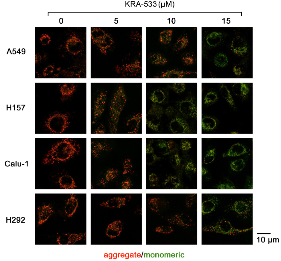
JC-1 purchased from MedChemExpress. Usage Cited in: Mol Cancer. 2019 Apr 10;18(1):85. [Abstract]
KRA-533 induces caspase 3 activation and reduces mitochondrial membrane potential in NSCLC cells. The measurement of mitochondrial membrane potential by JC-1 staining.
-
Adv Mater
Signal Converter-Based Therapy Platform Promoting Aging Bone Healing by Improving Permeability of the Mitochondrial Membrane. [Abstract]2025 Apr 28:e2500156. PMID: 40289881 -
Adv Mater
2025 Jan 10:e2411595. PMID: 39797465 -
Bioact Mater
On-demand mild photothermal cascade platform reprogramming mitochondrial immunity for tendon rejuvenation. [Abstract]2026 Jan 7:59:642-661. PMID: 41560899 -
Bioact Mater
Biosynthesis of fungus-based oral selenium microcarriers for radioprotection and immuno-homeostasis shaping against radiation-induced heart disease. [Abstract]2024 Apr 23:37:393-406. PMID: 38689659 -
Bioact Mater
Wet adhesive hydrogel cardiac patch loaded with anti-oxidative, autophagy-regulating molecule capsules and MSCs for restoring infarcted myocardium. [Abstract]2022 Aug 11;21:20-31. PMID: 36017068 -
-
Cancer Res
Metabolic reprogramming driven by IGF2BP3 promotes acquired resistance to EGFR inhibitors in non-small cell lung cancer. [Abstract]2023 Jul 5;83(13):2187-2207. PMID: 37061993 -
ACS Nano
An Immunomodulating Regenerating Hydrogel That Rescues the Oxidative Microenvironment and Reverses Cell Senescence for Osteoporotic Bone Defects. [Abstract]2025 Aug 12;19(31):28353-28371. PMID: 40754989 -
ACS Nano
2025 Jan 21;19(2):2695-2714. PMID: 39787443 -
ACS Nano
Engineered Extracellular Vesicle-Delivered CRISPR/Cas9 for Radiotherapy Sensitization of Glioblastoma. [Abstract]2023 Sep 12;17(17):16432-16447. PMID: 37646615 -
ACS Nano
A Redox Homeostasis Modulatory Hydrogel with GLRX3+ Extracellular Vesicles Attenuates Disc Degeneration by Suppressing Nucleus Pulposus Cell Senescence. [Abstract]2023 Jul 25;17(14):13441-13460. PMID: 37432866 -
ACS Nano
Trauma-Responsive Scaffold Synchronizing Oncolysis Immunization and Inflammation Alleviation for Post-Operative Suppression of Cancer Metastasis. [Abstract]2022 Apr 26;16(4):6064-6079. PMID: 35344338
JC-1 purchased from MedChemExpress. Usage Cited in: ACS Nano. 2022 Apr 26;16(4):6064-6079. [Abstract]
To investigate the mitochondrial membrane potential, ACCO is stained with JC-1 for 30 min, washed with PBS, and analyzed by flow cytometry using 488 nm excitation with 530/30 and 585/42 nm band-pass filters.
-
Nat Commun
Sensight enables quantitative multivariate engineering of high-performance chemical imaging tools. [Abstract]2026 Jan 27;17(1):2061. PMID: 41593056 -
Nat Commun
Endothelial major vault protein alleviates vascular remodeling via promoting Parkin-mediated mitophagy. [Abstract]2025 May 10;16(1):4365. PMID: 40348769 -
Cell Death Differ
Inhibition of Drp1 SUMOylation by ALR protects the liver from ischemia-reperfusion injury. [Abstract]2021 Apr;28(4):1174-1192. PMID: 33110216 -
Autophagy
AMPK protects against alcohol-induced liver injury through UQCRC2 to up-regulate mitophagy. [Abstract]2021 Nov;17(11):3622-3643. PMID: 33719895 -
Adv Sci (Weinh)
A ROS-Responsive Lipid Nanoparticles Release Multifunctional Hydrogel Based on Microenvironment Regulation Promotes Infected Diabetic Wound Healing. [Abstract]2024 Sep 23:e2403219. PMID: 39308241 -
Adv Sci (Weinh)
Copper-loaded Milk-Protein Derived Microgel Preserves Cardiac Metabolic Homeostasis After Myocardial Infarction. [Abstract]2024 Jul 15:e2401527. PMID: 39007192 -
Adv Sci (Weinh)
Sonodynamic Therapy of NRP2 Monoclonal Antibody-Guided MOFs@COF Targeted Disruption of Mitochondrial and Endoplasmic Reticulum Homeostasis to Induce Autophagy-Dependent Ferroptosis. [Abstract]2023 Oct;10(30):e2303872. PMID: 37661565 -
-
Biomaterials
Transforming lipid nanoparticles into radio-activatable therapeutics through synergistic ferroptosis for enhanced cancer radiotherapy. [Abstract]2026 Jul:330:124002. PMID: 41570670 -
Biomaterials
Self-assembling heterodimeric prodrug nanoparticles for dual-targeted cancer therapy via the disruption of redox homeostasis and Pin1 degradation. [Abstract]2026 Feb:325:123616. PMID: 40803234 -
Exp Mol Med
NLRP3-dependent lipid droplet formation contributes to posthemorrhagic hydrocephalus by increasing the permeability of the blood-cerebrospinal fluid barrier in the choroid plexus. [Abstract]2023 Mar;55(3):574-586. PMID: 36869068 -
J Nanobiotechnology
Polyphenolic nanodot-integrated spindle nanoplatform heals oral mucositis via microbiota restoration and glycolysis inhibition. [Abstract]2026 Feb 16;24(1):259. PMID: 41699655 -
J Nanobiotechnology
Multifunctional hydrogel targeting senescence to accelerate diabetic wound healing through promoting angiogenesis. [Abstract]2025 Mar 6;23(1):177. PMID: 40050885 -
Sci Adv
Matrix mechanical remodeled carrier-free nanosystem for programmable closed-loop reversal of liver fibrosis via STING alkylation. [Abstract]2025 Nov 21;11(47):eadz4126. PMID: 41259513 -
J Biomed Sci
A secreted form of chorismate mutase (Rv1885c) in Mycobacterium bovis BCG contributes to pathogenesis by inhibiting mitochondria-mediated apoptotic cell death of macrophages. [Abstract]2023 Dec 18;30(1):95. PMID: 38110948 -
Small
A Ferrocene-Functionalized Covalent Organic Framework for Enhancing Chemodynamic Therapy via Redox Dyshomeostasis. [Abstract]2021 Aug;17(32):e2101368. PMID: 34216420
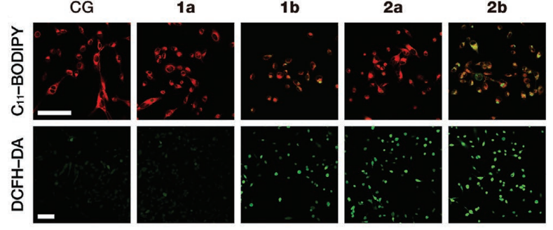
JC-1 purchased from MedChemExpress. Usage Cited in: Small. 2021 Aug;17(32):e2101368. [Abstract]
Laser scanning confocal fluorescence microscopy images of HT-1080 cells treated with different nanodrugs for the detection of intracellular lipid peroxidation and total ROS using C11-BODIPY and DCFH-DA as the corresponding fluorescence probes. Scale bar: 100 µm.
-
Small
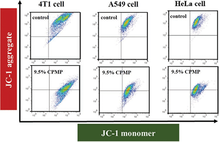
Multienzyme-Mimic Ultrafine Alloyed Nanoparticles in Metal Organic Frameworks for Enhanced Chemodynamic Therapy. [Abstract]2021 Feb;17(7):e2005865. PMID: 33502106
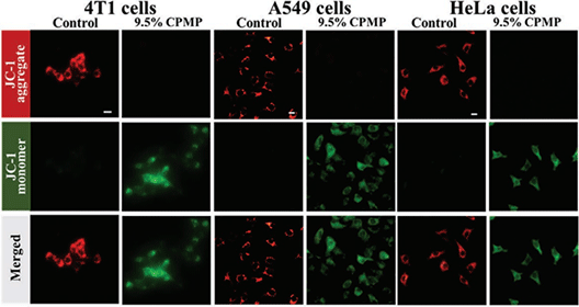
JC-1 purchased from MedChemExpress. Usage Cited in: Small. 2021 Feb;17(7):e2005865. [Abstract]
In 4T1 cells, A549 cells, and HeLa cells, cells are treated with JC-1 (5 µg/mL) for 20 min in the darkness, the fluorescence is observed by flow cytometry and the fluorescent color of the cells was observed with a fluorescence microscope, respectively
-
Small
Facile Nanolization Strategy for Therapeutic Ganoderma Lucidum Spore Oil to Achieve Enhanced Protection against Radiation-Induced Heart Disease. [Abstract]2019 Sep;15(36):e1902642. PMID: 31353836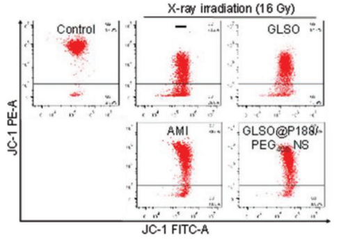
JC-1 purchased from MedChemExpress. Usage Cited in: Small. 2019 Sep;15(36):e1902642. [Abstract]
H9C2 cells are rinsed with pre-cooled PBS for 3 times and collected in PBS buffer containing 10 µg/mL JC-1 probe. After incubation for 20 min in dark place, cells are rinsed with PBS twice by centrifugation to remove the supernatant. Finally, H9C2 cell pellets suspended in PBS are analyzed by a flow cytometric analyzer.
-
Mol Ther
Autologous transplantation of mitochondria/rAAV IGF-I platforms in human osteoarthritic articular chondrocytes to treat osteoarthritis. [Abstract]2025 Jun 4;33(6):2900-2912. PMID: 39741406 -
Redox Biol
Ultrasmall Cu2-xSe nanoparticles alleviate vascular calcification through inhibiting oxidative stress and NF-κB/NLRP3-mediated inflammation. [Abstract]2025 Dec 3:89:103961. PMID: 41353801 -
Redox Biol
Extracellular vesicle-mediated delivery of mitochondrial circRNA MTCO2 protects against cerebral ischemia by modulating mPTP-dependent ferroptosis. [Abstract]2025 Aug 5:86:103806. PMID: 40768899 -
Redox Biol
Development of KEAP1-targeting PROTAC and its antioxidant properties: In vitro and in vivo. [Abstract]2023 Aug:64:102783. PMID: 37348157 -
J Control Release
Biofilm-camouflaged Prussian blue synergistic mitochondrial mass enhancement for Alzheimer's disease based on Cu2+ chelation and photothermal therapy. [Abstract]2024 Sep 13:375:269-284. PMID: 39245418 -
J Hazard Mater
Excessive mitochondrial stress response triggers neuronal injury through the persistent eIF2α phosphorylation in mice exposed to manganese. [Abstract]2025 Sep 15:496:139407. PMID: 40763529 -
J Hazard Mater
6PPD and 6PPDQ exposure promote the proliferation and migration of non-small cell lung cancer cells through PTEN dysfunction and ARG2-mediated metabolic reprogramming. [Abstract]2025 Jun 27:495:139088. PMID: 40602111 -
-
Research (Wash D C)
Targeting the Exonic Circular OGT RNA/O-GlcNAc Transferase/Forkhead Box C1 Axis Inhibits Asparagine- and Alanine-Mediated Ferroptosis Repression in Neuroblastoma Progression. [Abstract]2025 May 23:8:0703. PMID: 40416363 -
Research (Wash D C)
Mitochondrial-Oriented Injectable Hydrogel Microspheres Maintain Homeostasis of Chondrocyte Metabolism to Promote Subcellular Therapy in Osteoarthritis. [Abstract]2024 Jan 25:7:0306. PMID: 38274127 -
Pharmacol Res
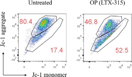
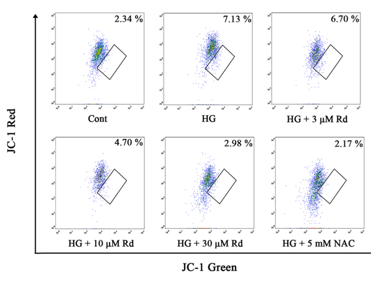
Ginsenoside Rd ameliorates high glucose-induced retinal endothelial injury through AMPK-STRT1 interdependence. [Abstract]2022 May;179:106123. PMID: 35150861
JC-1 purchased from MedChemExpress. Usage Cited in: Pharmacol Res. 2022 May;179:106123. [Abstract]
Mitochondrial membrane potential (Δψm) is measured with fluorochrome dye JC-1 (15 μM; 37 °C for 30 min in darkness). Flow cytometry was used to measure red (aggregation of JC-1) and green (monomeric JC-1) fluorescence intensity in HUVECs.
-
-
Mater Today Bio
Genetically engineered GSDME vesicle coated nanoparticle for immune activating and pyroptosis inducing synergetic therapy of fibrosarcoma. [Abstract]2025 Apr 26:32:101802. PMID: 40948580 -
Mater Today Bio
Enhanced anticancer effect of carfilzomib by codelivery of calcium peroxide nanoparticles targeting endoplasmic reticulum stress. [Abstract]2025 Mar 10:32:101649. PMID: 40160245 -
Cell Mol Biol Lett
Triptolide-induced cuproptosis is a novel antitumor strategy for the treatment of cervical cancer. [Abstract]2024 Aug 28;29(1):113. PMID: 39198750 -
Mater Today Bio
Drug-loaded oleic-acid grafted mesoporous silica nanoparticles conjugated with α-lactalbumin resembling BAMLET-like anticancer agent with improved biocompatibility and therapeutic efficacy. [Abstract]2022 Jun; 15: 100272. PMID: 35607417 -
Int J Biol Sci
Catalpol attenuates hepatic glucose metabolism disorder and oxidative stress in triptolide-induced liver injury by regulating the SIRT1/HIF-1α pathway. [Abstract]2024 Aug 1;20(10):4077-4097. PMID: 39113710 -
J Colloid Interface Sci
Redox-driven hybrid nanoenzyme dynamically activating ferroptosis and disulfidptosis for hepatocellular carcinoma theranostics. [Abstract]2025 Apr 15:693:137611. PMID: 40253866 -
Adv Healthc Mater
Constructing Mg-Based Hydrogen Container for Mitochondrial Dysfunction and Neuronal Ferroptosis in TCAR-Induced Cerebral Ischemia/Reperfusion Injury. [Abstract]2025 Dec 28:e02482. PMID: 41457679 -
Adv Healthc Mater
Cerium Dioxide Nanoparticles-Based Inspector Enhances Mitochondrial Quality Control to Maintain Chondrocyte Homeostasis in Osteoarthritis Therapy. [Abstract]2025 Apr;14(10):e2405069. PMID: 40033885 -
Adv Healthc Mater
Engineered Cell Membrane-Coated Keratin Nanoparticles Attenuated Intervertebral Disc Degeneration by Remodeling the Disc Microenvironment. [Abstract]2025 Jan 28:e2404173. PMID: 39876590 -
Cell Death Dis
DDAH-1 maintains endoplasmic reticulum-mitochondria contacts and protects dopaminergic neurons in Parkinson's disease. [Abstract]2024 Jun 7;15(6):399. PMID: 38849335 -
Cell Death Dis
Targeted inhibition of protein synthesis renders cancer cells vulnerable to apoptosis by unfolded protein response. [Abstract]2023 Aug 26;14(8):561. PMID: 37626037 -
Cell Death Dis
BCR-ABL triggers a glucose-dependent survival program during leukemogenesis through the suppression of TXNIP. [Abstract]2023 Apr 24;14(4):287. PMID: 37095099 -
Cell Death Dis
Bromodomain-containing protein 4 (BRD4) as an epigenetic regulator of fatty acid metabolism genes and ferroptosis. [Abstract]2022 Oct 29;13(10):912. PMID: 36309482 -
Cell Death Dis
RNA binding protein NKAP protects glioblastoma cells from ferroptosis by promoting SLC7A11 mRNA splicing in an m6A-dependent manner. [Abstract]2022 Jan 21;13(1):73. PMID: 35064112 -
Cell Death Dis
SESN2 protects against denervated muscle atrophy through unfolded protein response and mitophagy. [Abstract]2021 Aug 24;12(9):805. PMID: 34429398 -
Cell Death Dis
2021 Jan 21;12(1):110. PMID: 33479200 -
-
NPJ Biofilms Microbiomes
Akkermansia muciniphila ameliorates doxorubicin-induced cardiotoxicity by regulating PPARα-dependent mitochondrial biogenesis. [Abstract]2025 May 23;11(1):86. PMID: 40410194 -
Proc Natl Acad Sci U S A
ER-tethered stress sensor CREBH regulates mitochondrial unfolded protein response to maintain energy homeostasis. [Abstract]2024 Dec 3;121(49):e2410486121. PMID: 39589874 -
J Pharm Anal
Naringenin boosts Parkin-mediated mitophagy via estrogen receptor alpha to maintain mitochondrial quality control and heal diabetic foot ulcer. [Abstract]2025 Dec;15(12):101333. PMID: 41487142 -
Int J Biol Macromol
Peroxisome proliferator-activated receptor gamma (PPARγ) promotes the morula-to-blastocyst transition in Tibetan sheep embryos by enhancing mitophagy through suppression of the AKT/mTOR pathway. [Abstract]2025 Oct 17;331(Pt 2):148371. PMID: 41110574 -
Int J Biol Macromol
The naringin/carboxymethyl chitosan/sodium hyaluronate/silk fibroin scaffold facilitates the healing of diabetic wounds by restoring the ROS-related dysfunction of vascularization and macrophage polarization. [Abstract]2024 Mar;260(Pt 1):129348. PMID: 38219943 -
Acta Pharmacol Sin
Alpha lipoamide inhibits diabetic kidney fibrosis via improving mitochondrial function and regulating RXRα expression and activation. [Abstract]2023 May;44(5):1051-1065. PMID: 36347997 -
Phytomedicine
Brusatol induced ferroptosis in osteosarcoma cells by modulating the Keap1/Nrf2/SLC7A11 signaling pathway. [Abstract]2025 May 30:144:156912. PMID: 40494017 -
Phytomedicine
Hernandezine acts as a CDK4 suppressor inhibiting tumor growth by the CDK4/PKM2/NRF2 axis in colon cancer. [Abstract]2024 May 28:131:155775. PMID: 38838401 -
-
ACS Appl Mater Interfaces
Tumor Microenvironment-Activated and ROS-Augmented Nanoplatform Amplified PDT against Colorectal Cancer through Impairing GPX4 To Induce Ferroptosis. [Abstract]2025 Jul 23;17(29):41586-41596. PMID: 40629874 -
ACS Appl Mater Interfaces
Cerium-Organic Framework and Resveratrol Composite Hydrogel Scaffold with Dual Antioxidant Activity for Enhanced Bone Regeneration. [Abstract]2025 Jun 4;17(22):31828-31842. PMID: 40401746 -
Free Radic Biol Med
Role of ferroptosis mediated by abnormal membrane structure in DEHP-induced reproductive injury. [Abstract]2025 Apr 28:235:150-161. PMID: 40306442 -
Free Radic Biol Med
Ferroptosis and PANoptosis under hypoxia pivoting on the crosstalk between DHODH and GPX4 in corneal epithelium. [Abstract]2025 Feb 16:228:173-182. PMID: 39761766 -
Free Radic Biol Med
USP14 modulates cell pyroptosis and ameliorates doxorubicin-induced cardiotoxicity by deubiquitinating and stabilizing SIRT3. [Abstract]2024 Oct 28:225:741-757. PMID: 39490774 -
Free Radic Biol Med
Potassium ion efflux induces exaggerated mitochondrial damage and non-pyroptotic necrosis when energy metabolism is blocked. [Abstract]2024 Feb 20:212:117-132. PMID: 38151213 -
ACS Appl Mater Interfaces
Thermo-Chemical Resistance to Combination Therapy of Glioma Depends on Cellular Energy Level. [Abstract]2023 Aug 23;15(33):39053-39063. PMID: 37552210 -
Free Radic Biol Med
Targeting the Na+/K+ ATPase DR-region with DR-Ab improves doxorubicin-induced cardiotoxicity. [Abstract]2023 Aug 1:204:38-53. PMID: 37100355 -
ACS Appl Mater Interfaces
Thermosensitive Hydrogel Incorporating Prussian Blue Nanoparticles Promotes Diabetic Wound Healing via ROS Scavenging and Mitochondrial Function Restoration. [Abstract]2022 Mar 30;14(12):14059-14071. PMID: 35298140 -
Free Radic Biol Med
Sevoflurane induced neurotoxicity in neonatal mice links to a GSK3β/Drp1-dependent mitochondrial fission and apoptosis. [Abstract]2022 Mar:181:72-81. PMID: 35122996 -
Chemosphere
PCB126 impairs human sperm functions by affecting post-translational modifications and mitochondrial functions. [Abstract]2024 Jan:346:140532. PMID: 37918541 -
-
Mucosal Immunol
Gut-derived short-chain fatty acids modulate skin barrier integrity by promoting keratinocyte metabolism and differentiation. [Abstract]2022 May;15(5):908-926. PMID: 35672452 -
J Transl Med
S1PR3 inhibition protects against LPS-induced ARDS by inhibiting NF-κB and improving mitochondrial oxidative phosphorylation. [Abstract]2024 Jun 5;22(1):535. PMID: 38840216 -
Biomed Pharmacother
Tongmai formula improves cardiac function via regulating mitochondrial quality control in the myocardium with ischemia/reperfusion injury. [Abstract]2020 Dec;132:110897. PMID: 33113431 -
Environ Pollut
Monobutyl phthalate (MBP) induces energy metabolism disturbances in the gills of adult zebrafish (Danio rerio). [Abstract]2020 Nov;266(Pt 1):115288. PMID: 32795888 -
Cell Death Discov
GPX1 confers resistance to metabolic stress in BCR/ABL-T315I mutant chronic myeloid leukemia cells. [Abstract]2025 May 9;11(1):229. PMID: 40346054 -
Cell Death Discov
NOXA exacerbates endoplasmic-reticulum-stress-induced intervertebral disc degeneration by activating apoptosis and ECM degradation. [Abstract]2025 May 28;11(1):257. PMID: 40436859 -
Cell Death Discov
Cysteine dioxygenase 1 attenuates the proliferation via inducing oxidative stress and integrated stress response in gastric cancer cells. [Abstract]2022 Dec 16;8(1):493. PMID: 36526626 -
J Med Chem
Phytohormone Strigolactams Overcomes Oxaliplatin Resistance in Colorectal Cancer by Blocking the Late-Stage Autophagy to Tip the Metabolic Balance toward Ferroptosis. [Abstract]2025 Nov 27;68(22):24546-24559. PMID: 41208142 -
J Med Chem
Rational Design of a Novel Class of Human ClpP Agonists through a Ring-Opening Strategy with Enhanced Antileukemia Activity. [Abstract]2024 Apr 25;67(8):6769-6792. PMID: 38620134 -
Neural Regen Res
Bromocriptine protects perilesional spinal cord neurons from lipotoxicity after spinal cord injury. [Abstract]2024 May;19(5):1142-1149. PMID: 37862220 -
Antioxidants (Basel)
Fruit of Gardenia jasminoides Induces Mitochondrial Activation and Non-Shivering Thermogenesis through Regulation of PPARγ. [Abstract]2021 Sep 5;10(9):1418. PMID: 34573050 -
Mol Med
TOP2A deficiency leads to human recurrent spontaneous abortion and growth retardation of mouse pre-implantation embryos. [Abstract]2022 Dec 30;28(1):165. PMID: 36585615 -
-
J Agric Food Chem
Extraction, Purification, Structural Characterization, and Antitumor Effects of Water-Soluble Intracellular Polysaccharide (IPSW-1) from Phellinus igniarius Mycelia. [Abstract]2024 Sep 11;72(36):19721-19732. PMID: 39205635 -
EMBO Rep
Lysosomal K+ channel TMEM175 promotes apoptosis and aggravates symptoms of Parkinson's disease. [Abstract]2022 Sep 5;23(9):e53234. PMID: 35913019 -
J Agric Food Chem
Asparanin A from Asparagus officinalis L. Induces G0/G1 Cell Cycle Arrest and Apoptosis in Human Endometrial Carcinoma Ishikawa Cells via Mitochondrial and PI3K/AKT Signaling Pathways. [Abstract]2020 Jan 8;68(1):213-224. PMID: 31861958 -
Ecotoxicol Environ Saf
Proteomic analysis reveals that cigarette smoke exposure diminishes ovarian reserve in mice by disrupting the CREB1-mediated ovarian granulosa cell proliferation-apoptosis balance. [Abstract]2024 Feb:271:115989. PMID: 38242047 -
Cell Biol Toxicol
HUWE1 regulates mitophagy to protect dopaminergic neurons from 6-OHDA- and MPP⁺-induced neurotoxicity. [Abstract]2026 Feb 5;42(1):34. PMID: 41639490 -
Cell Biol Toxicol
Syringaresinol protects against diabetic nephropathy by inhibiting pyroptosis via NRF2-mediated antioxidant pathway. [Abstract]2023 Jun;39(3):621-639. PMID: 36640193 -
-
Chin Med
Oleuropein alleviates myocardial ischemia-reperfusion injury by suppressing oxidative stress and excessive autophagy via TLR4/MAPK signaling pathway. [Abstract]2024 Apr 8;19(1):59. PMID: 38589925 -
J Invest Dermatol
Epidermal stem cell derived exosomes alleviate excessive autophagy induced endothelial cell apoptosis by delivering miR200b-3p to diabetic wounds. [Abstract]2024 May;144(5):1134-1147.e2. PMID: 37838331 -
Bioeng Transl Med
Synergistic ferroptosis-starvation therapy for bladder cancer based on hyaluronic acid modified metal-organic frameworks. [Abstract]2023 Mar 22;8(3):e10515. PMID: 37206228 -
Colloids Surf B Biointerfaces
Drug-reinforced metal-organic framework nanozyme for combined treatment of ischemia/reperfusion acute kidney injury. [Abstract]2026 Jun:262:115514. PMID: 41671821 -
Colloids Surf B Biointerfaces
Combining sodium chloride nanoparticle osmolarity spikes with irinotecan chemotherapy to amplify oxidative stress in the treatment of colorectal cancer. [Abstract]2025 Nov 19:259:115294. PMID: 41297221 -
Biochem Pharmacol
Icariin inhibits autophagy and promotes apoptosis by regulating mitochondrial division in Triple-negative breast cancer. [Abstract]2026 Jan;243(Pt 2):117549. PMID: 41242617 -
Colloids Surf B Biointerfaces
Injectable DMM/GelMA hydrogel for diabetic wound healing via regulating mitochondrial metabolism and macrophage repolarization. [Abstract]2025 Apr:248:114488. PMID: 39765076 -
Colloids Surf B Biointerfaces
In-situ growth of CeO2 on biofilms: Innovative nanoparticles for photothermal therapy & multi-pronged attack on Alzheimer's disease. [Abstract]2024 Jun:238:113887. PMID: 38581835 -
Biochem Pharmacol
Celecoxib and sulindac sulfide elicit anticancer effects on PIK3CA-mutated head and neck cancer cells through endoplasmic reticulum stress, reactive oxygen species, and mitochondrial dysfunction. [Abstract]2024 Jun:224:116221. PMID: 38641308 -
Biochem Pharmacol
Design, synthesis and anti-tumor activity of novel benzothiophenonaphthalimide derivatives targeting mitochondrial DNA (mtDNA) G-quadruplex. [Abstract]2022 Apr 30;201:115062. PMID: 35504316 -
Chem Biol Interact
Cigarette smoke exposure diminishes ovarian reserve in mice by regulating granulosa cells redox homeostasis imbalance through Wnt10b-ERβ feedback loop. [Abstract]2026 Jan 5:423:111811. PMID: 41202950 -
J Ethnopharmacol
Preservation of mitochondrial homeostasis is responsible for the ameliorative effects of Suhuang antitussive capsule on non-resolving inflammation via inhibition of NF-κB signaling and NLRP3 inflammasome activation. [Abstract]2021 May 10:271:113827. PMID: 33460751 -
-
Int J Pharm
Photoactive Parietin-loaded nanocarriers as an efficient therapeutic platform against triple-negative breast cancer. [Abstract]2023 Aug 25:643:123217. PMID: 37429562 -
Cancer Immunol Immunother
Spautin-1 inhibits the growth of diffuse large B-cell lymphoma by inducing mitochondrial damage-mediated PANoptosis and anti-tumor immunity. [Abstract]2025 Aug 25;74(9):293. PMID: 40853486 -
Stem Cell Reports
2024 Nov 20:S2213-6711(24)00296-0. PMID: 39577429 -
Life Sci
Loss of Ninjurin1 alleviates acetaminophen-induced liver injury via enhancing AMPKα-NRF2 pathway. [Abstract]2024 Jun 5:122782. PMID: 38848941 -
Life Sci
Oleic acid protects saturated fatty acid mediated lipotoxicity in hepatocytes and rat of non-alcoholic steatohepatitis. [Abstract]2018 Jun 15:203:291-304. PMID: 29709653 -
Inflammation
2024 Feb;47(1):285-306. PMID: 37759136 -
Int J Mol Sci
Baloxavir Acid-Induced Mitochondrial Toxicity and Cell Cycle Arrest Contribute to Its Adverse Effects. [Abstract]2026 Mar 25;27(7):2967. PMID: 41977157 -
Int J Mol Sci
Ischemic Rescue Potential of Conditioned Medium Derived from Skeletal Muscle Cells-Seeded Electrospun Fiber-Coated Human Amniotic Membrane Scaffolds. [Abstract]2024 Oct 30;25(21):11697. PMID: 39519249 -
Int J Oncol
α‑hederin overcomes hypoxia‑mediated drug resistance in colorectal cancer by inhibiting the AKT/Bcl2 pathway. [Abstract]2023 Mar;62(3):33. PMID: 36704835 -
Front Pharmacol
Selective anti-cancer effects of cannabidiol and Δ9-tetrahydrocannabinol via PI3K/AKT/mTOR inhibition and PTEN restoration in ovarian cancer cells. [Abstract]2025 Dec 15:16:1693129. PMID: 41472794 -
Front Bioeng Biotechnol
2022 May 24:10:892015. PMID: 35685086 -
Front Cell Infect Microbiol
Dysregulation of Cytosolic c-di-GMP in Edwardsiella piscicida Promotes Cellular Non-Canonical Ferroptosis. [Abstract]2022 Feb 4;12:825824. PMID: 35186798 -
Front Pharmacol
Icariin Ameliorates Diabetic Cardiomyopathy Through Apelin/Sirt3 Signalling to Improve Mitochondrial Dysfunction. [Abstract]2020 Mar 19;11:256. PMID: 32265695 -
Int Immunopharmacol
Quercetin alleviates imatinib-induced premature ovarian insufficiency by regulating mitophagy via the ROS/JNK/c-JUN pathway. [Abstract]2026 Apr 15:175:116405. PMID: 41722539 -
Eur J Pharmacol
Ertugliflozin induces vasodilation by inhibiting AMPK-mediated mitochondrial fission in vascular smooth muscle cells. [Abstract]2025 May 30:177784. PMID: 40451567 -
Eur J Pharmacol
Dioscin improves hypertrophic scars by inducing apoptosis and ferroptosis of scar fibroblasts through mitochondrial oxidative stress damage. [Abstract]2025 May 22:177759. PMID: 40412745 -
Int Immunopharmacol
Extracellular ATP contributes to the reactive oxygen species burst and exaggerated mitochondrial damage in D-galactosamine and lipopolysaccharide-induced fulminant hepatitis. [Abstract]2024 Mar 30:130:111680. PMID: 38368772 -
Am J Physiol Cell Physiol
RTA 408 ameliorates diabetic cardiomyopathy by activating Nrf2 to regulate mitochondrial fission and fusion and inhibiting NF-κB-mediated inflammation. [Abstract]2024 Feb 1;326(2):C331-C347. PMID: 38047307 -
ACS Appl Bio Mater
Mild-Temperature Photothermal Effect Enhanced by Functional Conjugated Polymer Nanoparticles through Enzyme-Mediated Starvation. [Abstract]2022 Jun 20;5(6):2536-2542. PMID: 35535955 -
Toxicology
Perfluorooctanoic acid induces mitochondrial dysfunction-driven apoptosis and PINK1/Parkin-mediated mitophagy in testicular cells. [Abstract]2026 Jun 23:526:154530. PMID: 42336259 -
RSC Adv
2026 Apr 8;16(21):18745-18756. PMID: 41959546 -
Toxicology
Polystyrene microplastics induce skeletal muscle atrophy through disruption of anabolic signaling and mitochondrial function. [Abstract]2026 Mar 20:154452. PMID: 41865969 -
Molecules
Benzoxazole Derivative K313 Induces Cell Cycle Arrest, Apoptosis and Autophagy Blockage and Suppresses mTOR/p70S6K Pathway in Nalm-6 and Daudi Cells. [Abstract]2020 Feb 21;25(4):971. PMID: 32098126 -
Mitochondrion
Hypoxia improves self-renew and migration of urine-derived stem cells by upregulating autophagy and mitochondrial function through ERK signal pathway. [Abstract]2023 Nov:73:1-9. PMID: 37678426 -
J Mol Biol
The phosphorylation status of Hsp82 regulates mitochondrial homeostasis during glucose sensing in Saccharomyces cerevisiae. [Abstract]2023 Jul 1;435(13):168106. PMID: 37068581 -
Eur J Pharm Biopharm
Enhanced photodynamic therapy of curcumin using biodegradable PLGA coated mesoporous silica nanoparticles. [Abstract]2024 Sep 18:114503. PMID: 39303950 -
Exp Gerontol
Overexpression of solute carrier family 6 member 12 promotes cell injury in Parkinson's disease via MAPK signaling pathway. [Abstract]2024 Jun 13:194:112484. PMID: 38871234 -
Exp Gerontol
Acoustic stimulation during sleep improves cognition and ameliorates Alzheimer's disease pathology in APP/PS1 mice. [Abstract]2023 Oct 15:182:112299. PMID: 37776987 -
J Physiol Biochem
Lipoxin A4 attenuated dexamethasone-induced muscle atrophy via activation of PGC-1α/Nrf2/TFAM pathway. [Abstract]2023 Feb;79(1):107-115. PMID: 36125698 -
-
Biochim Biophys Acta Mol Basis Dis
PGC-1α role in rescuing ferroptosis in cerebral ischemia/reperfusion injury through promoting mitochondrial biogenesis and UCP2 expression. [Abstract]2025 Apr 26;1871(6):167874. PMID: 40294850 -
FASEB J
Mitophagy induced by UMI-77 preserves mitochondrial fitness in renal tubular epithelial cells and alleviates renal fibrosis. [Abstract]2022 Jun;36(6):e22342. PMID: 35524750 -
J Cell Mol Med
Inhibition of the long non-coding RNA ZFAS1 attenuates ferroptosis by sponging miR-150-5p and activates CCND2 against diabetic cardiomyopathy. [Abstract]2021 Nov;25(21):9995-10007. PMID: 34609043 -
Transl Oncol
Tubeimoside I induces mitophagy by activating the PINK1/Parkin/Mfn2 signaling pathway in acute myeloid leukemia cells. [Abstract]2025 May:55:102355. PMID: 40112502 -
Neurochem Int
Retinoic acid signaling regulates astrocyte reactivity by modulating MAPK/NF-κB pathways and mitochondrial integrity. [Abstract]2026 Feb 20:106134. PMID: 41724387 -
-
J Cell Physiol
Telomere Attrition-Induced Senescence in Human Pluripotent Stem Cell-Derived Astrocytes: Distinct Cellular and Functional Characteristics. [Abstract]2025 Dec;240(12):e70118. PMID: 41340457 -
Sci Rep
DH_27, a right-half derivative of renieramycin T, induce apoptosis through inhibition of mTOR signaling for lung cancer suppression. [Abstract]2026 May 6. PMID: 42092109 -
Ann Clin Transl Neurol
2026 Mar 24. PMID: 41876403 -
Sci Rep
Proteomics analysis reveals progesterone receptor induced mitochondria-mediated apoptosis in breast cancer cells. [Abstract]2025 Dec 24;15(1):44446. PMID: 41444289 -
Sci Rep
Substituents introduction of methyl and methoxy functional groups on resveratrol stabilizes mTOR binding for autophagic cell death induction. [Abstract]2025 Apr 26;15(1):14675. PMID: 40287470 -
J Biol Chem
BL-918 activates PINK1/Parkin signaling pathway to ameliorate the progression of Parkinson's disease. [Abstract]2024 Jul 9:107543. PMID: 38992440 -
Chem Res Toxicol
Polybrominated Diphenyl Ethers Quinone Induced Parthanatos-like Cell Death through a Reactive Oxygen Species-Associated Poly(ADP-ribose) Polymerase 1 Signaling. [Abstract]2018 Nov 19;31(11):1164-1171. PMID: 30295471 -
Cell Signal
Leptin attenuates diabetic cardiomyopathy-induced cardiac remodeling via regulating cGAS/STING signaling and Opa1-mediated mitochondrial fusion. [Abstract]2025 Apr 15:111805. PMID: 40246132 -
Mol Cell Biochem
Erythropoietin promotes energy metabolism to improve LPS-induced injury in HK-2 cells via SIRT1/PGC1-α pathway. [Abstract]2023 Mar;478(3):651-663. PMID: 36001204 -
Stem Cells
Downregulation of augmenter of liver regeneration impairs the therapeutic efficacy of liver epithelial progenitor cells against acute liver injury by enhancing mitochondrial fission. [Abstract]2021 Nov;39(11):1546-1562. PMID: 34310799 -
Exp Cell Res
Mechanical stretch-triggered EGR1 overexpression enhances macrophage M2 polarization and drives mitochondrial fission. [Abstract]2025 May 31:114621. PMID: 40456378 -
Toxicol Appl Pharmacol
2025 Dec 8:507:117682. PMID: 41371369 -
Toxicol Appl Pharmacol
Comparative assessment of female reproductive toxicity from PFOA and its alternative GenX in mice and human granulosa cells. [Abstract]2026 Jan:506:117639. PMID: 41232729 -
Nanotoxicology
The nanomaterial-induced bystander effects reprogrammed macrophage immune function and metabolic profile. [Abstract]2020 Oct;14(8):1137-1155. PMID: 32916084 -
Anim Reprod Sci
Effects of mitoquinone (MitoQ) supplementation during boar semen cryopreservation on sperm quality, antioxidant status and mitochondrial proteomics. [Abstract]2022 Dec:247:107099. PMID: 36306716 -
J Pharm Pharmacol
Taoren Honghua Jian improves ferroptosis-mediated myocardial damage following myocardial infarction via Nrf2-dependent enhancement of GPX4/FSP1. [Abstract]2025 Oct 17:rgaf075. PMID: 41105727 -
Cell Stress Chaperones
The role of Atp2a2-mediated calcium imbalance and endoplasmic reticulum stress in hydrocortisone-induced neurotoxicity. [Abstract]2025 Sep 17:100112. PMID: 40972936 -
Front Biosci (Landmark Ed)
P110 Inhibits DRP1/FIS1-Mediated Mitochondrial Fission to Alleviate Uric Acid-Induced Apoptosis in HK-2 Cells. [Abstract]2025 Nov 26;30(11):46700. PMID: 41351411 -
Cardiovasc Drugs Ther
Hyperglycemia-Induced Overexpression of PH Domain Leucine-Rich Repeat Protein Phosphatase 1 (PHLPP1) Compromises the Cardioprotective Effect of Ischemic Postconditioning Via Modulation of the Akt/Mst1 Pathway Signaling. [Abstract]2023 Dec;37(6):1087-1101. PMID: 35715527 -
J Leukoc Biol
IFNγ-mediated repression of system xc- drives vulnerability to induced ferroptosis in hepatocellular carcinoma cells. [Abstract]2021 Aug;110(2):301-314. PMID: 34318944 -
Curr Res Toxicol
The role of autophagy in microwave radiation induced toxicity in iPSC-derived cardiomyocytes. [Abstract]2026 Mar 10:10:100288. PMID: 41847319 -
J Bioenerg Biomembr
Astragaloside IV ameliorates cardiomyocyte injury and heart failure through hif/rho/rock pathway regulation: In vitro and in vivo insights. [Abstract]2025 Oct 14. PMID: 41085879 -
J Bioenerg Biomembr
Blockage of PHLPP1 protects against myocardial ischemia/reperfusion injury in diabetic mice via activation of STAT3 signaling. [Abstract]2023 Oct;55(5):325-339. PMID: 37584737 -
Toxicol Lett
2024 Apr:394:76-91. PMID: 38428544 -
Pflugers Arch
Ulinastatin ameliorated streptozotocin-induced diabetic nephropathy: Potential effects via modulating the components of gut-kidney axis and restoring mitochondrial homeostasis. [Abstract]2023 Oct;475(10):1161-1176. PMID: 37561129 -
Mol Brain
Ferroptosis participates in neuron damage in experimental cerebral malaria and is partially induced by activated CD8+ T cells. [Abstract]2022 Jun 20;15(1):57. PMID: 35725567 -
Reprod Toxicol
Co-exposure to polystyrene nanoplastics and hexachlorocyclohexane induces enhanced human sperm toxicity in vitro. [Abstract]2026 Apr:141:109201. PMID: 41730440 -
Integr Cancer Ther
Unveiling the Anticancer Potential and Molecular Mechanisms of Fangchinoline Against Cholangiocarcinoma Using FTIR Microspectroscopy, In Vitro and In Silico Approaches. [Abstract]2025 Jan-Dec:24:15347354251396513. PMID: 41261767 -
Reprod Toxicol
Removal and toxic intervention of polystyrene microplastics and nanoplastics by magnetic nano-Fe3O4 in spermatogonial GC-1 cells. [Abstract]2025 Jul 31:109020. PMID: 40752657 -
Theriogenology
Proteomic analysis reveals the potential positive effects of Mito-TEMPO on ram sperm motility and fertility during cryopreservation. [Abstract]2023 Jul 15:205:27-39. PMID: 37084501 -
Mol Biotechnol
Berberine Alleviates the Damage, Oxidative Stress and Mitochondrial Dysfunction of PC12 Cells Induced by High Glucose by Activating the KEAP1/Nrf2/ARE Pathway. [Abstract]2023 Oct;65(10):1632-1643. PMID: 36737555 -
Reprod Sci
Heat Shock Protein B7 Inhibits the Progression of Endometrial Carcinoma by Inhibiting PI3K/AKT/mTOR Pathway. [Abstract]2023 Feb;30(2):590-600. PMID: 35859224 -
Tissue Cell
Tetratricopeptide repeat domain 36 protects renal tubular cells from cisplatin-induced apoptosis potentially via maintaining mitochondrial homeostasis. [Abstract]2022 Jun:76:101749. PMID: 35176677 -
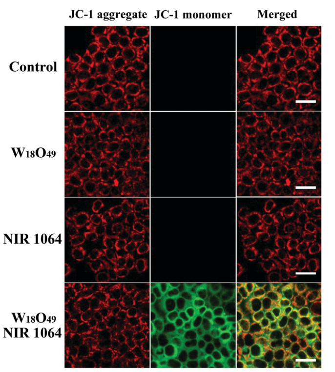
JC-1 purchased from MedChemExpress. Usage Cited in: New J Chem. 2017 41(23).
Measurement of mitochondria membrane potential by JC-1 staining. HeLa cells are incubated with or without WOs for 24 h, followed by NIR irradiation for NIR and WOs + NIR groups.
-
BMC Nephrol
Statins attenuate cholesterol-induced ROS via inhibiting NOX2/NOX4 and mitochondrial pathway in collecting ducts of the kidney. [Abstract]2022 May 13;23(1):184. PMID: 35562673 -
Biochem Biophys Res Commun
Tangeretin protects against Aβ1-42-induced toxicity and exploring mitochondria-lysosome interactions in HT22 cells. [Abstract]2025 Apr 6:762:151769. PMID: 40220719 -
Leuk Lymphoma
FDA-approved disulfiram induces ferroptosis via alteration of redox balance and lipid peroxidation and overcomes carfilzomib resistance in multiple myeloma. [Abstract]2024 Nov 11:1-12. PMID: 39527722 -
Biochem Biophys Res Commun
Cholic acid activation of GPBAR1 does not induce or exacerbate acute pancreatitis but promotes exocrine pancreatic secretion. [Abstract]2024 Nov 26:735:150825. PMID: 39426134 -
Biochem Biophys Res Commun
Sympathetic β2-adrenergic receptor blockade overcomes docetaxel resistance in prostate cancer. [Abstract]2023 May 21:657:69-79. PMID: 36989842 -
World Neurosurg
Activated Phosphoinositide 3-Kinase/Akt/Mammalian Target of Rapamycin Signal and Suppressed Autophagy Participate in Protection Offered by Licochalcone A Against Amyloid-β Peptide Fragment 25-35-Induced Injury in SH-SY5Y Cells. [Abstract]2022 Jan:157:e390-e400. PMID: 34662660 -
Biol Pharm Bull
Neuroprotective Effects of Tetrahydroxystilbene Glucoside against Rotenone-Induced Toxicity in PC12 Cells. [Abstract]2022 Jan 1;45(1):143-149. PMID: 34707025 -
-
J Obstet Gynaecol Res
Crizotinib-induced anti-cancer activity in human cervical carcinoma cells via ROS-dependent mitochondrial depolarization and induction of apoptotic pathway. [Abstract]2021 Nov;47(11):3923-3930. PMID: 34482598 -
-
-
-
-
-
-
-
-
-
-
-
-
-
-
-
Oxid Med Cell Longev
Inhibition of TRPA1 Ameliorates Periodontitis by Reducing Periodontal Ligament Cell Oxidative Stress and Apoptosis via PERK/eIF2 α/ATF-4/CHOP Signal Pathway. [Abstract]2022 Jun 10:2022:4107915. PMID: 35720191 -
-
-
-
Oxid Med Cell Longev
Programmed NP Cell Death Induced by Mitochondrial ROS in a One-Strike Loading Disc Degeneration Organ Culture Model. [Abstract]2021 Aug 31;2021:5608133. PMID: 34512867 -
-
-


Solvent & Solubility
DMSO : 5 mg/mL (7.67 mM; ultrasonic and warming and heat to 60°C; Hygroscopic DMSO has a significant impact on the solubility of product, please use newly opened DMSO)
H2O : < 0.1 mg/mL (insoluble)
Please refer to the solubility information to select the appropriate solvent. Once prepared, please aliquot and store the solution to prevent product inactivation from repeated freeze-thaw cycles.
Storage method and period of stock solution: -80°C, 6 months; -20°C, 1 month (sealed storage, away from moisture and light). When stored at -80°C, please use it within 6 months. When stored at -20°C, please use it within 1 month.
Concentration (start) × Volume (start) = Concentration (final) × Volume (final)


Purity & Documentation
-
Data Sheet (281 KB)
-
SDS (538 KB)
- English - EN (538 KB)
- Français - FR (538 KB)
- Deutsch - DE (538 KB)
- Norwegian - NO (538 KB)
- Español - ES (538 KB)
- Swedish - SV (538 KB)
- Italian - IT (538 KB)
- Korean - KR (538 KB)
- Portuguese - PT (538 KB)
-
Handling Instructions (2659 KB)
References
[1]. A Perelman, et al. JC-1: alternative excitation wavelengths facilitate mitochondrial membrane potential cytometry. Cell Death Dis. 2012 Nov 22;3:e430. [Content Brief]
[2]. Vera C. Keil, et al. Ratiometric high-resolution imaging of JC-1 fluorescence reveals the subcellular heterogeneity of astrocytic mitochondria. Pflügers Archiv - European Journal of Physiology. 2011,462(5): 693-708. [Content Brief]
[4]. Salvioli S, et al. JC-1, but not DiOC6(3) or rhodamine 123, is a reliable fluorescent probe to assess delta psi changes in intact cells: implications for studies on mitochondrial functionality during apoptosis. FEBS Lett. 1997 Jul 7;411(1):77-82. [Content Brief]


Complete Stock Solution Preparation Table
Please refer to the solubility information to select the appropriate solvent. Once prepared, please aliquot and store the solution to prevent product inactivation from repeated freeze-thaw cycles.
Storage method and period of stock solution: -80°C, 6 months; -20°C, 1 month (sealed storage, away from moisture and light). When stored at -80°C, please use it within 6 months. When stored at -20°C, please use it within 1 month.
| Optional Solvent | Concentration Solvent Mass | 1 mg | 5 mg | 10 mg | 25 mg |
|---|---|---|---|---|---|
| DMSO | 1 mM | 1.5332 mL | 7.6660 mL | 15.3320 mL | 38.3300 mL |
| 5 mM | 0.3066 mL | 1.5332 mL | 3.0664 mL | 7.6660 mL |


 Powered by Bioz