1. Signaling Pathways
  2. Apoptosis
  3. TNF Receptor

TNF Receptor

Tumor Necrosis Factor Receptor; TNFR

Tumor necrosis factor (TNF) is a major mediator of apoptosis as well as inflammation and immunity, and it has been implicated in the pathogenesis of a wide spectrum of human diseases, including sepsis, diabetes, cancer, osteoporosis, multiple sclerosis, rheumatoid arthritis, and inflammatory bowel diseases.

TNF-α is a 17-kDa protein consisting of 157 amino acids that is a homotrimer in solution. In humans, the gene is mapped to chromosome 6. Its bioactivity is mainly regulated by soluble TNF-α–binding receptors. TNF-α is mainly produced by activated macrophages, T lymphocytes, and natural killer cells. Lower expression is known for a variety of other cells, including fibroblasts, smooth muscle cells, and tumor cells. In cells, TNF-α is synthesized as pro-TNF (26 kDa), which is membrane-bound and is released upon cleavage of its pro domain by TNF-converting enzyme (TACE).

Many of the TNF-induced cellular responses are mediated by either one of the two TNF receptors, TNF-R1 and TNF-R2, both of which belong to the TNF receptor super-family. In response to TNF treatment, the transcription factor NF-κB and MAP kinases, including ERK, p38 and JNK, are activated in most types of cells and, in some cases, apoptosis or necrosis could also be induced. However, induction of apoptosis or necrosis is mainly achieved through TNFR1, which is also known as a death receptor. Activation of the NF-κB and MAPKs plays an important role in the induction of many cytokines and immune-regulatory proteins and is pivotal for many inflammatory responses.

Cat. No. Product Name Effect Purity Chemical Structure
  • HY-11109
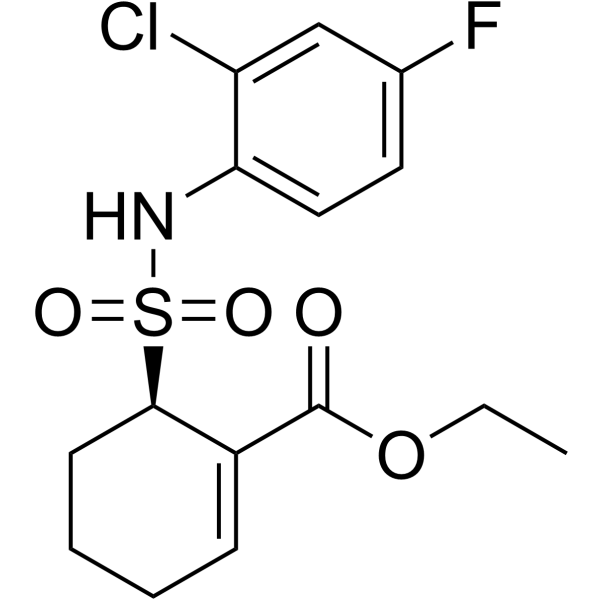
    Resatorvid
    Inhibitor 99.95%
    Resatorvid (TAK-242) is a selective Toll-like receptor 4 (TLR4) inhibitor. Resatorvid inhibits NO, TNF-α and IL-6 production with IC50s of 1.8 nM, 1.9 nM and 1.3 nM, respectively. Resatorvid downregulates expression of TLR4 downstream signaling molecules MyD88 and TRIF. Resatorvid inhibits autophagy and plays pivotal role in various inflammatory diseases.
    Resatorvid
  • HY-N0822
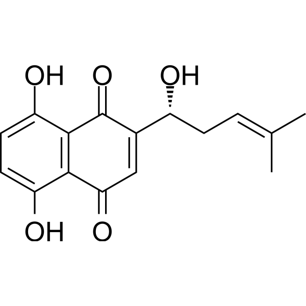
    Shikonin
    Inhibitor 99.80%
    Shikonin is a major component of a Chinese herbal medicine named zicao. Shikonin is a potent TMEM16A chloride channel inhibitor with an IC50 of 6.5 μM. Shikonin is a specific pyruvate kinase M2 (PKM2) inhibitor and can also inhibit TNF-α and NF-κB pathway. Shikonin decreases exosome secretion through the inhibition of glycolysis. Shikonin inhibits AIM2 inflammasome activation.
    Shikonin
  • HY-P9970
    Infliximab
    Inhibitor
    Infliximab (Avakine) is a chimeric monoclonal IgG1 antibody that specifically binds to TNF-α. Infliximab prevents the interaction of TNF-α with TNF-α receptor (TNFR1 and TNFR2). Infliximab has the potential for autoimmune, chronic inflammatory diseases and diabetic neuropathy research.
    Infliximab
  • HY-N0182
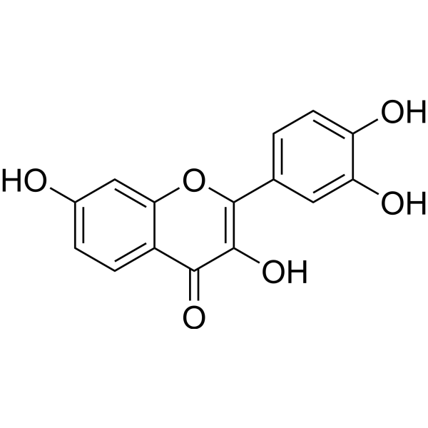
    Fisetin
    Inhibitor 98.39%
    Fisetin is a natural flavonol found in many fruits and vegetables with various benefits, such as antioxidant, anticancer, neuroprotection effects.
    Fisetin
  • HY-P9908
    Adalimumab
    Inhibitor 99.62%
    Adalimumab is a human monoclonal IgG1 antibody targeting tumour necrosis factor α (TNF-α).
    Adalimumab
  • HY-P990007
    Tulisokibart
    Inhibitor
    Tulisokibart (PRA023) is a humanized IgG1-κ monoclonal antibody targeting to TNFSF15/TL1A. Tulisokibart can be used to study a variety of inflammatory/fibrotic diseases, such as Crohn's Disease (CD).
    Tulisokibart
  • HY-P990008
    Atrosab
    Antagonist
    Atrosab is a humanized IgG1 antagonistic anti-TNFR1 antibody. Atrosab inhibits TNF-mediated apoptosis induction and IL-6 and IL-8 production. Atrosab can be used for research of inflammatory disease.
    Atrosab
  • HY-P99796
    Ozoralizumab
    Inhibitor 99.80%
    Ozoralizumab (ATN-103) is an anti-TNFα humanized antibody. Ozoralizumab is a humanized trivalent nanobody compound that consists of two anti-human TNFα nanobodies and an anti-human serum albumin (HSA) nanobody. Ozoralizumab can be used in research of arthritis.
    Ozoralizumab
  • HY-108847
    Etanercept
    Inhibitor 99.20%
    Etanercept, a dimeric fusion protein that binds TNF, acts as a TNF inhibitor. Etanercept competitively inhibits the binding of both TNF-α and TNF-β to cell surface TNF receptors, rendering TNF biologically inactive. Etanercept shows efficacy against rheumatoid arthritis, juvenile idiopathic arthritis, and plaque psoriasis.
    Etanercept
  • HY-P0224
    N-Formyl-Met-Leu-Phe
    Inhibitor 99.83%
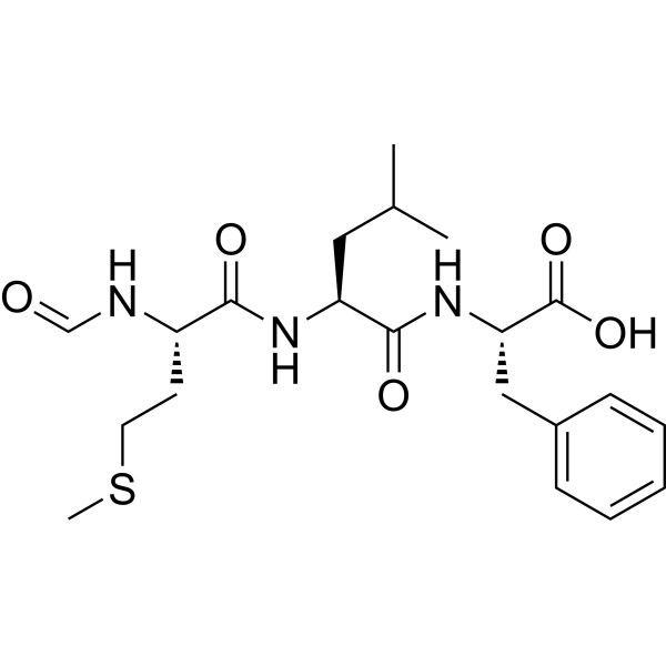
    N-Formyl-Met-Leu-Phe (fMLP; N-Formyl-MLF) is a chemotactic peptide and a specific ligand of N-formyl peptide receptor (FPR). N-Formyl-Met-Leu-Ph is reported to inhibit TNF-alpha secretion.
    N-Formyl-Met-Leu-Phe
  • HY-13812
    QNZ
    Inhibitor 99.51%
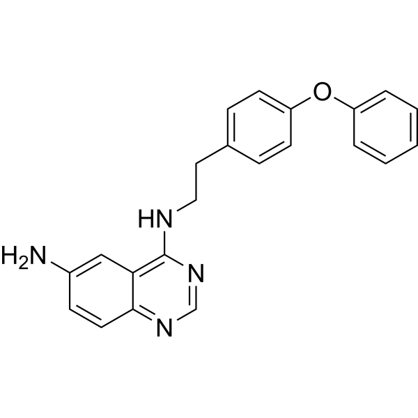
    QNZ (EVP4593) shows strong inhibitory effects on NF-κB transcriptional activation and TNF-α production with IC50s of 11 and 7 nM, respectively. QNZ (EVP4593) is a neuroprotective inhibitor of SOC channel.
    QNZ
  • HY-12085
    Apremilast
    Inhibitor 99.91%
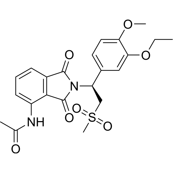
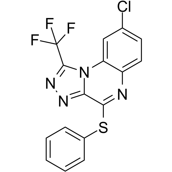
    Apremilast (CC-10004) is an orally available inhibitor of type-4 cyclic nucleotide phosphodiesterase (PDE-4) with an IC50 of 74 nM. Apremilast inhibits TNF-α release by lipopolysaccharide (LPS) with an IC50 of 104 nM.
    Apremilast
  • HY-B0809
    Theophylline
    Inhibitor 99.89%
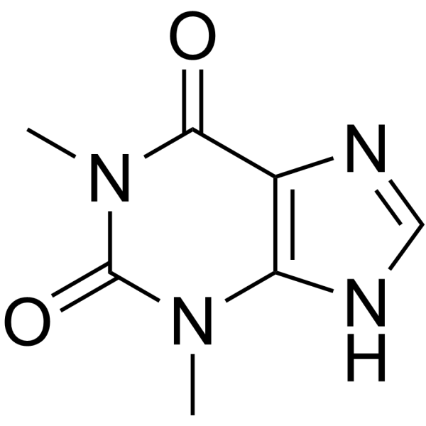
    Theophylline (1,3-Dimethylxanthine) is a potent phosphodiesterase (PDE) inhibitor, adenosine receptor antagonist, and histone deacetylase (HDAC) activator. Theophylline (1,3-Dimethylxanthine) inhibits PDE3 activity to relax airway smooth muscle. Theophylline (1,3-Dimethylxanthine) has anti-inflammatory activity by increase IL-10 and inhibit NF-κB into the nucleus. Theophylline (1,3-Dimethylxanthine) induces apoptosis. Theophylline (1,3-Dimethylxanthine) can be used for asthma and chronic obstructive pulmonary disease (COPD) research.
    Theophylline
  • HY-110203
    R-7050
    Antagonist 98.55%
    R-7050 (TNF-α Antagonist III) is a tumor necrosis factor receptor (TNFR) antagonist with greater selectivity toward TNFα.
    R-7050
  • HY-B0190A
    Nafamostat mesylate
    98.06%
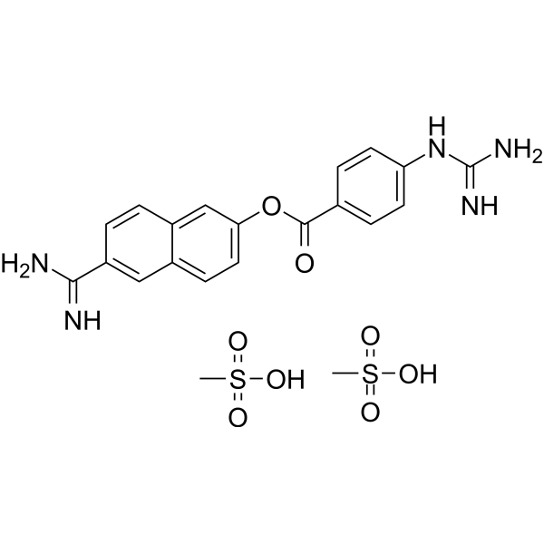
    Nafamostat mesylate (FUT-175), an anticoagulant, is a synthetic serine protease inhibitor. Nafamostat mesylate has anticancer and antivirus effect. Nafamostat mesylate induce apoptosis by up-regulating the expression of tumor necrosis factor receptor-1 (TNFR1). Nafamostat mesylate can be used in the development of the pathological thickening of the arterial wall3].
    Nafamostat mesylate
  • HY-120934
    C25-140
    Inhibitor 99.84%
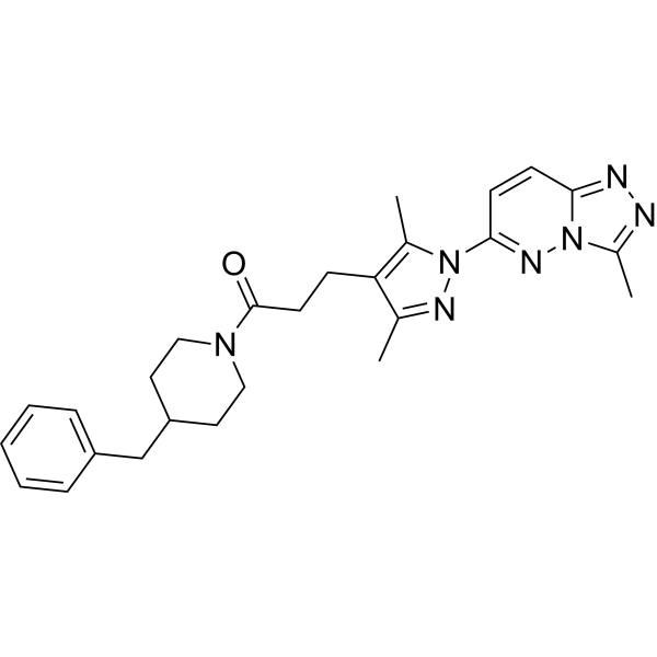
    C25-140, a first-in-class, orally active, and fairly selective TRAF6-Ubc13 inhibitor, directly binds to TRAF6, and blocks the interaction of TRAF6 with Ubc13. C25-140 lowers TRAF6 activity, reduces NF-κB activation, and combats autoimmunity.
    C25-140
  • HY-123942
    Diprovocim
    Agonist 98.35%
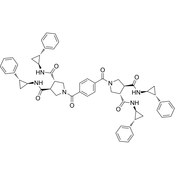
    Diprovocim is a potent TLR1/TLR2 agonist. Diprovocim elicits full agonist activity in human THP-1 cells (EC50=110 pM). Diprovocim stimulates the release of TNF-α from mouse macrophages (EC50=1.3 nM). Diprovocim activates downstream MAPK and NF-κB signaling pathway. Diprovocim displays strong adjuvant activity in mice, particularly abetting cellular immune responses.
    Diprovocim
  • HY-133122
    UCB-9260
    Inhibitor 99.96%
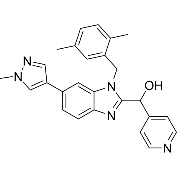
    UCB-9260, an orally active compound, inhibits TNF signaling by stabilising an asymmetric form of the trimer. UCB-9260 is selective for TNF over other superfamily members, and binds TNF with a similar Kd of 13 nM.
    UCB-9260
  • HY-P99107
    Brentuximab vedotin
    Inhibitor 98.00%
    Brentuximab vedotin (cAC10-vcMMAE) is an antibody-drug conjugate (ADC) comprising an anti-CD30 antibody and the cytotoxic agent Monomethyl auristatin E (MMAE). Brentuximab vedotin inhibits CD30-positive cells with an IC50 of 2.5 ng/mL. Brentuximab vedotin can be used for the research of relapsed and refractory Hodgkin lymphoma.
    Brentuximab vedotin
  • HY-N0633
    Muscone
    Inhibitor ≥98.0%
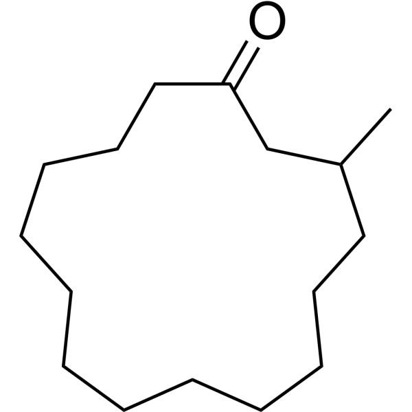
    Muscone is the main active monomer of traditional Chinese medicine musk. Muscone inhibits NF-κB and NLRP3 inflammasome activation. Muscone remarkably decreases the levels of inflammatory cytokines (IL-1β, TNF-α and IL-6), and ultimately improves cardiac function and survival rate.
    Muscone
Cat. No. Product Name / Synonyms Species Source

Following the binding of TNF to TNF receptors, TNFR1 binds to TRADD, which recruits RIPK1, TRAF2/5 and cIAP1/2 to form TNFR1 signaling complex I; TNFR2 binds to TRAF1/2 directly to recruit cIAP1/2. Both cIAP1 and cIAP2 are E3 ubiquitin ligases that add K63 linked polyubiquitin chains to RIPK1 and other components of the signaling complex. The ubiquitin ligase activity of the cIAPs is needed to recruit the LUBAC, which adds M1 linked linear polyubiquitin chains to RIPK1. K63 polyubiquitylated RIPK1 recruits TAB2, TAB3 and TAK1, which activate signaling mediated by JNK and p38, as well as the IκB kinase complex. The IKK complex then activates NF-κB signaling, which leads to the transcription of anti-apoptotic factors-such as FLIP and Bcl-XL-that promote cell survival. 


The formation of TNFR1 complex IIa and complex IIb depends on non-ubiquitylated RIPK1. For the formation of complex IIa, ubiquitylated RIPK1 in complex I is deubiquitylated by CYLD. This deubiquitylated RIPK1 dissociates from the membrane-bound complex and moves into the cytosol, where it interacts with TRADD, FADD, Pro-caspase 8 and FLIPL to form complex IIa. By contrast, complex IIb is formed when the RIPK1 in complex I is not ubiquitylated owing to conditions that have resulted in the depletion of cIAPs, which normally ubiquitylate RIPK1. This non-ubiquitylated RIPK1 dissociates from complex I, moves into the cytosol, and assembles with FADD, Pro-caspase 8, FLIPL and RIPK3 (but not TRADD) to form complex IIb. For either complex IIa or complex IIb to prevent necroptosis, both RIPK1 and RIPK3 must be inactivated by the cleavage activity of the Pro-caspase 8-FLIPL heterodimer or fully activated caspase 8. The Pro-caspase 8 homodimer generates active Caspase 8, which is released from complex IIa and complex IIb. This active Caspase 8 then carries out cleavage reactions to activate downstream executioner caspases and thus induce classical apoptosis. 


Formation of the complex IIc (necrosome) is initiated either by RIPK1 deubiquitylation mediated by CYLD or by RIPK1 non-ubiquitylation due to depletion of cIAPs, similar to complex IIa and complex IIb formation. RIPK1 recruits numerous RIPK3 molecules. They come together to form amyloid microfilaments called necrosomes. Activated RIPK3 phosphorylates and recruits MLKL, eventually leading to the formation of a supramolecular protein complex at the plasma membrane and necroptosis [1][2].


Reference:
[1]. Brenner D, et al. Regulation of tumour necrosis factor signalling: live or let die.Nat Rev Immunol. 2015 Jun;15(6):362-74. 
[2]. Conrad M, et al. Regulated necrosis: disease relevance and therapeutic opportunities.Nat Rev Drug Discov. 2016 May;15(5):348-66.