


Propidium Iodide
Based on 156 publication(s) in Google Scholar
Propidium Iodide (PI) is a nuclear staining agent that stains DNA. Propidium Iodide is an analogue of ethidine bromide that emits red fluorescence upon embedding in double-stranded DNA. Propidium Iodide cannot pass through living cell membranes, but it can pass through damaged cell membranes to stain the nucleus. Propidium Iodide has a fluorescence wavelength of 493/617 nm and a wavelength of 536/635 nm after Mosaic with DNA. Propidium Iodide is commonly used in the detection of apoptosis (apoptosis) or necrosis (necrosis), and is often used in flow cytometry analysis.
For research use only. We do not sell to patients.
- Purity: 99.77%
- CAS No.: 25535-16-4
- Formula: C27H34I2N4
- Molecular Weight:668.39
-
Storage:
4°C, sealed storage, away from moisture and light
* In solvent : -80°C, 2 years; -20°C, 1 year (sealed storage, away from moisture and light)

To place orders, for customer services and technical support, please contact: MedChemExpress USA
Tel: 609-228-6898 E-mail: [email protected] [email protected]


-
 Biological Activity
-
Chemical Information
-
Solvent & Solubility
- Protocol
- Purity & Documentation
- References
-
 Help & FAQs


Publications Citing Use of MedChemExpress (MCE) Propidium Iodide
More- Adv Mater. 2025 Jan 23:e2414437. [Abstract]
- Cell Res. 2025 Jul;35(7):483-496. [Abstract]
- Bioact Mater. 2022 Aug 11;21:20-31. [Abstract]
- Adv Funct Mater. 2025 Sep 23.
- Adv Funct Mater. 2025 Jan 25.
- Adv Funct Mater. 2024 May 1.
- Cancer Res. 2023 Oct 2;83(19):3220-3236. [Abstract]
- ACS Nano. 2025 Dec 16;19(49):41540-41556. [Abstract]
- ACS Nano. 2023 Feb 28;17(4):3334-3345. [Abstract]
- Nat Commun. 2026 Mar 24;17(1):4360. [Abstract]
- Nat Commun. 2026 Feb 9;17(1):1441. [Abstract]
- Nat Commun. 2025 May 30;16(1):5006. [Abstract]
- Nat Commun. 2025 Feb 25;16(1):1774. [Abstract]
- Nat Commun. 2024 Oct 26;15(1):9261. [Abstract]
- Acta Pharm Sin B. 2025 Aug;15(8):4014-4029. [Abstract]
- Autophagy. 2023 Feb;19(2):632-643. [Abstract]
- Adv Sci (Weinh). 2026 Jul;13(39):e20095.
- Adv Sci (Weinh). 2025 Dec 14:e14191. [Abstract]
- Adv Sci (Weinh). 2024 Jul;11(28):e2400527. [Abstract]
- Nat Chem Biol. 2025 Aug 22. [Abstract]
- Chem Eng J. 2025 Nov 3;525:170400.
- Chem Eng J. 2025 Jul 1.
- Chem Eng J. 2025 Apr 15.
- J Adv Res. 2025 Jan 18:S2090-1232(25)00045-1. [Abstract]
- J Adv Res. 2022 Nov:41:205-218. [Abstract]
- Biomaterials. 2025 May 5:322:123391. [Abstract]
- Sci Adv. 2025 Sep 12;11(37):eadw9299. [Abstract]
- Small. 2022 Jul;18(30):e2202002. [Abstract]
- Redox Biol. 2025 Sep 22:87:103877. [Abstract]
- Redox Biol. 2024 Sep:75:103270. [Abstract]
- Redox Biol. 2022 Feb;49:102207. [Abstract]
- J Control Release. 2025 Jan 8:379:45-58. [Abstract]
- J Control Release. 2024 Jan:365:176-192. [Abstract]
- J Hazard Mater. 2025 Mar 18:492:137969. [Abstract]
- EBioMedicine. 2025 May 13:116:105738. [Abstract]
- Mater Today Bio. 2025 Jan 15:31:101495. [Abstract]
- Clin Cancer Res. 2023 Nov 14;29(22):4644-4659. [Abstract]
- Cancer Lett. 2025 Sep 25:634:218064. [Abstract]
- Cell Death Dis. 2026 Jan 7;17(1):8. [Abstract]
- Cell Death Dis. 2022 Dec 12;13(12):1034. [Abstract]
- Cell Death Dis. 2021 Oct 2;12(10):902. [Abstract]
- NPJ Biofilms Microbiomes. 2025 Jun 21;11(1):113. [Abstract]
- Int J Biol Macromol. 2025 Apr 7:142867. [Abstract]
- Int J Biol Macromol. 2024 Jun;269(Pt 2):131795. [Abstract]
- Int J Biol Macromol. 2024 May;266(Pt 1):130637. [Abstract]
- Phytomedicine. 2024 Aug 19:134:155959. [Abstract]
- ACS Appl Mater Interfaces. 2025 Dec 17;17(50):67595-67608. [Abstract]
- ACS Appl Mater Interfaces. 2025 Oct 1;17(39):54423-54436. [Abstract]
- ACS Appl Mater Interfaces. 2023 Jun 28;15(25):29841-29853. [Abstract]
- J Orthop Translat. 2025 Jun 2:53:12-25. [Abstract]
- Br J Pharmacol. 2026 Aug;183(16):4989-5017.
- J Transl Med. 2026 May 22.
- Biomed Pharmacother. 2024 Aug 5:178:116992. [Abstract]
- Environ Pollut. 2025 Sep 10:385:127111. [Abstract]
- Cell Death Discov. 2024 Mar 12;10(1):134. [Abstract]
- Cell Rep. 2025 Sep 2;44(9):116215. [Abstract]
- Cell Rep. 2025 Jul 26;44(8):116076. [Abstract]
- Microbiol Res. 2024 Nov 5:290:127964. [Abstract]
- Cell Rep. 2023 Dec 27;43(1):113591. [Abstract]
- Cell Rep. 2022 Aug 23;40(8):111240. [Abstract]
- Anal Chem. 2025 Aug 5;97(30):16142-16150. [Abstract]
- Int J Pharm X. 2025 Oct 9:10:100413. [Abstract]
- Phytother Res. 2026 Jun;40(6):3595-3612.
- J Agric Food Chem. 2025 Oct 22. [Abstract]
- J Agric Food Chem. 2025 Sep 3;73(35):21976-21986. [Abstract]
- Biomater Adv. 2026 Jun:183:214761. [Abstract]
- Eur J Med Chem. 2023 Jan 5;245(Pt 2):114919. [Abstract]
- Food Biosci. 11 December 2021, 101501.
- Biochem Pharmacol. 2026 Aug;250(Pt 1):117992.
- Biochem Pharmacol. 2026 Jun:248:117843. [Abstract]
- Biochem Pharmacol. 2024 May:223:116197. [Abstract]
- Virulence. 2026 Dec 31;17(1):2646808. [Abstract]
- Mar Drugs. 2025 Jun 24;23(7):264. [Abstract]
- J Enzyme Inhib Med Chem. 2025 Dec;40(1):2468852. [Abstract]
- Int J Food Microbiol. 2026 Oct 2:459:111897.
- Foods. 2026 Apr 21;15(8):1451.
- Cancer Immunol Immunother. 2025 Dec 22;75(1):23. [Abstract]
- CNS Neurosci Ther. 2026 Jun;32(6):e70854.
- Inflammation. 2025 Jan 30. [Abstract]
- CNS Neurosci Ther. 2023 Oct;29(10):2787-2799. [Abstract]
- Nat Prod Bioprospect. 2026 May 12;16(1):64.
- Int J Mol Sci. 2023 Mar 8;24(6):5174. [Abstract]
- Int J Mol Sci. 2023 Jan 11;24(2):1416. [Abstract]
- Pharmaceuticals (Basel). 2025 Sep 16;18(9):1378. [Abstract]
- Pharmaceuticals. 2022 Aug 26;15(9):1059. [Abstract]
- Int Immunopharmacol. 2026 Jul 15:181:116705.
- Int Immunopharmacol. 2025 Sep 5:165:115437. [Abstract]
- Eur J Pharmacol. 2024 Dec 31:177247. [Abstract]
- Int Immunopharmacol. 2024 Jun 24:138:112518. [Abstract]
- Eur J Pharmacol. 2022 Apr 5;920:174830. [Abstract]
- ACS Appl Bio Mater. 2021 Jan 6.
- Hepatol Commun. 2025 Jul 21;9(8):e0759. [Abstract]
- Toxicology. 2024 Aug 6:153906. [Abstract]
- Molecules. 2022 Oct 9;27(19):6733. [Abstract]
- Front Microbiol. 2025 Sep 18:16:1670356. [Abstract]
- Genes Immun. 2025 Aug;26(4):365-375. [Abstract]
- Eur J Pharm Biopharm. 2026 Jul:224:115081.
- J Cell Mol Med. 2025 Mar;29(5):e70486. [Abstract]
- Plants. 2026 Jan 9;15(2):211. [Abstract]
- iScience. 2025 Nov 10;28(12):114003. [Abstract]
- Oncol Res. 2025 Aug 28;33(9):2491-2506. [Abstract]
- APL Bioeng. 2025 May 27;9(2):026119. [Abstract]
- iScience. 2024 Dec 1;27(12):111517. [Abstract]
- iScience. 2024 Dec 3;27(12):111513. [Abstract]
- J Funct Foods. 2024 Oct.
- J Funct Foods. 2024 Aug.
- ACS Synth Biol. 2025 Sep 19;14(9):3767-3783. [Abstract]
- Sci Rep. 2025 Aug 19;15(1):30411. [Abstract]
- Sci Rep. 2025 Jun 4;15(1):19590. [Abstract]
- ACS Chem Neurosci. 2025 Feb 5;16(3):374-383. [Abstract]
- Langmuir. 2025 Jan 21;41(2):1260-1270. [Abstract]
- J Biol Chem. 2024 Jul 15:107570. [Abstract]
- Aging (Albany NY). 2018 Nov 28;10(11):3353-3370. [Abstract]
- Microbiol Spectr. 2026 Apr 13;14(5):e0172225.
- J Virol. 2026 Apr 21;100(4):e0209725. [Abstract]
- ACS Infect Dis. 2026 Feb 13;12(2):623-636. [Abstract]
- Cell Signal. 2025 Dec 19:139:112333. [Abstract]
- Heliyon. 2024 Aug 30;10(17):e37227. [Abstract]
- Mol Med Rep. 2026 May;33(5):156.
- Exp Cell Res. 2024 Oct 1;442(2):114274. [Abstract]
- Mol Med Rep. 2019 Jan;19(1):41-50. [Abstract]
- Stem Cells Int. 2022 Dec 1:2022:6795573. [Abstract]
- J Anim Sci Technol. 2025.
- Mol Carcinog. 2025 Mar 18. [Abstract]
- Naunyn Schmiedebergs Arch Pharmacol. 2026 Jun;399(9):14157-14175.
- Front Biosci (Landmark Ed). 2025 Jan 16;30(1):26186. [Abstract]
- Mol Immunol. 2026 May:193:123-134.
- Curr Issues Mol Biol. 2025 Jul 4;47(7):516. [Abstract]
- FEBS Lett. 2025 Feb;599(3):367-380. [Abstract]
- J Pharmacol Sci. 2022 May;149(1):27-36. [Abstract]
- Clin Transl Radiat Oncol. 2025 Jan 15:51:100920. [Abstract]
- BMC Pharmacol Toxicol. 2024 Nov 20;25(1):89. [Abstract]
- Blood Sci. 2020 Jul 25;2(3):89-99. [Abstract]
- Arch Microbiol. 2025 Nov 1;207(12):339. [Abstract]
- Thorac Cancer. 2022 May;13(9):1299-1310. [Abstract]
- Oncol Lett. 2026 Mar 26;31(5):196. [Abstract]
- Biochim Biophys Acta Gen Subj. 2026 Mar;1870(3):130901. [Abstract]
- Biochem Biophys Res Commun. 2025 Sep 8:778:152383. [Abstract]
- Brain Inj. 2026 May;40(7):619-630.
- Cytotechnology. 2024 Dec;76(6):817-832. [Abstract]
- STAR Protoc. 2023 Sep 19;4(4):102553. [Abstract]
- Biocell. 2025 Sep 25;49(9):1711-1731.
- bioRxiv. 2025 Oct 31:2025.10.31.685607. [Abstract]
- bioRxiv. 2025 Oct 20.
- SSRN. 2025 Apr 3.
- SSRN. 2025 Mar 21.
- bioRxiv. 2025 March 26.
- SSRN. 2024 Nov 11.
- bioRxiv. 2024 November 25.
- Research Square Preprint. 2024 Sep 10.
- Res Sq. 2024 Aug 11.
- Research Square Preprint. 2021 Sep.
- Oxid Med Cell Longev. 2021 Aug 31;2021:5608133. [Abstract]
- Preprints. 2020, 2020090120.
- Research Square Preprint. 2020 Jul.
- Chinese Pharmacological Bulletin. 2018 May; 34(5): 620-626.
 Customer Validation & Images

-
IF
-
IF
-
Flow Cytometry
-
Flow Cytometry
-
IF
All DNA/RNA Synthesis Isoforms
More

Biological Activity
|
Cell Line
|
Type | Value | Description | References |
|---|---|---|---|---|
| HEK293 | IC50 |
1.1 μM
Compound: propidium iodide
|
Inhibition of wild type mouse AChE expressed in HEK293 cells using ATCh as substrate
Inhibition of wild type mouse AChE expressed in HEK293 cells using ATCh as substrate
|
[PMID: 23984975] |
Guide (The following is our recommended solution. This solution is merely a guideline and should be modified according to your specific needs.)
1. Prepare the storage solution and working solution
1.1 Prepare the storage solution
Prepare the 1 mg/mL stock solution using DDH2O.
1.2 Preparation of working solution
Dilute the stored solution with pre-heated serum-free cell culture medium or PBS to prepare a 20-50 μg/mL working solution of Propidium Iodide.
Note: Please adjust the concentration of the Propidium Iodide working solution according to the actual situation, and prepare it as needed.
2. Cell Staining (Suspension Cells)
2.1 Centrifuge to collect the cells, then add PBS for two washes, each for 5 minutes. The cell density is 1×10⁶ per mL.
2.2 Add 1 mL of the working solution and incubate at room temperature for 5 to 10 minutes.
2.3 400 grams. Centrifuge for 3-4 minutes. Discard the supernatant.
2.4 Add PBS to wash the cells twice, for 5 minutes each time.
2.5 After resuspending the cells in 1 mL of serum-free medium or PBS, observe them using a fluorescence microscope or a flow cytometer.
3. Cell Staining (Monolayer Cells)
3.1 Place the monolayer cells on a sterile cover slip.
3.2 Remove the cover glass from the culture medium and aspirate away the excess medium.
3.3 Add 100 μL of the dye working solution and gently shake to ensure complete coverage of the cells. Incubate for 5 to 30 minutes.
3.4 Remove the dye working solution and wash with the culture medium 2-3 times, for 5 minutes each time. Observe under a fluorescence microscope.
Note: If flow cytometry is to be used for detection, the cells should first be digested with trypsin, resuspended, and then stained.
Notes
1. Please adjust the concentration of Propidium Iodide working solution and the incubation time according to the actual situation.
2. Only the nuclei of dead cells will be stained red.
3. This product is exclusively for professional scientific research purposes only. It shall not be used for clinical diagnosis or treatment, nor for food or medicine.
3. Propidium Iodide is carcinogenic. For your safety and health, please wear a laboratory coat and use disposable gloves when operating.
MedChemExpress (MCE) has not independently confirmed the accuracy of these methods. They are for reference only.
615
535


Chemical Information
-
CAS No. 25535-16-4
-
Appearance Solid
-
Molecular Weight 668.39
-
Formula C27H34I2N4
-
Color Brown to red
-
SMILES
C[N+](CCC[N+]1=C(C2=CC=CC=C2)C3=CC(N)=CC=C3C4=C1C=C(N)C=C4)(CC)CC.[I-].[I-]
-
Shipping
Room temperature in continental US; may vary elsewhere.
-
Storage
4°C, sealed storage, away from moisture and light
* In solvent : -80°C, 2 years; -20°C, 1 year (sealed storage, away from moisture and light)


Publications (156)
-
Journal Impact Factor
-
Most Recent
-
Adv Mater
Directional Freeze-Casting Cryogel Loaded with Quaternized Chitosan Modified Gallium Metal-Organic Frameworks to Capture and Eradicate the Resistant Bacteria for Guided Regeneration in Infected Bone Defects. [Abstract]2025 Jan 23:e2414437. PMID: 39846310 -
Cell Res
A thermosensor FUST1 primes heat-induced stress granule formation via biomolecular condensation in Arabidopsis. [Abstract]2025 Jul;35(7):483-496. PMID: 40360668 -
Bioact Mater
Wet adhesive hydrogel cardiac patch loaded with anti-oxidative, autophagy-regulating molecule capsules and MSCs for restoring infarcted myocardium. [Abstract]2022 Aug 11;21:20-31. PMID: 36017068 -
-
-
-
Cancer Res
2023 Oct 2;83(19):3220-3236. PMID: 37463119 -
ACS Nano
Sequential Mitochondrial Transplantation for Myocardial Ischemia-Reperfusion Injury Treatment. [Abstract]2025 Dec 16;19(49):41540-41556. PMID: 41320927 -
ACS Nano
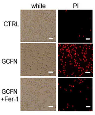
A Ferroptosis-Inducing and Leukemic Cell-Targeting Drug Nanocarrier Formed by Redox-Responsive Cysteine Polymer for Acute Myeloid Leukemia Therapy. [Abstract]2023 Feb 28;17(4):3334-3345. PMID: 36752654
Propidium Iodide purchased from MedChemExpress. Usage Cited in: ACS Nano. 2023 Feb 28;17(4):3334-3345. [Abstract]
Propidium iodide (PI) staining for cell death with GCFN and ferroptosis inhibitor ferrostatin-1 treatment for 48 h.
-
Nat Commun
KIF11 prevents retinal endothelial ferroptosis in familial exudative vitreoretinopathy by inhibiting phosphorylation-driven PRDX1 phase separation. [Abstract]2026 Mar 24;17(1):4360. PMID: 41872221 -
Nat Commun
Triple targeting of STING, TGF-β, and PD-L1 boosts CXCL16-CXCR6 signaling for potent antitumor response. [Abstract]2026 Feb 9;17(1):1441. PMID: 41663428 -
Nat Commun
2025 May 30;16(1):5006. PMID: 40442064 -
Nat Commun
Attenuated growth factor signaling during cell death initiation sensitizes membranes towards peroxidation. [Abstract]2025 Feb 25;16(1):1774. PMID: 40000627 -
Nat Commun
TNF inhibitors target a mevalonate metabolite/TRPM2/calcium signaling axis in neutrophils to dampen vasculitis in Behçet's disease. [Abstract]2024 Oct 26;15(1):9261. PMID: 39461948 -
Acta Pharm Sin B
The protein arginine methyltransferase PRMT1 ameliorates cerebral ischemia-reperfusion injury by suppressing RIPK1-mediated necroptosis and apoptosis. [Abstract]2025 Aug;15(8):4014-4029. PMID: 40893679 -
Autophagy
Autophagy loss impedes cancer-associated fibroblast activation via downregulating proline biosynthesis. [Abstract]2023 Feb;19(2):632-643. PMID: 35786294 -
-
Adv Sci (Weinh)
PDIA3 Inhibition Facilitates Sensitivity of IKE-Induced Ferroptosis via STAT3/LCN2 Axis to Improve Glioblastoma Therapy. [Abstract]2025 Dec 14:e14191. PMID: 41391030 -
Adv Sci (Weinh)
Polyacrylic Acid-Coated Selenium-Doped Carbon Dots Inhibit Ferroptosis to Alleviate Chemotherapy-Associated Acute Kidney Injury. [Abstract]2024 Jul;11(28):e2400527. PMID: 38689508 -
Nat Chem Biol
2025 Aug 22. PMID: 40846996 -
-
-
-
J Adv Res
SCARB1 links cholesterol metabolism-mediated ferroptosis inhibition to radioresistance in tumor cells. [Abstract]2025 Jan 18:S2090-1232(25)00045-1. PMID: 39832721 -
J Adv Res
Metformin suppresses vascular smooth muscle cell senescence by promoting autophagic flux. [Abstract]2022 Nov:41:205-218. PMID: 36328749 -
Biomaterials
Radiation-triggerable bioreactors enable bioenergetic reprograming of cancer stem cell plasticity via targeted arginine metabolism disruption for augmented radio-immunotherapy. [Abstract]2025 May 5:322:123391. PMID: 40344881 -
Sci Adv
Temporal biphasic regulation of photoreceptor degeneration by microglial TREM2: A metabolic-immune nexus in retinitis pigmentosa. [Abstract]2025 Sep 12;11(37):eadw9299. PMID: 40938987 -
Small
Versatile Protein Coronation Approach with Multiple Depleted Serum for Creating Biocompatible, Precision Nanomedicine. [Abstract]2022 Jul;18(30):e2202002. PMID: 35775952 -
Redox Biol
Synthetic anticoagulant octaparin targets mitochondrial cardiolipin-GSDMD axis to rescue redox homeostasis in sepsis. [Abstract]2025 Sep 22:87:103877. PMID: 40992080 -
Redox Biol
2024 Sep:75:103270. PMID: 39047638 -
Redox Biol
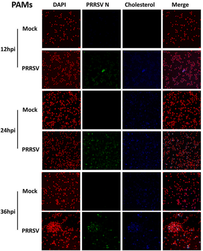
Porcine reproductive and respiratory syndrome virus nsp4 positively regulates cellular cholesterol to inhibit type I interferon production. [Abstract]2022 Feb;49:102207. PMID: 34911669
Propidium Iodide purchased from MedChemExpress. Usage Cited in: Redox Biol. 2022 Feb;49:102207. [Abstract]
PAMs cells were washed with phosphate-buffered saline (PBS), and then incubated with the nuclear dye propidium iodide for 15 min. The nuclei were counterstained with propidium iodide (red). Fluorescent images were acquired with a confocal laser scanning microscope.
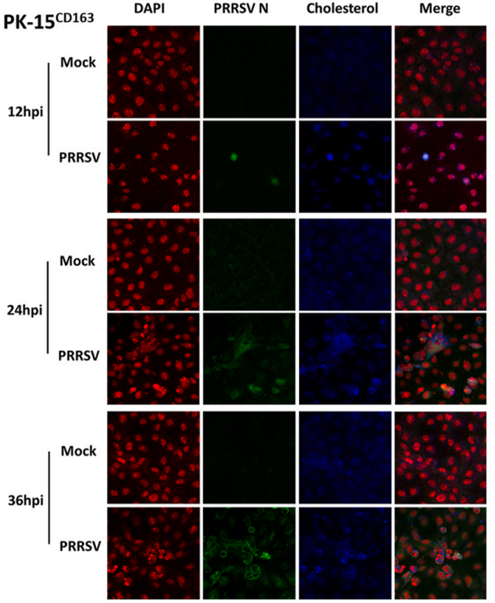
Propidium Iodide purchased from MedChemExpress. Usage Cited in: Redox Biol. 2022 Feb;49:102207. [Abstract]
PK-15CD163 cells were washed with phosphate-buffered saline (PBS), and then incubated with the nuclear dye propidium iodide for 15 min. The nuclei were counterstained with propidium iodide (red). Fluorescent images were acquired with a confocal laser scanning microscope.
-
J Control Release
Combating cisplatin-resistant lung cancer using a coiled-coil lipopeptides modified membrane fused drug delivery system. [Abstract]2025 Jan 8:379:45-58. PMID: 39756686 -
J Control Release
Biofunctional coacervate-based artificial protocells with membrane-like and cytoplasm-like structures for the treatment of persistent hyperuricemia. [Abstract]2024 Jan:365:176-192. PMID: 37992873 -
J Hazard Mater
Isobavachalcone confers protection against Cryptococcus neoformans-induced ferroptosis in Caenorhabditis elegans via lifespan extension and GSH-GPX-1 axis modulation. [Abstract]2025 Mar 18:492:137969. PMID: 40154123 -
EBioMedicine
Influenza A virus dissemination and infection leads to tissue resident cell injury and dysfunction in viral sepsis. [Abstract]2025 May 13:116:105738. PMID: 40367638 -
Mater Today Bio
Okra juice used for rapid wound healing through its bioadhesive and antioxidant capabilities. [Abstract]2025 Jan 15:31:101495. PMID: 39896277 -
Clin Cancer Res
Entinostat enhances the efficacy of chemotherapy in small cell lung cancer through S-phase arrest and decreased base excision repair. [Abstract]2023 Nov 14;29(22):4644-4659. PMID: 37725585 -
Cancer Lett
Tumor-derived CCL5 recruits CCR1+ macrophages to suppress apoptosis and drive proliferation in duodenal adenocarcinoma. [Abstract]2025 Sep 25:634:218064. PMID: 41015267 -
Cell Death Dis
Post-irradiation dietary restriction impairs hematopoiesis via inhibition of the pentose phosphate pathway in hematopoietic stem and progenitor cells. [Abstract]2026 Jan 7;17(1):8. PMID: 41500989 -
Cell Death Dis
Idarubicin combats abiraterone and enzalutamide resistance in prostate cells via targeting XPA protein. [Abstract]2022 Dec 12;13(12):1034. PMID: 36509750 -
Cell Death Dis
Metformin sensitises hepatocarcinoma cells to methotrexate by targeting dihydrofolate reductase. [Abstract]2021 Oct 2;12(10):902. PMID: 34601503 -
NPJ Biofilms Microbiomes
Lactobacillus amylovorus extracellular vesicles mitigate mammary gland ferroptosis via the gut-mammary gland axis. [Abstract]2025 Jun 21;11(1):113. PMID: 40544167 -
Int J Biol Macromol
3β-hydroxysteroid-Δ24 reductase integrates cholesterol metabolism and innate immune to promote PRRSV replication. [Abstract]2025 Apr 7:142867. PMID: 40203946 -
Int J Biol Macromol
A synergistically antimicrobial and antioxidant hyaluronic acid hydrogel for infected wounds. [Abstract]2024 Jun;269(Pt 2):131795. PMID: 38670175 -
Int J Biol Macromol
Engineering of VCAM-1-targeted nanostructured lipid carriers for delivery of melatonin against acute lung injury through SIRT1/NLRP3 mediated pyroptosis signaling pathway. [Abstract]2024 May;266(Pt 1):130637. PMID: 38490396 -
Phytomedicine
β,β-Dimethylacrylalkannin, a key component of Zicao, induces cell cycle arrest and necrosis in hepatocellular carcinoma cells. [Abstract]2024 Aug 19:134:155959. PMID: 39178682 -
ACS Appl Mater Interfaces
Synergistic Proteolysis Targeting Chimera Chemotherapy Conjugate for Potent Non-small Cell Lung Cancer Treatment. [Abstract]2025 Dec 17;17(50):67595-67608. PMID: 41348354 -
ACS Appl Mater Interfaces
Recombinant Trichosanthin-Loaded Nanoparticles with Tumor-Targeting and Cell-Penetrating Capabilities for Activatable Antitumor Therapy. [Abstract]2025 Oct 1;17(39):54423-54436. PMID: 40985241 -
ACS Appl Mater Interfaces
Wound-Healing Material with Antibacterial and Antioxidant Functions, Constructed Using Keratin, Hyperbranched Polymers, and MnO2. [Abstract]2023 Jun 28;15(25):29841-29853. PMID: 37338013 -
J Orthop Translat
Activation of LAMP1-mediated lipophagy by sulforaphane inhibits cellular senescence and intervertebral disc degeneration. [Abstract]2025 Jun 2:53:12-25. PMID: 40525097 -
-
-
Biomed Pharmacother
Targeted-lung delivery of bardoxolone methyl using PECAM-1 antibody-conjugated nanostructure lipid carriers for the treatment of lung inflammation. [Abstract]2024 Aug 5:178:116992. PMID: 39106709 -
Environ Pollut
UV-aged biodegradable and non-biodegradable microplastics further enhance horizontal transfer of antibiotic resistance plasmids both in vitro and in intestinal flora. [Abstract]2025 Sep 10:385:127111. PMID: 40939716 -
Cell Death Discov
Arnicolide D induces endoplasmic reticulum stress-mediated oncosis via ATF4 and CHOP in hepatocellular carcinoma cells. [Abstract]2024 Mar 12;10(1):134. PMID: 38472168 -
Cell Rep
2025 Sep 2;44(9):116215. PMID: 40906555 -
Cell Rep
The Hippo pathway kinase MST1 mediates a feedback loop to maintain NLRP3 inflammasome homeostasis. [Abstract]2025 Jul 26;44(8):116076. PMID: 40716062 -
Microbiol Res
Extracellular Hsp90 of Candida albicans contributes to the virulence of the pathogen by activating the NF-κB signaling pathway and inducing macrophage pyroptosis. [Abstract]2024 Nov 5:290:127964. PMID: 39522202 -
Cell Rep
Oral fecal transplantation enriches Lachnospiraceae and butyrate to mitigate acute liver injury. [Abstract]2023 Dec 27;43(1):113591. PMID: 38153838 -
Cell Rep
Krüppel-like factor 5 rewires NANOG regulatory network to activate human naive pluripotency specific LTR7Ys and promote naive pluripotency. [Abstract]2022 Aug 23;40(8):111240. PMID: 36001968 -
Anal Chem
Chiral Carbon Dots as Optical Probes: Selective Detection of Acetylcholinesterase via Enhanced Photoluminescence. [Abstract]2025 Aug 5;97(30):16142-16150. PMID: 40711814 -
Int J Pharm X
2025 Oct 9:10:100413. PMID: 41142964 -
-
J Agric Food Chem
Profiling Class-Wide Bioactivities of Flavonoids While Minimizing Compound-Specific Effects Using an Equimolar Mixture Strategy. [Abstract]2025 Oct 22. PMID: 41126446 -
J Agric Food Chem
Postbiotics Suppress Helicobacter pylori Adhesion and Survival through a Coaggregation Mechanism. [Abstract]2025 Sep 3;73(35):21976-21986. PMID: 40833828 -
Biomater Adv
A neutrophil membrane-biomimetic drug delivery system enhances the antifungal and anti-inflammatory efficacy of natamycin for fungal keratitis. [Abstract]2026 Jun:183:214761. PMID: 41671926 -
Eur J Med Chem
Development of novel oridonin analogs as specifically targeted NLRP3 inflammasome inhibitors for the treatment of dextran sulfate sodium-induced colitis. [Abstract]2023 Jan 5;245(Pt 2):114919. PMID: 36399877 -
-
-
Biochem Pharmacol
Cabozantinib inhibits necroptosis by targeting MLKL oligomerization and alleviates psoriasis in vivo. [Abstract]2026 Jun:248:117843. PMID: 41747872 -
Biochem Pharmacol
KLF4 suppresses anticancer effects of brusatol via transcriptional upregulating NCK2 expression in melanoma. [Abstract]2024 May:223:116197. PMID: 38583810 -
Virulence
Antibacterial efficacy and mechanism of the novel antimicrobial peptide lachnospirin-1 against Acinetobacter baumannii. [Abstract]2026 Dec 31;17(1):2646808. PMID: 41838520 -
Mar Drugs
Lipotrichaibol A and Trichoderpeptides A-D: Five New Peptaibiotics from a Sponge-Derived Trichoderma sp. GXIMD 01001. [Abstract]2025 Jun 24;23(7):264. PMID: 40710489 -
J Enzyme Inhib Med Chem
Discovery of a selective PI3Kα inhibitor via structure-based virtual screening for targeted colorectal cancer therapy. [Abstract]2025 Dec;40(1):2468852. PMID: 39992303 -
-
-
Cancer Immunol Immunother
FUBP3 mediates MXI1 stability to silence RRAS and hinder MAPK signaling in acute megakaryoblastic leukemia progression. [Abstract]2025 Dec 22;75(1):23. PMID: 41428087 -
-
Inflammation
Exploring the Role of TRAF6-TAK1 Pathway in Podocyte Pyroptosis and Its Implications for Primary Membranous Nephropathy Therapy. [Abstract]2025 Jan 30. PMID: 39883393 -
CNS Neurosci Ther
The neurotrophic activities of brain-derived neurotrophic factor are potentiated by binding with apigenin, a common flavone in vegetables, in stimulating the receptor signaling. [Abstract]2023 Oct;29(10):2787-2799. PMID: 37101380 -
-
Int J Mol Sci
C-Terminal Truncated HBx Facilitates Oncogenesis by Modulating Cell Cycle and Glucose Metabolism in FXR-Deficient Hepatocellular Carcinoma. [Abstract]2023 Mar 8;24(6):5174. PMID: 36982249 -
Int J Mol Sci
Trichosanthin Promotes Anti-Tumor Immunity through Mediating Chemokines and Granzyme B Secretion in Hepatocellular Carcinoma. [Abstract]2023 Jan 11;24(2):1416. PMID: 36674931 -
Pharmaceuticals (Basel)
Puerarin Inhibits Proliferation, Migration and Invasion of Colon Cancer Cells and Induces Apoptosis via Suppression of the PI3K/AKT Signaling Pathway. [Abstract]2025 Sep 16;18(9):1378. PMID: 41011246 -
Pharmaceuticals
2022 Aug 26;15(9):1059. PMID: 36145280 -
-
Int Immunopharmacol
Crocin facilitates peripheral nerve regeneration through modulation of the STAT3/Bcl-2/Beclin-1 signaling axis-mediated autophagic pathway. [Abstract]2025 Sep 5:165:115437. PMID: 40913861 -
Eur J Pharmacol
Targeting the ALKBH5-NLRP3 positive feedback loop alleviates cardiomyocyte pyroptosis after myocardial infarction. [Abstract]2024 Dec 31:177247. PMID: 39746531 -
Int Immunopharmacol
Targeting lipid peroxidation-associated ferroptosis suppresses lung carcinoma progression by regulating cell cycle arrest. [Abstract]2024 Jun 24:138:112518. PMID: 38917528 -
Eur J Pharmacol
GSK-3β-mediated activation of NLRP3 inflammasome leads to pyroptosis and apoptosis of rat cardiomyocytes and fibroblasts. [Abstract]2022 Apr 5;920:174830. PMID: 35182545
Propidium Iodide purchased from MedChemExpress. Usage Cited in: Eur J Pharmacol. 2022 Apr 5;920:174830. [Abstract]
RCMs were stained with Propidium Iodide (PI, 2 μg/mL; 2 h) and analyzed using a Nikon Ti2 microscope.
-
-
Hepatol Commun
Hepatocyte-derived Pumilio1-enriched exosomes inhibit HSC activation by suppressing tropomyosin-4 translation. [Abstract]2025 Jul 21;9(8):e0759. PMID: 40689527 -
Toxicology
Proteasome inhibition induces apoptosis through simultaneous inactivation of MCL-1/BCL-XL by NOXA independent of CHOP and JNK pathways. [Abstract]2024 Aug 6:153906. PMID: 39117261 -
Molecules
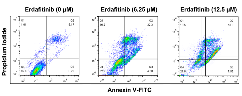
Erdafitinib Inhibits Tumorigenesis of Human Lung Adenocarcinoma A549 by Inducing S-Phase Cell-Cycle Arrest as a CDK2 Inhibitor. [Abstract]2022 Oct 9;27(19):6733. PMID: 36235266
Propidium Iodide purchased from MedChemExpress. Usage Cited in: Molecules. 2022 Oct 9;27(19):6733. [Abstract]
A549 cells were treated with erdafitinib (0, 6.25 and 12.5 μM, 24 h) and cell apoptosis was analyzed by flow cytometry and statistically analyzed. The Annexin V+/PI- and Annexin V+/PI+ cells were considered as early and late apoptotic cells, respectively, and the sum of the above two was calculated as apoptotic cells. Propidium iodide (PI, 50 μg/mL) was added for incubation for 30 min in the dark.
-
Front Microbiol
Cholesterol in viral envelope determines infectivity of SARS-CoV-2 and other coronaviruses. [Abstract]2025 Sep 18:16:1670356. PMID: 41048493 -
Genes Immun
S-nitrosylated NEDD4 exacerbates gouty arthritis by upregulating NOD1 to induce pyroptosis. [Abstract]2025 Aug;26(4):365-375. PMID: 40594913 -
-
J Cell Mol Med
2025 Mar;29(5):e70486. PMID: 40052646 -
Plants
2026 Jan 9;15(2):211. PMID: 41600018 -
iScience
Nitroxoline as an antimicrobial synergist reverses colistin resistance in multidrug-resistant Escherichia coli. [Abstract]2025 Nov 10;28(12):114003. PMID: 41377659 -
Oncol Res
SORBS1 Knockdown Resists S/G2 Arrest and Apoptosis Caused by Polyphyllin H-Induced DNA Damage in Pancreatic Cancer. [Abstract]2025 Aug 28;33(9):2491-2506. PMID: 40918460 -
APL Bioeng
2025 May 27;9(2):026119. PMID: 40438388 -
iScience
The mesenteric adipokine SFRP5 alleviated intestinal epithelial apoptosis improving barrier dysfunction in Crohn's disease. [Abstract]2024 Dec 1;27(12):111517. PMID: 39759008 -
iScience
Zinc finger protein 593 promotes breast cancer development by ensuring DNA damage repair and cell-cycle progression. [Abstract]2024 Dec 3;27(12):111513. PMID: 39758980 -
-
-
ACS Synth Biol
Signal Peptide-Guided Delivery of a Mucin-Like Collagen Analogue for Periplasmic Barrier Reinforcement: A Platform for Enhancing Microbial Survival. [Abstract]2025 Sep 19;14(9):3767-3783. PMID: 40924575 -
Sci Rep
HIIT and MICT mitigate endothelial dysfunction in early atherosclerotic mice via PCSK9 inhibition. [Abstract]2025 Aug 19;15(1):30411. PMID: 40830352 -
Sci Rep
The hydroxamate based HDAC inhibitor WMJ-J-09 induces colorectal cancer cell death by targeting tubulin and downregulating survivin. [Abstract]2025 Jun 4;15(1):19590. PMID: 40467895 -
ACS Chem Neurosci
2025 Feb 5;16(3):374-383. PMID: 39800970 -
Langmuir
2025 Jan 21;41(2):1260-1270. PMID: 39772538 -
J Biol Chem
Her4.3+ radial glial cells maintain the brain vascular network through activation of Wnt signaling. [Abstract]2024 Jul 15:107570. PMID: 39019216 -
Aging (Albany NY)
LOC134466 methylation promotes oncogenesis of endometrial carcinoma through LOC134466/hsa-miR-196a-5p/TAC1 axis. [Abstract]2018 Nov 28;10(11):3353-3370. PMID: 30485833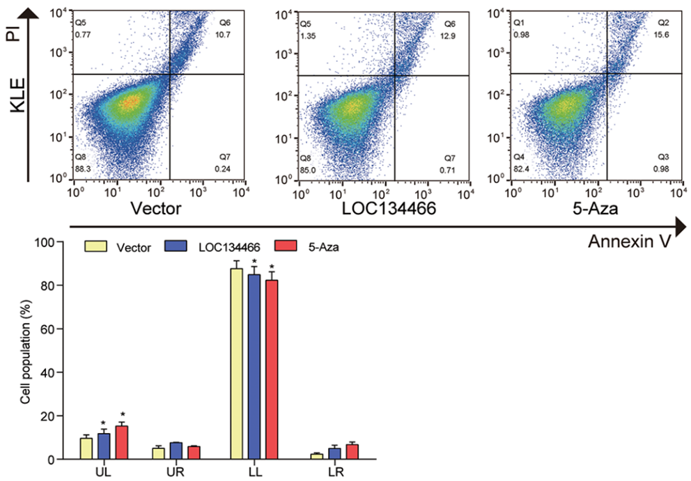
Propidium Iodide purchased from MedChemExpress. Usage Cited in: Aging (Albany NY). 2018 Nov 28;10(11):3353-3370. [Abstract]
KLE cells were stained by 25 μg/mL Propidium Iodide (PI) in fluorescence-activated cell sorting buffer, incubated cells for 30 min in dark environment at room temperature. Cell apoptosis was determined by Annexin V/PI double staining and percentages of cells in each phase (LL, viable; LR, early apoptotic; UL and UR, late apoptotic/necrotic cell) were calculated.
-
-
J Virol
PEX19 restricts porcine deltacoronavirus replication through farnesylation-dependent and -independent mechanisms. [Abstract]2026 Apr 21;100(4):e0209725. PMID: 41874194 -
ACS Infect Dis
Repurposing GSK2018682 Confers Dual Antibacterial and Antibiofilm Activity against Staphylococcus aureus. [Abstract]2026 Feb 13;12(2):623-636. PMID: 41525229 -
Cell Signal
PERK-eIF2alpha-mediated translational inhibition of MCL-1 contributes to potential 2-deoxy-D-glucose and BAD mimetic combinatorial cancer therapy. [Abstract]2025 Dec 19:139:112333. PMID: 41423011 -
Heliyon
Molecular basis of CX-5461-induced DNA damage response in primary vascular smooth muscle cells. [Abstract]2024 Aug 30;10(17):e37227. PMID: 39296007 -
-
Exp Cell Res
Leptin promotes tendon stem/progenitor cell senescence through the AKT-mTOR signaling pathway. [Abstract]2024 Oct 1;442(2):114274. PMID: 39393753 -
Mol Med Rep
Hyperoside decreases the apoptosis and autophagy rates of osteoblast MC3T3‑E1 cells by regulating TNF‑like weak inducer of apoptosis and the p38mitogen activated protein kinase pathway. [Abstract]2019 Jan;19(1):41-50. PMID: 30387825 -
Stem Cells Int
Asymptomatic Hyperuricemia Is Associated with Achilles Tendon Rupture through Disrupting the Normal Functions of Tendon Stem/Progenitor Cells. [Abstract]2022 Dec 1:2022:6795573. PMID: 36504525 -
-
Mol Carcinog
CtBP2 Regulates Wnt Signal Through EGR1 to Influence the Proliferation and Apoptosis of DLBCL Cells. [Abstract]2025 Mar 18. PMID: 40099624 -
-
Front Biosci (Landmark Ed)
Inhibition of p70 Ribosomal S6K1 Protects the Myocardium against Ischemia/Reperfusion-Induced Necrosis through Downregulation of RIP3. [Abstract]2025 Jan 16;30(1):26186. PMID: 39862085 -
-
Curr Issues Mol Biol
Inhibiting the Interaction Between Phospholipase A2 and Phospholipid Serine as a Potential Therapeutic Method for Pneumonia. [Abstract]2025 Jul 4;47(7):516. PMID: 40728986 -
FEBS Lett
Identification of novel anti-leishmanials targeting glutathione synthetase of the parasite: a drug repurposing approach. [Abstract]2025 Feb;599(3):367-380. PMID: 39266470 -
J Pharmacol Sci
Circular RNA circPSAP functions as an efficient miR-331-3p sponge to regulate proliferation, apoptosis and bortezomib sensitivity of human multiple myeloma cells by upregulating HDAC4. [Abstract]2022 May;149(1):27-36. PMID: 35369902 -
Clin Transl Radiat Oncol
Live-cell imaging and analysis of 3D spheroids in hypoxia- and radiotherapy-related research. [Abstract]2025 Jan 15:51:100920. PMID: 39898333 -
BMC Pharmacol Toxicol
Pulrodemstat, a selective inhibitor of KDM1A, suppresses head and neck squamous cell carcinoma growth by triggering apoptosis. [Abstract]2024 Nov 20;25(1):89. PMID: 39567962 -
Blood Sci
BECN1 modulates hematopoietic stem cells by targeting Caspase-3-GSDME-mediated pyroptosis. [Abstract]2020 Jul 25;2(3):89-99. PMID: 35402821 -
Arch Microbiol
Bile and short-chain fatty acid salts affect survival and virulence of Klebsiella Oxytoca of mussel origin. [Abstract]2025 Nov 1;207(12):339. PMID: 41175196 -
Thorac Cancer
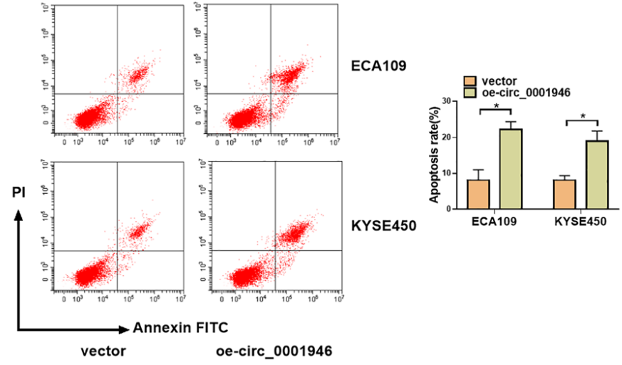
Identification of a novel circ_0001946/miR-1290/SOX6 ceRNA network in esophageal squamous cell cancer. [Abstract]2022 May;13(9):1299-1310. PMID: 35411716
Propidium Iodide purchased from MedChemExpress. Usage Cited in: Thorac Cancer. 2022 May;13(9):1299-1310. [Abstract]
KYSE450 and ECA109 cells after 96 h various transfections were washed three times in cold PBS, stained with Propidium iodide (PI, 50 μg/mL) and Annexin V‐FITC (25 μg/mL) for 20 min in the dark, and analyzed within 1 h.
-
Oncol Lett
2026 Mar 26;31(5):196. PMID: 41947899 -
Biochim Biophys Acta Gen Subj
Targeting the IGF1/Twist1 axis: A novel mechanism for β-elemene-induced anoikis and EMT inhibition in breast cancer cells. [Abstract]2026 Mar;1870(3):130901. PMID: 41490592 -
Biochem Biophys Res Commun
Mitochondrial phosphoenolpyruvate carboxykinase 2 counteracts ferroptosis via catalytic activity independent of mitochondrial stress. [Abstract]2025 Sep 8:778:152383. PMID: 40712389 -
-
Cytotechnology
E2F8-TPX2 axis regulates glycolysis and angiogenesis to promote progression and reduce chemosensitivity of liver cancer. [Abstract]2024 Dec;76(6):817-832. PMID: 39435417 -
STAR Protoc
2023 Sep 19;4(4):102553. PMID: 37729057 -
-
bioRxiv
HCMV infection depends on EGLN1-mediated mitochondrial activation to increase dNTP pools for viral DNA replication. [Abstract]2025 Oct 31:2025.10.31.685607. PMID: 41279967 -
-
-
-
-
-
-
-
-
-
Oxid Med Cell Longev
Programmed NP Cell Death Induced by Mitochondrial ROS in a One-Strike Loading Disc Degeneration Organ Culture Model. [Abstract]2021 Aug 31;2021:5608133. PMID: 34512867
Propidium Iodide purchased from MedChemExpress. Usage Cited in: Oxid Med Cell Longev. 2021 Aug 31;2021:5608133. [Abstract]
The NP tissue slices are incubated with a culture medium containing 1 μg/ml Calcein AM and 1 μg/ml PI for one hour at 37°C. After washing with PBS, the slices are immediately viewed using an inverted confocal laser scanning microscope.
-
-
-


Solvent & Solubility
DMSO : 100 mg/mL (149.61 mM; Need ultrasonic; Hygroscopic DMSO has a significant impact on the solubility of product, please use newly opened DMSO)
H2O : 3.57 mg/mL (5.34 mM; ultrasonic and warming and heat to 60°C)
Please refer to the solubility information to select the appropriate solvent. Once prepared, please aliquot and store the solution to prevent product inactivation from repeated freeze-thaw cycles.
Storage method and period of stock solution: -80°C, 2 years; -20°C, 1 year (sealed storage, away from moisture and light). When stored at -80°C, please use it within 2 years. When stored at -20°C, please use it within 1 year.
* Note: If you choose water as the stock solution, please dilute it to the working solution, then filter and sterilize it with a 0.22 μm filter before use.
Concentration (start) × Volume (start) = Concentration (final) × Volume (final)

Protocol
Flow cytometric analysis: Propidium iodide is prepared in in 0.1% sodium citrate plus 0.1% Triton X-100 (50 μg/mL). The 200 ×g centrifuged cell pellet is gently resuspended in 1.5 mL hypotonlc fluorochrome solution (Propidium iodide 50 μg/mL), in 12×75 polypropylene tubes. The tubes are placed at 4°C in the dark overnight before the flow cytometric analysis. The propidium Iodide fluorescence of individual nuclei is measured using a FACScan flow cytometer. The nuclei traverses the light beam of a 488 nm Argon laser. A 560 nm dichrolc nurror (DM 570) and a 600 nm band pass filter (bandwidth 35 nm) are used for collecting the red fluorescence due to propidium Iodide staining of DNA and the data are registered on a logarithmic scale[2].
MedChemExpress (MCE) has not independently confirmed the accuracy of these methods. They are for reference only.


Purity & Documentation
-
Data Sheet (279 KB)
-
SDS (419 KB)
- English - EN (419 KB)
- Français - FR (419 KB)
- Deutsch - DE (419 KB)
- Norwegian - NO (419 KB)
- Español - ES (419 KB)
- Swedish - SV (419 KB)
- Italian - IT (419 KB)
- Korean - KR (419 KB)
- Portuguese - PT (419 KB)
-
Handling Instructions (2659 KB)
References
[1]. Hezel M, et al. Propidium iodide staining: a new application in fluorescence microscopy for analysis of cytoarchitecture in adult and developing rodent brain. Micron. 2012 Oct;43(10):1031-8. [Content Brief]
[2]. Nicoletti I, et al. A rapid and simple method for measuring thymocyte apoptosis by propidium iodide staining and flow cytometry. J Immunol Methods. 1991 Jun 3;139(2):271-9. [Content Brief]
[3]. A rapid and simple method for measuring thymocyte apoptosis by propidium iodidestaining and flow cytometry. J Immunol Methods. 1991 Jun 3;139(2):271-9. [Content Brief]


Complete Stock Solution Preparation Table
Please refer to the solubility information to select the appropriate solvent. Once prepared, please aliquot and store the solution to prevent product inactivation from repeated freeze-thaw cycles.
Storage method and period of stock solution: -80°C, 2 years; -20°C, 1 year (sealed storage, away from moisture and light). When stored at -80°C, please use it within 2 years. When stored at -20°C, please use it within 1 year.
| Optional Solvent | Concentration Solvent Mass | 1 mg | 5 mg | 10 mg | 25 mg |
|---|---|---|---|---|---|
| H2O / DMSO | 1 mM | 1.4961 mL | 7.4807 mL | 14.9613 mL | 37.4033 mL |
| 5 mM | 0.2992 mL | 1.4961 mL | 2.9923 mL | 7.4807 mL | |
| DMSO | 10 mM | 0.1496 mL | 0.7481 mL | 1.4961 mL | 3.7403 mL |
| 15 mM | 0.0997 mL | 0.4987 mL | 0.9974 mL | 2.4936 mL | |
| 20 mM | 0.0748 mL | 0.3740 mL | 0.7481 mL | 1.8702 mL | |
| 25 mM | 0.0598 mL | 0.2992 mL | 0.5985 mL | 1.4961 mL | |
| 30 mM | 0.0499 mL | 0.2494 mL | 0.4987 mL | 1.2468 mL | |
| 40 mM | 0.0374 mL | 0.1870 mL | 0.3740 mL | 0.9351 mL | |
| 50 mM | 0.0299 mL | 0.1496 mL | 0.2992 mL | 0.7481 mL | |
| 60 mM | 0.0249 mL | 0.1247 mL | 0.2494 mL | 0.6234 mL | |
| 80 mM | 0.0187 mL | 0.0935 mL | 0.1870 mL | 0.4675 mL | |
| 100 mM | 0.0150 mL | 0.0748 mL | 0.1496 mL | 0.3740 mL |
* Note: If you choose water as the stock solution, please dilute it to the working solution, then filter and sterilize it with a 0.22 μm filter before use.


 Powered by Bioz