



VF 647A-Annexin V/PI Apoptosis Detection Kit
Based on 1 publication(s) in Google Scholar
MCE VF 647A-Annexin V/PI Apoptosis Detection Kit can be utilized to detect the level of cell apoptosis.
-
Storage :
4℃, 2 year. Keep away from light, do not freeze.


To place orders, for customer services and technical support, please contact: MedChemExpress USA
Tel: 609-228-6898 E-mail: [email protected] [email protected]



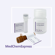


Description & Advantages
Apoptosis is a form of programmed cell death. Based on the cellular changes during programmed cell death, apoptosis is often classified into early, middle, and later stages. The later stages of apoptosis are characterized by changes in nuclear morphology, chromatin condensation, nuclear envelope degradation, and DNA fragmentation.
Annexin V is a 35-36 kDa Ca2+ - dependent phospholipid-binding protein that has a high affinity for PS. In normal live cells, Phosphatidylserine (PS) is located on the inner side of the cell membrane. Upon initiation of apoptosis, PS translocates from the inner to the outer leaflet of the membrane and binds with Annexin V. Therefore, Annexin V serves as one of the sensitive indicator of early-stage apoptosis. Once labeled with a fluorescent probe, Annexin V can be used to detect the early-stage apoptosis using fluorescence microscopy or flow cytometry. Propidium Iodide (PI) is a cell-membrane impermeable dye that can stain late-stage apoptosis cells and necrosis cells with red fluorescence.
MCE VF 647A-Annexin V/PI Apoptosis Detection Kit can be utilized to detect the level of cell apoptosis. After co-staining with Annexin V (labeled with a far-red fluorescent probe (VF 647A)) and PI, normal cells are essentially non-fluorescent (Annexin V-/PI-), early-stage apoptosis cells show far-red fluorescence (Annexin V+/PI-), late-stage apoptosis cells and necrosis cells show red fluorescence (Annexin V+/PI+).


Protocol
1. Cell staining
1. Collect approximately 1 × 10⁵-5 × 10⁵ cells, centrifuge at 300 × g for 5 min at 4°C, discard the supernatant, and wash once with pre-cooled PBS.
2. Gently resuspend cells in 100 μL 1× Annexin V Binding Buffer.
3. Add 5 μL VF 647A-Annexin V and 10 μL PI Staining Solution and mix gently.
4. Incubate for 15 min at room temperature protected from light.
5. Add 400 μL 1× Annexin V Binding Buffer and mix gently.
6. Analyze by flow cytometry preferably within 1 h.
Interpretation
Viable cells are Annexin V-negative/counterstain-negative; early apoptotic cells are Annexin V-positive/counterstain-negative; late apoptotic or necrotic cells are positive for both Annexin V and PI Staining Solution.
Note: Protect stained cells from light and analyze promptly. For blood-derived samples, remove platelets thoroughly because platelet phosphatidylserine may bind Annexin V and interfere with the result.


Publications
-
Journal Impact Factor
-
Most Recent
Storage
4℃, 2 year.
Keep away from light, do not freeze.


Components
| Components | HY-K1093-50 T | HY-K1093-100 T |
|---|---|---|
| Annexin V Binding Buffer | 50 mL | 50 mL × 2 |
| VF 647A-Annexin V | 250 μL | 500 μL |
| PI Staining Solution | 500 μL | 1 mL |


 Please enter the correct Lot Number
