



Anti-Flag Affinity Gel
Based on 6 publication(s) in Google Scholar
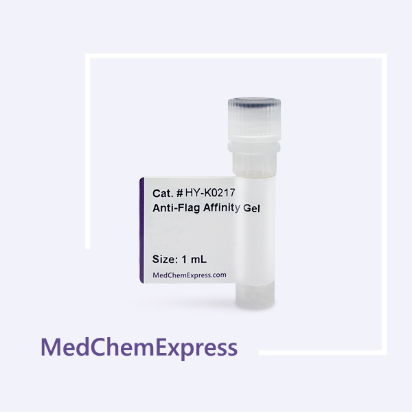
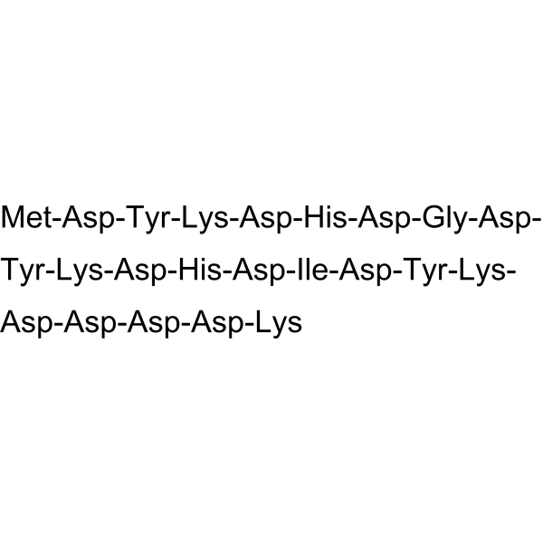
MCE Anti-Flag Affinity Gel is used for immunoprecipitation (IP) or protein purification of specific Flag-tagged (DYKDDDDK) proteins expressed in bacterial and mammalian cells and in vitro expression systems.
-
Storage :
-20°C, 2 years.

To place orders, for customer services and technical support, please contact: MedChemExpress USA
Tel: 609-228-6898 E-mail: [email protected] [email protected]



-
HY-P0319
3X FLAG peptide -
HY-K0207
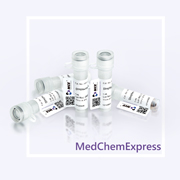
Anti-Flag Magnetic Beads -
HY-K0208
Streptavidin Magnetic Beads -

HY-K0232
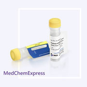
Anti-HA Affinity Gel -
HY-K0010
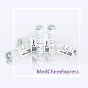
Protease Inhibitor Cocktail (EDTA-Free, 100× in DMSO) -
HY-K0201
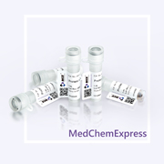
Anti-HA Magnetic Beads -
HY-K0202
Protein A/G Magnetic Beads -
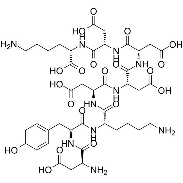
HY-P0223
FLAG peptide -
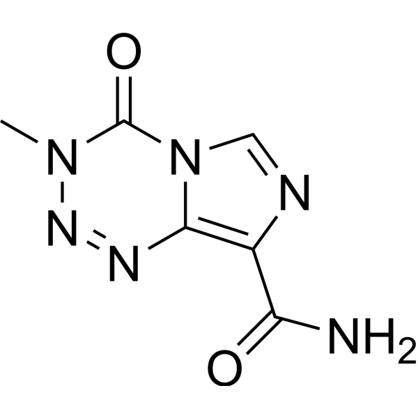
HY-17364
Temozolomide -

HY-10583
Y-27632 dihydrochloride -
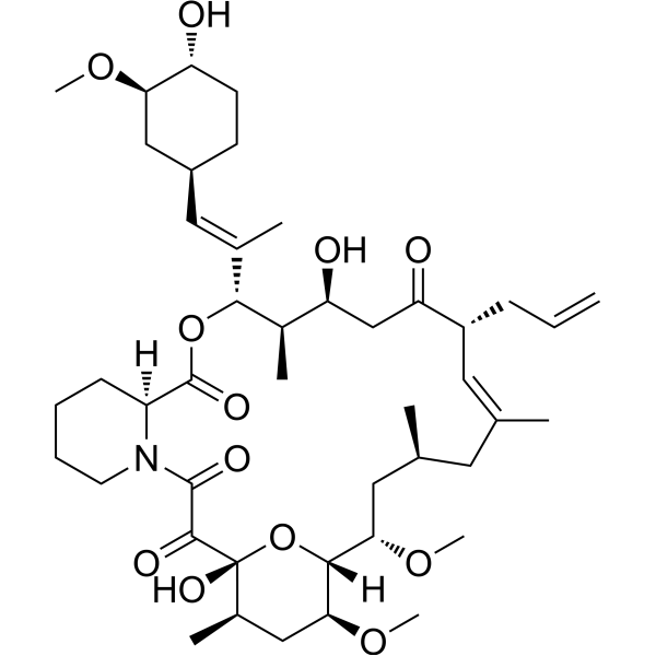
HY-13756
Tacrolimus -

HY-10071
Y-27632


Description & Advantages
MCE Anti-Flag Affinity Gel is a purified mouse IgG2b monoclonal antibody covalently attached to agarose Sepharose 4B. With high protein-binding capacity (>1.1 mg protein/mL) and stability, this product is ideal for high performance purification or immunoprecipitation (IP) of Flag-tagged (DYKDDDDK) proteins expressed in E.coli, yeast, insect and mammalian expression systems.
Features:
1. Convenient and time saving.
2. Low non-specific binding.
3. Minimal samples loss.
4. Protein binding capacity up to 1.1 mg/mL.
The specifications of the product correspond to the actual resin volume, with the resin content of 50%.


Protocol
1. Preparation of Anti-Flag Affinity Gel
1.1 Thoroughly suspend the Anti-Flag Affinity Gel. Transfer 10 μL of the gel suspension (about 5 μL of settled gel) to a clean tube.
1.2 Add 600 μL of Wash Buffer to resuspend the gel. Centrifuge at 10,000 rpm for 30 seconds. Remove the supernatant carefully. Be sure that most of the wash buffer is removed and no gel is discarded. Repeat 3-4 times.
2. Protein Binding
2.1 Add 500 μL of cell lysate (the sample containing Flag-tagged protein) to the cleaned gel from step 1.2. Incubate for 2 hours at 4°C while gently rotating the tube. For higher binding efficiency, please incubate overnight.
2.2 Centrifuge at 10,000 rpm for 30 seconds, and transfer the supernatant to a new cube (the supernatant can be used to determine whether there is residual Flag-tagged protein).
3. Washing
Wash the gel with 500 μL of Wash Buffer, centrifuge at 10,000 rpm for 30 seconds. Remove the supernatant carefully. Be sure that most of the wash buffer is removed and no gel is discarded. Wash until the OD280 of the supernatant liquid<0.05.
4. Elution/Detection
Three elution methods are recommended according to protein characteristics or further usage:
4.1 Elution with SDS-PAGE Loading Buffer for gel electrophoresis and immuoblotting.
Add 50 μL of 1× SDS-PAGE Loading Buffer to each tube. Mix well and boil for 5 minutes. Centrifuge at 10,000 rpm for 30 seconds. Keep the supernatant for SDS-PAGE analysis.
4.2 Elution with Elution Buffer A under acidic condition.
Add 50 μL of Elution Buffer A to each tube. Mix well and incubate for 10 minutes at room temperature. Centrifuge at 10,000 rpm for 30 seconds and transfer the supernatant to a new tube. Add 25 μL of Neutralization Buffer for each 50 μL of eluate to neutralize the low pH, which may help preserve bioactivity of target proteins.
4.3 Elution with Elution Buffer B under native condition.
Add 30-50 μL of Elution Buffer B to each tube. Incubate with gentle shaking or on a rotator for 1 hour at room temperature or 2 hours at 4°C. Centrifuge at 10,000 rpm for 30 seconds and transfer the supernatant to a new tube.
NOTE: For immediate use, store the eluates at 4°C, or store at -20°C for long term storage.


Publications
-
Journal Impact Factor
-
Most Recent
-
Adv Sci (Weinh)Unraveling the Role of MDK-SDC4 Interaction in Pancreatic Cancer-Associated New-Onset Diabetes by Single-Cell Transcriptomic Analysis. [Abstract]2025 Jul 25:e09987. PMID: 40709672
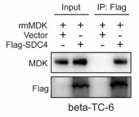
Anti-Flag Affinity Gel purchased from MedChemExpress. Usage Cited in: Adv Sci (Weinh). 2025 Jul 25:e09987. [Abstract]
The co-IP assay analyzed the interaction between SDC4 and MDK.beta‐TC‐6 cells were lysed using RIPA lysis buffer (50 mm Tris‐Base, 150 mm NaCl, 5 mm EDTA, 1% NP‐40, 0.1% SDS, pH 7.4). The cell lysates were then incubated with Anti‐Flag Affinity Gel overnight at 4 °C
-
Adv Sci (Weinh)Branched-Chain Amino Acids Deficiency Promotes Diabetic Neuropathic Pain Through Upregulating LAT1 and Inhibiting Kv1.2 Channel. [Abstract]2024 Sep;11(33):e2402086. PMID: 38946582
Anti-Flag Affinity Gel purchased from MedChemExpress. Usage Cited in: Adv Sci (Weinh). 2024 Sep;11(33):e2402086. [Abstract]
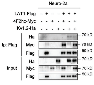
The interactions between Kv1.2 and LAT1, and between 4F2hc and LAT1 in Neuro‐2a cells were examined by co‐immunoprecipitation. the Neuro‐2a cells were lysed with 0.5% NP‐40 buffer (50 mm Tris‐HCl (pH 7.5), 150 mm NaCl, 0.5% NP‐40, 1 µg mL−1 aprotinin, 1 µg mL−1 leupeptin, 1 µg mL−1 pepstatin and 1 mm PMSF). Cell lysates were incubated with Anti‐Flag Affinity Gel (HY‐K0217, MedChemExpress) for 12 h at 4 °C.
-
Prog NeurobiolTranslated retained intron 11 sequence confers pathological properties to Tau in Alzheimer's disease. [Abstract]2026 Feb:257:102872. PMID: 41422990
Anti-Flag Affinity Gel purchased from MedChemExpress. Usage Cited in: Prog Neurobiol. 2026 Feb:257:102872. [Abstract]
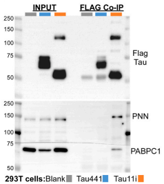
Immunoblot of Flag co-IP from 293 T cells expressing either Flag-Tau441 or Flag-Tau11i. “Blank” indicates non-transfected cells. The input lysate and Co-IP fractions were probed with Flag, PNN and PABPC1 antibodies.
-
Open BiolRNA binding protein AUF1/HNRNPD regulates nuclear export, stability and translation of SNCA transcripts. [Abstract]2023 Nov;13(11):230158. PMID: 37989221
-
Anti-Flag Affinity Gel purchased from MedChemExpress. Usage Cited in: SSRN. 2025 Jun 14.
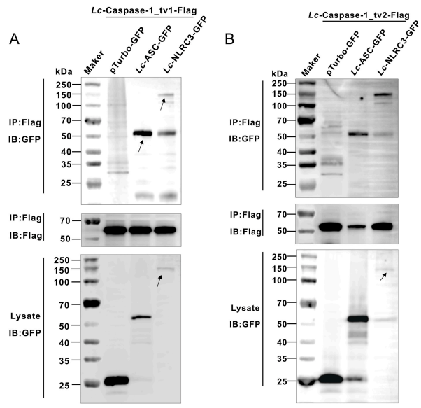
Co-immunoprecipitation (Co-IP) analysis of the interaction between Lc-Caspase-1 transcripts and Lc-ASC/Lc-NLRC3. HEK 293T cells transfected with Flag-Caspase-1_tv1, Flag-Caspase-1_tv2, pTurbo-ASC-GFP, pTurbo-NLRC3-GFP, or pTurboGFP-N (vector control) were lysed, and cell lysates were immunoprecipitated with Anti-Flag agarose beads. Precipitates were subjected to Western blotting analysis using Anti-GFP antibody (upper panels in A and B) and Anti-Flag antibody (middle panels in A and B). Ten percent of cell lysates were used to verify protein expression.
Storage
-20°C, 2 years.


Components
| Contents | HY-K0217-1 mL | HY-K0217-5 mL | HY-K0217-10 mL |
|---|---|---|---|
| MCE Anti-Flag Affinity Gel | 1 mL | 5 mL | 10 mL |


 Please enter the correct Lot Number
