



Mitochondria Isolation Kit for Tissue
Based on 10 publication(s) in Google Scholar
MCE Mitochondria Isolation Kit for Tissue enables the fast and efficient isolation of intact mitochondria from tissue using differential centrifugation.
-
Storage :
-20°C, 1 year.

To place orders, for customer services and technical support, please contact: MedChemExpress USA
Tel: 609-228-6898 E-mail: [email protected] [email protected]



-
HY-K1060
Mitochondria Isolation Kit for Cultured Cells -
HY-B0496
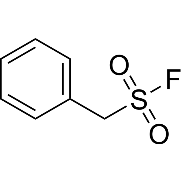
PMSFCathepsin Inhibitor -

HY-K0601
JC-1 Mitochondrial Membrane Potential Assay Kit -
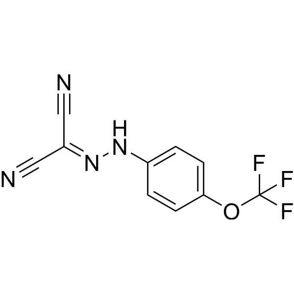
HY-100410
FCCPOXPHOS Uncoupler -
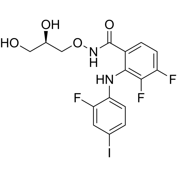
HY-10254
MirdametinibMEK Inhibitor -
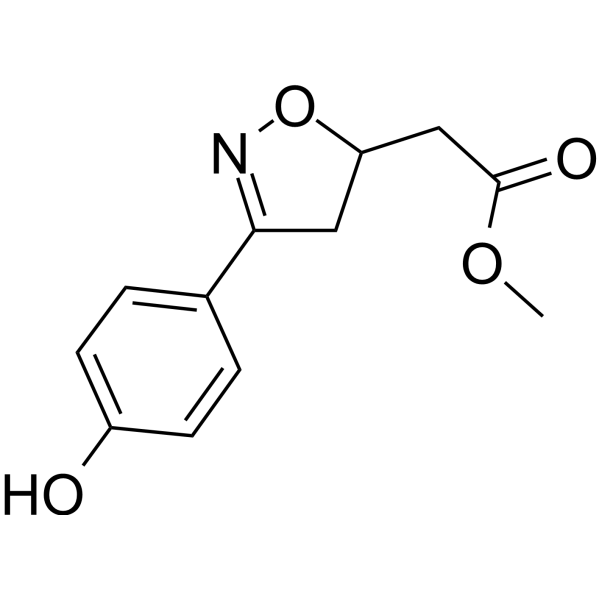
HY-16692
ISO-1MIF Antagonist -

HY-10182
LaduviglusibGSK-3 Inhibitor -
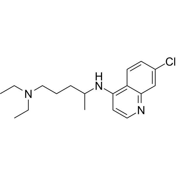
HY-17589A
ChloroquineAntimalarial Agent -
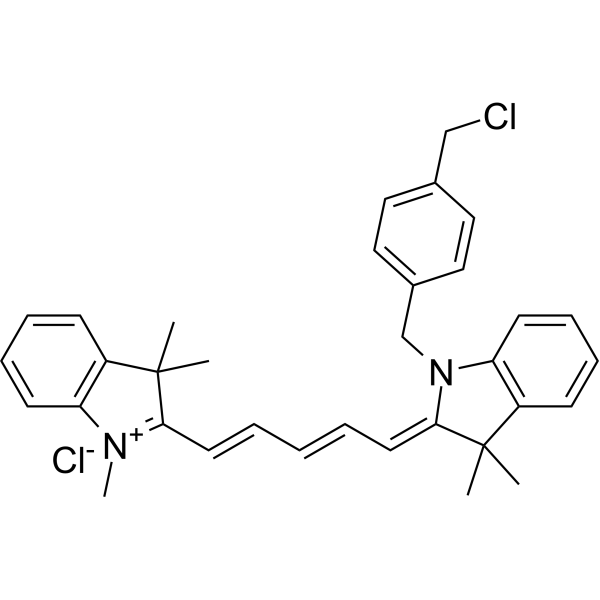
HY-D1783
MitoTracker Deep Red FMRed Fluorescent Dye -

HY-K0314
ATP Assay Kit -
HY-K0010
Protease Inhibitor Cocktail (EDTA-Free, 100× in DMSO)


Description & Advantages
Mitochondria, the site of most energy production in eukaryotic cells, have a double membrane structure: an outer membrane and a folded inner membrane. The key to the preparation of mitochondria is to ensure the integrity and purity of mitochondria.
MCE Mitochondria Isolation Kit for Tissue enables the fast and efficient isolation of mitochondria from tissue using differential centrifugation. Most of the isolated mitochondria have intact inner and outer membranes, as well as physiological functions. In addition, this kit can also be used to extract mitochondrial proteins and mitochondria-free cytoplasmic protein. This kit contains enough reagents for 50-100 isolation procedures from 50-100 mg tissue.


Protocols
Preparation of mitochondria from soft tissues (liver or brain)
1. Use fresh tissue obtained within 1 h of sacrifice and keep it on ice. Do not freeze.
2. Weigh 50-100 mg tissue and wash with PBS.
3. Cut the tissue into the smallest possible pieces and suspend in 10 volumes of pre-cooled Mitochondria Isolation Reagent A containing 1 mM PMSF. For example, add 500 μL Reagent A to 50 mg tissue.
4. Homogenize on ice for approximately 10 strokes.
5. Centrifuge at 600 × g for 5 min at 4°C. For higher-purity mitochondria, 1,000 × g may be used, with a lower mitochondrial yield.
6. Carefully transfer the supernatant to a fresh tube and centrifuge at 11,000 × g for 10 min at 4°C. To reduce lysosome and peroxisome contamination, 3,500 × g may be used, with a lower mitochondrial yield.
7. Carefully remove the supernatant; the pellet contains mitochondria. To obtain mitochondria-free cytoplasmic protein, centrifuge the supernatant further at 12,000 × g for 10 min at 4°C and collect the supernatant for BCA or Bradford analysis.
Preparation of mitochondria from hard tissues (heart or skeletal muscle)
1. Use fresh tissue obtained within 1 h of sacrifice and keep it on ice. Do not freeze.
2. Weigh 50-100 mg tissue and wash with PBS.
3. Cut into small pieces, suspend in 10 volumes of pre-cooled PBS, incubate on ice for 3 min, centrifuge at 600 × g for 10-20 sec, and discard the supernatant.
4. Suspend in 8 volumes of pre-cooled Trypsin Buffer, incubate on ice for 20 min, centrifuge at 600 × g for 10-20 sec, and discard the supernatant.
5. Suspend in 2 volumes of the appropriate pre-cooled Mitochondria Isolation Reagent, centrifuge at 600 × g for 10-20 sec, and discard the supernatant. Use Reagent A for heart muscle, Reagent B for skeletal muscle, and Reagent A for other tissues.
6. Suspend in 8 volumes of the appropriate Mitochondria Isolation Reagent containing 1 mM PMSF and homogenize on ice for 20-30 strokes.
7. Centrifuge at 600 × g for 5 min at 4°C.
8. Carefully transfer the supernatant to a fresh tube and centrifuge at 11,000 × g for 10 min at 4°C.
9. Carefully remove the supernatant; the pellet contains mitochondria. Obtain mitochondria-free cytoplasmic protein as described for soft tissues.
Application of mitochondria
a. For applications requiring intact mitochondria, add 40 μL Mitochondria Storage Buffer per 100 mg tissue.
b. For mitochondrial protein characterization or functional assays, add 150-200 μL Mitochondria Lysis Buffer containing 1 mM PMSF per 50-100 mg tissue. The expected protein concentration is approximately 10-20 mg/mL. Before protein determination, centrifuge at 12,000 × g for 3-5 min at 4°C.


Publications
-
Journal Impact Factor
-
Most Recent
-
Cell Stem CellENPP1 blockade with a humanized monoclonal antibody enhances renal repair after acute kidney injury. [Abstract]2026 Jun 16:S1934-5909(26)00203-1. PMID: 42302792
-
Nat CommunCLPX acquires an iron-sulfur cluster to sustain mitochondrial proteostasis in cancer cells. [Abstract]2026 Jun 9;17(1):5072. PMID: 42265110
-
J Adv ResATOX1 deficiency induces memory impairment via promoting cuproptosis in Alzheimer's disease. [Abstract]2026 Jun 5:S2090-1232(26)00462-5. PMID: 42250721
-
Cell Rep MedA preclinical candidate of cyclophilin D inhibition improves alcohol-associated liver injury. [Abstract]2026 Mar 17;7(3):102654. PMID: 41850244
-
Int J Mol SciThe Effect of Prohibitins on Mitochondrial Function during Octopus tankahkeei Spermiogenesis. [Abstract]2023 Jun 12;24(12):10030. PMID: 37373178
Storage
-20°C, 1 year.


Components
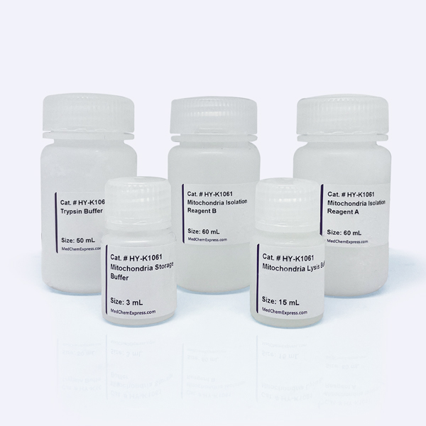
| Components | HY-K1061 |
|---|---|
| Mitochondria Isolation Reagent A | 60 mL |
| Mitochondria Isolation Reagent B | 60 mL |
| Trypsin Buffer | 50 mL |
| Mitochondria Storage Buffer | 3 mL |
| Mitochondria Lysis Buffer | 15 mL |


 Please enter the correct Lot Number
