



ATP Assay Kit
Based on 42 publication(s) in Google Scholar
MCE ATP Assay Kit provides a rapid method to measure intracellular ATP. The kit is suitable for ATP determination in cells, tissue, and other biological samples.
-
Storage :
-20°C, 6 months, -80°C, 1 year. Keep away from light and avoid repeat freeze-thaw cycles.


To place orders, for customer services and technical support, please contact: MedChemExpress USA
Tel: 609-228-6898 E-mail: [email protected] [email protected]



-
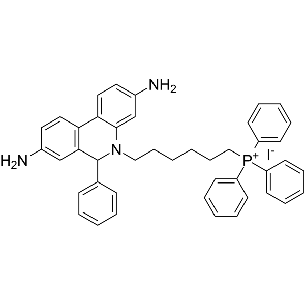
HY-D1055
MitoSOX Red -

HY-K0601
JC-1 Mitochondrial Membrane Potential Assay Kit -
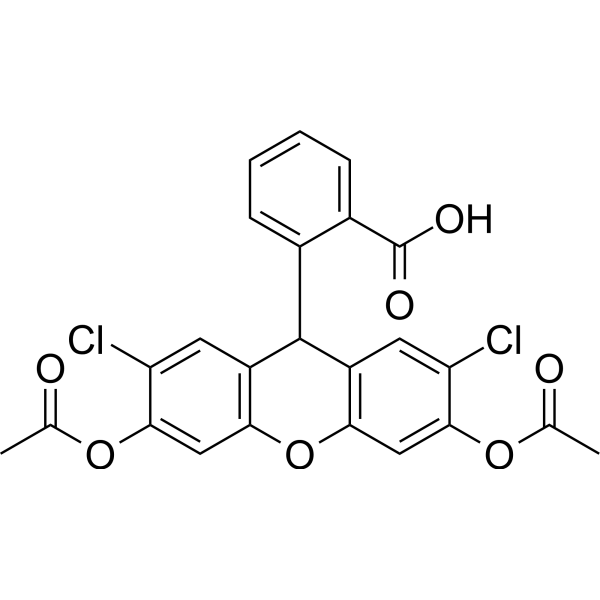
HY-D0940
H2DCFDA -

HY-100558
Bafilomycin A1 -
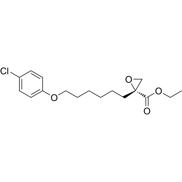
HY-50202
Etomoxir -
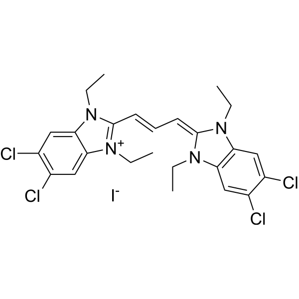
HY-15534
JC-1 -

HY-K0301
Cell Counting Kit-8 -

HY-K0313
NAD+/NADH Assay Kit (WST-8) -
HY-K0010
Protease Inhibitor Cocktail (EDTA-Free, 100× in DMSO) -

HY-100579
Ferrostatin-1 -

HY-B0215
Acetylcysteine -

HY-K1090
Cytotoxicity LDH Assay Kit


Description & Advantages
MCE ATP Assay Kit provides a rapid method to measure intracellular ATP. The principle is that ATP reacts with the substrate luciferin under the action of Firefly Luciferase to produce fluorescence. When both Luciferase and luciferin are in excess, the fluorescence intensity is proportional to the ATP concentration within a certain range. By measuring the fluorescence intensity, the ATP concentration in the sample can be determined.
The kit is suitable for ATP determination in cells, tissue, and other biological samples. A single package of this kit (100T) is sufficient to test at least 100 samples.


Protocol
Reagent Preparation
1. 1× ATP Lysis Buffer: Dilute an appropriate amount of ATP Detection Reagent C (10×) 10-fold with 1× ATP Detection Buffer to prepare 1× ATP Lysis Buffer.
2. ATP Detection Buffer: Thaw the required reagents on ice in advance. Based on 100 μL ATP Detection Working Solution required for each sample or standard, dilute an appropriate amount of ATP Detection Buffer (20×) 20-fold with ultrapure water to prepare 1× ATP Detection Buffer. Keep the prepared buffer on ice.
3. ATP Detection Working Solution: Prepare the working solution by adding 40 μL ATP Detection Reagent A (100×), 40 μL ATP Detection Reagent D (100×), and 400 μL ATP Detection Solution B (10×) to every 3520 μL ATP Detection Buffer. Mix gently and keep on ice protected from light. Prepare fresh according to the actual number of wells and use immediately.
4. ATP Standard Solutions: Serially dilute the 0.5 mM ATP Standard Solution with 1× ATP Lysis Buffer to prepare ATP standards at 0, 0.01, 0.03, 0.1, 0.3, 1, 3, and 10 μM.
Sample Preparation
1. Adherent Cells: Remove the culture medium and wash the cells once with PBS. For cells cultured in a 6-well plate, add 150-200 μL of pre-chilled 1× ATP Lysis Buffer to each well. Lyse the cells thoroughly and collect the lysate.
2. Suspension Cells: Collect the cells and centrifuge at 600 × g for 5 min. Discard the supernatant. Add 200 μL of pre-chilled 1× ATP Lysis Buffer per 1 × 10⁶ cells and lyse thoroughly.
3. Tissue Samples: Weigh 10-30 mg of tissue. Add 100-200 μL of pre-chilled 1× ATP Lysis Buffer per 10 mg of tissue and homogenize thoroughly on ice.
4. Centrifuge the cell or tissue lysates at 12,000 × g for 5 min at 4°C. Collect the supernatant and keep it on ice for subsequent detection.
Detection Procedure
1. Equilibrate the ATP Detection Working Solution and samples to the temperature required for detection. Add 100 μL of ATP Detection Working Solution to each well of a white opaque 96-well plate.
2. Incubate at room temperature for 3-5 min to allow the background luminescence to stabilize.
3. Add 10 μL of ATP standard solution or sample supernatant to each well and mix immediately. It is recommended to test each sample in 2-3 replicate wells.
4. Immediately measure the luminescence using a chemiluminescence microplate reader. The integration time may be set to 0.5-1 s, and identical detection settings should be used for all wells.
Data Analysis
Plot the ATP standard concentration on the x-axis and the blank-subtracted luminescence values on the y-axis to generate a standard curve, and calculate the ATP concentration of each sample. Multiply the calculated value by the sample dilution factor, and normalize the results to cell number, tissue weight, or protein content as appropriate.


Publications
-
Journal Impact Factor
-
Most Recent
-
Adv MaterSelf-Manipulating Sodium Ion Gradient-Based Endogenic Electrical Stimulation Dressing for Wound Repair. [Abstract]2025 Apr;37(13):e2419149. PMID: 39951003
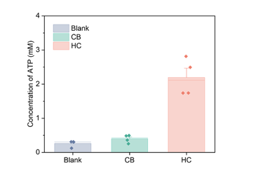
ATP Assay Kit purchased from MedChemExpress. Usage Cited in: Adv Mater. 2025 Apr;37(13):e2419149. [Abstract]
ATP content in HaCaT cells.
-
TheranosticsHilnc- mediated UCP1 translation repression contributes to thermogenesis and energy expenditure. [Abstract]2026 Jan 1;16(7):3263-3285. PMID: 41608567
-
Mater Today BioSLC7A11-independent disulfidptosis induced by a two-pronged delivery strategy for bladder cancer chemotherapy and cisplatin-resistance reverse. [Abstract]2025 Nov 13:35:102547. PMID: 41322137
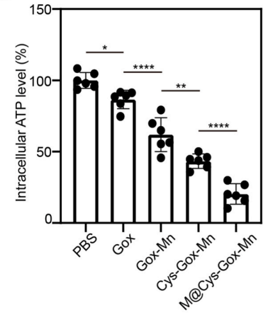
ATP Assay Kit purchased from MedChemExpress. Usage Cited in: Mater Today Bio. 2025 Nov 13:35:102547. [Abstract]
The ATP levels in the T24 cells after treated by Gox, Gox-Mn NPs, Cys-Gox-Mn NPs, and M@Cys-Gox-Mn NPs, respectively. Gox-equivalent dose: 0.2 μg/ml, 2 h incubation.
-
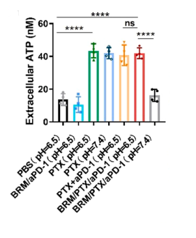
ATP Assay Kit purchased from MedChemExpress. Usage Cited in: Nano Today. 2025 Jun.
Results of extracellular ATP released by 4T1 cells incubated with different formulations for 8 h.
-
J Hazard Mater6PPD and 6PPDQ exposure promote the proliferation and migration of non-small cell lung cancer cells through PTEN dysfunction and ARG2-mediated metabolic reprogramming. [Abstract]2025 Sep 5:495:139088. PMID: 40602111
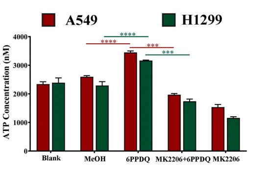
ATP Assay Kit purchased from MedChemExpress. Usage Cited in: J Hazard Mater. 2025 Sep 5:495:139088. [Abstract]
Changes in ATP levels in cells were detected using the luciferase method after 24 h of MK2206 and 6PPDQ treatments on A549 and H1299 cells.
-
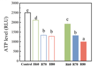
ATP Assay Kit purchased from MedChemExpress. Usage Cited in: Innov Food Sci Emerg Technol. 2025 Nov 21;107:104375.
ATP level of A. niger during heat and RF treatment.
-
Curr Res ToxicolThe role of autophagy in microwave radiation induced toxicity in iPSC-derived cardiomyocytes. [Abstract]2026 Mar 10:10:100288. PMID: 41847319
-
PLoS OneEpimedin C enhances mitochondrial energy supply by regulating the interaction between MIC25 and UBC in rodent model. [Abstract]2025 May 28;20(5):e0325031. PMID: 40435285
Storage
-20°C, 6 months, -80°C, 1 year.
Keep away from light and avoid repeat freeze-thaw cycles.


Components
| Components | HY-K0314-100 T |
|---|---|
| ATP Detection Reagent A (100×) | 100 μL |
| ATP Detection Reagent B (10×) | 1 mL |
| ATP Detection Reagent C (10×) | 3 mL |
| ATP Detection Reagent D (100×) | 100 μL |
| ATP Standard Solution (0.5 mM) | 250 μL |
| ATP Detection Solution (20×) | 1.8 mL |


 Please enter the correct Lot Number
