



Protein G Agarose
Based on 11 publication(s) in Google Scholar
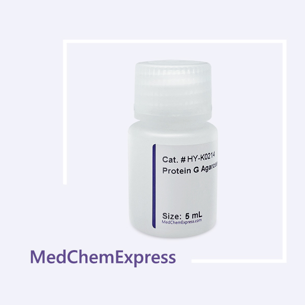
MCE Protein G Agarose, a 4% highly cross-linked agarose reagent coupled with recombinant Protein G, effectively purifies mammalian monoclonal and polyclonal antibodies, such as human IgG3 and rat IgG2a.
-
Storage :
Store at 2-8°C, and is stable for at least 2 years. Do not dry or freeze.

To place orders, for customer services and technical support, please contact: MedChemExpress USA
Tel: 609-228-6898 E-mail: [email protected] [email protected]



-
HY-K0213
Protein A Agarose -

HY-K0230
Protein A/G Agarose -
HY-K0202
Protein A/G Magnetic Beads -

HY-18739
Phorbol 12-myristate 13-acetatePKC Activator -
HY-K0215
Protein L Agarose -
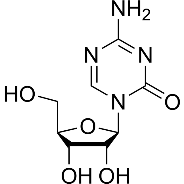
HY-10586
5-AzacytidineDNMT Inhibitor -
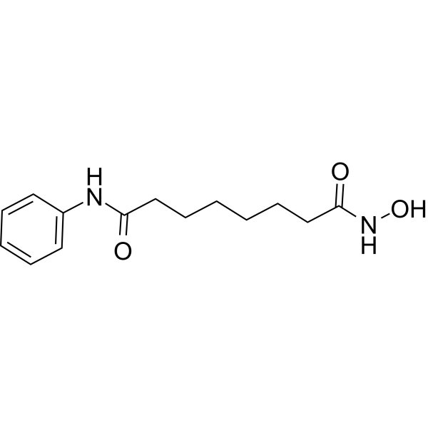
HY-10221
VorinostatHDAC Inhibitor -
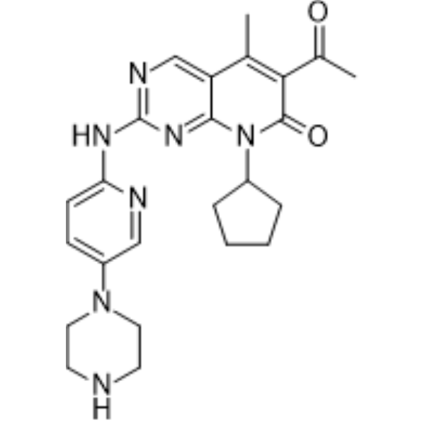
HY-50767
PalbociclibCDK4/6 Inhibitor -

HY-13259
MG-132Proteasome Inhibitor -

HY-10431
SB-431542TGF-β Receptor Kinase Inhibitor -
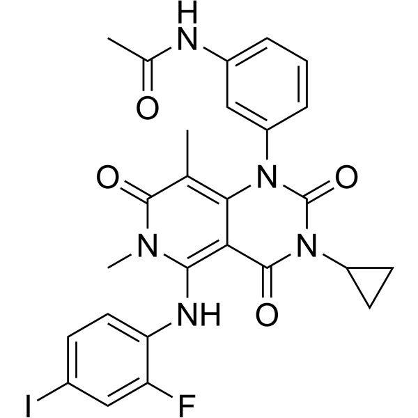
HY-10999
TrametinibMEK Inhibitor -
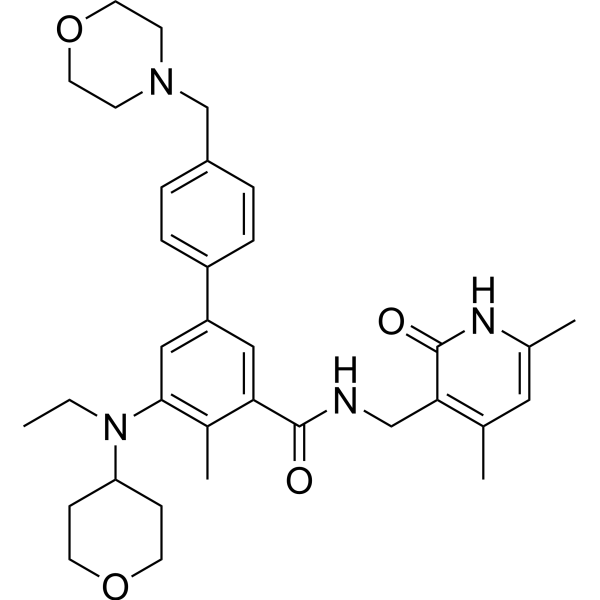
HY-13803
TazemetostatEZH2 Inhibitor


Description & Advantages
Protein G is a bacterial cell wall protein isolated from group G Streptococci and binds to most mammalian IgGs mainly through Fc regions. Native Protein G contains 3 IgG binding domains and sites for albumin and cell surface binding. The latter have been eliminated from recombinant Protein G to reduce nonspecific binding.
MCE Protein G Agarose, a 4% highly cross-linked agarose reagent coupled with recombinant Protein G, effectively purifies mammalian monoclonal and polyclonal antibodies, such as human IgG3 and rat IgG2a.
| Characteristics | |
|---|---|
| Composition | 4% cross-linked agarose |
| Degree of Substitution | 2 mg Recombinant Protein G/mL Settled Resin |
| Bead Diameter | 45-165 μm |
| Binding Capacity | 20 mg Human IgG/mL Settled Resin |
| Storage Solution | 50% slurry in 1× PBS containing 20% ethanol |
The specifications of the product correspond to the actual resin volume, with the resin content of 50%.


Protocols
1. Preparation of Buffers :
| Equilibration Buffer | 20 mM Na2HPO4, 0.15 M NaCl, pH 7.0 |
| Elution Buffer | 0.1 M Glycine, pH 2.5 |
| Neutralization Buffer | 1 M Tris-HCl, pH 8.5 |
It is recommended to prepare buffers using high‑purity water and sterilize them by filtration through a 0.45 μm membrane after preparation.
2. Sample Processing:
(1) To ensure efficient binding of antibody samples to Protein G Agarose, dilute serum, ascites, etc. with equilibration buffer at a ratio of at least 1:1; alternatively, dialyze the sample overnight in equilibration buffer.
(2) Centrifuge or filter the sample through a 0.45 μm membrane before loading to reduce impurities, improve purification efficiency, and prevent column clogging.
3. Packing of Protein G Agarose:
(1) Rinse the bottom frit of the column with deionized water to ensure no air bubbles remain. Close the column outlet and leave 1–2 cm of deionized water at the bottom.
(2) Mix the Protein G Agarose slurry thoroughly and carefully pour the suspension into the column. Using a glass rod to guide the slurry along the column wall can help reduce bubble formation.
(3) Open the column outlet and initially allow the buffer to flow slowly through the column. Gradually increase the flow rate to 1 mL/min to avoid hydraulic shock to the forming column bed and achieve optimal packing. Mark the column bed height.
4. Sample Purification:
(1) Equilibration: Use at least 5 column volumes of equilibration buffer to equilibrate the column. Allow the buffer to flow out slowly at approximately 1 mL/min.
(2) Sample loading: Load the sample using a pump or syringe. Allow the sample to flow out slowly at approximately 0.5–1 mL/min. Collect the flow‑through if necessary. Repeated or cyclic loading may improve yield.
(3) Washing: Wash the column with equilibration buffer at approximately 2 mL/min until the OD280 UV absorbance reaches a stable baseline (typically 15–30 column volumes). Collect the flow‑through.
(4) Elution: After washing, elute the column with 10–15 column volumes of elution buffer at approximately 1 mL/min. Collect the eluted fractions and immediately add neutralization buffer (1/10 of the elution volume) to adjust the pH to 7.4.
(3) SDS-PAGE analysis: Analyze the purified antibody samples (including flow‑through, wash fractions, and elution fractions) as well as the original sample by SDS‑PAGE to evaluate purification efficiency.
5. Resin Regeneration:
Wash the resin with 10 column volumes of hydrochloric acid solution (6 M, pH 8.0) or elution buffer, then immediately wash with 5 column volumes of equilibration buffer. Protein G Agarose can be regenerated up to 10 times without significant loss of binding capacity.
6. Resin Storage:
Protein G Agarose should be stored in equilibration buffer containing 20% ethanol at 2–8°C. Do not freeze.


Publications
-
Journal Impact Factor
-
Most Recent
-
Adv Sci (Weinh)2025 Nov 30:e11683. PMID: 41319268
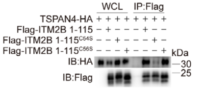
Protein G Agarose purchased from MedChemExpress. Usage Cited in: Adv Sci (Weinh). 2025 Nov 30:e11683. [Abstract]
ITM2B truncation or its point mutants and TSPAN4 were expressed in 786‐O cells, their interactions were detected. Protein G Agarose was used in this study (3 h).
-
Int J Biol MacromolIdentification and epitope mapping of monoclonal antibodies against p150 protein of African swine fever virus. [Abstract]2025 Oct 16;331(Pt 2):148358. PMID: 41109379
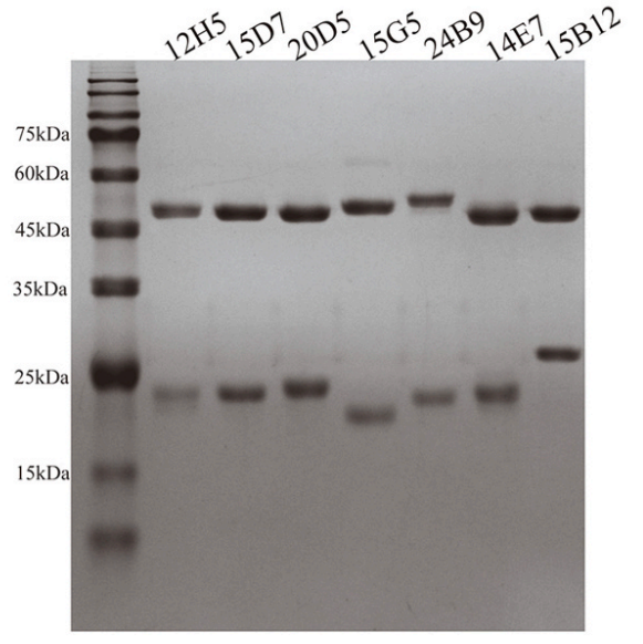
Protein G Agarose purchased from MedChemExpress. Usage Cited in: Int J Biol Macromol. 2025 Oct 16;331(Pt 2):148358. [Abstract]
Sodium dodecyl sulfate–polyacrylamide gel electrophoresis (SDS–PAGE) analysis of purified monoclonal antibodies. The results demonstrated that all seven mAbs were purified from mouse ascites via protein G affinity chromatography, yielding highly pure antibodies. In this experiment, Protein G Agarose (HY-K0214) was used for the purification of mouse ascites.
-
Int J Biol MacromolDuck plague virus UL13 hijacks an RNA-binding protein, interferon induced protein with tetratricopeptide repeats 5, to promote replication. [Abstract]2025 Sep 9;328(Pt 1):147576. PMID: 40935035
Protein G Agarose purchased from MedChemExpress. Usage Cited in: Int J Biol Macromol. 2025 Sep 9;328(Pt 1):147576. [Abstract]
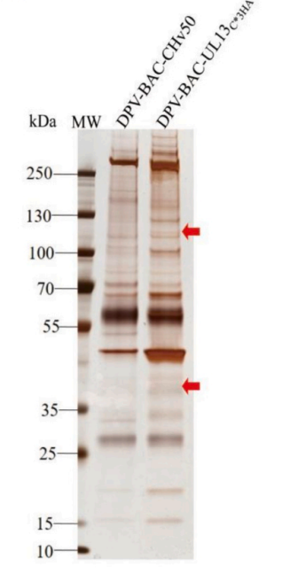
DPV-BAC-CHv50 and DPV-BAC-UL13C3⁎HA were used to infect DEFs, and silver staining was performed 36 h later to detect specific bands. Protein G Agarose (HY-K0214, 2 h) was used in this experiment.
Protein G Agarose purchased from MedChemExpress. Usage Cited in: Int J Biol Macromol. 2025 Sep 9;328(Pt 1):147576. [Abstract]
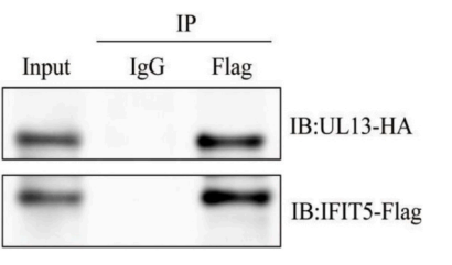
pCAGGS-IFIT5-Flag and pCAGGS-UL13-HA were transfected into duck embryo fibroblasts, and the interaction between IFIT5-Flag and UL13-HA was verified by co-immunoprecipitation. Protein G Agarose (HY-K0214, 2 h) was used in this experiment.
-
Int Immunopharmacol(+)-a-Longipinene alleviates murine laser-induced choroidal neovascularization: Evidence from in vivo and in vitro experiments. [Abstract]2025 Jun 26:159:114913. PMID: 40412129
Protein G Agarose purchased from MedChemExpress. Usage Cited in: Int Immunopharmacol. 2025 Jun 26:159:114913. [Abstract]
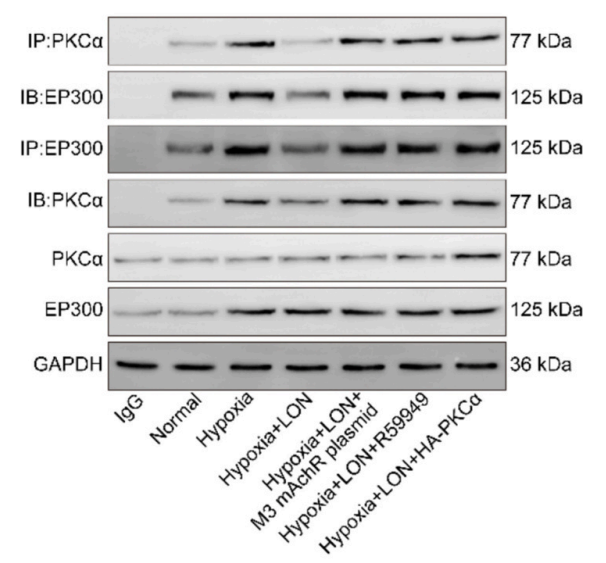
Immunoprecipitation (IP) was performed using a PKCα or a EP300 antibody to observe the interaction of PKCα and EP300. PKCα and EP300 protein levels in the lysates of HCECs were detected using WB. Protein G Agarose (HY-K0214) was used in this experiment.
Storage
Store at 2-8°C, and is stable for at least 2 years.
Do not dry or freeze.


Components
| Components | HY-K0214-5 mL | HY-K0214-10 mL | HY-K0214-50 mL |
|---|---|---|---|
| Protein G Agarose (Settled Resin) | 5 mL | 10 mL | 50 mL |


 Please enter the correct Lot Number
