



ROS Assay Kit
Based on 14 publication(s) in Google Scholar
MCE ROS Assay Kit utilizes the fluorescent probe DCFH-DA to detect ROS.
-
Storage :
-20℃, 1 year. Avoid repeated freeze-thaw cycles.


To place orders, for customer services and technical support, please contact: MedChemExpress USA
Tel: 609-228-6898 E-mail: [email protected] [email protected]



-

HY-K0601
JC-1 Mitochondrial Membrane Potential Assay Kit -

HY-K0319
Lipid Peroxidation (MDA) Assay Kit -

HY-K0311
GSH/GSSG Assay Kit -

HY-K0314
ATP Assay Kit -

HY-100579
Ferrostatin-1 -

HY-K0301
Cell Counting Kit-8 -

HY-K1090
Cytotoxicity LDH Assay Kit -
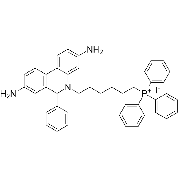
HY-D1055
MitoSOX Red -
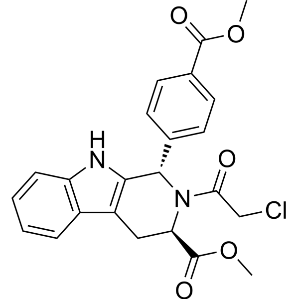
HY-100218A
RSL3 -
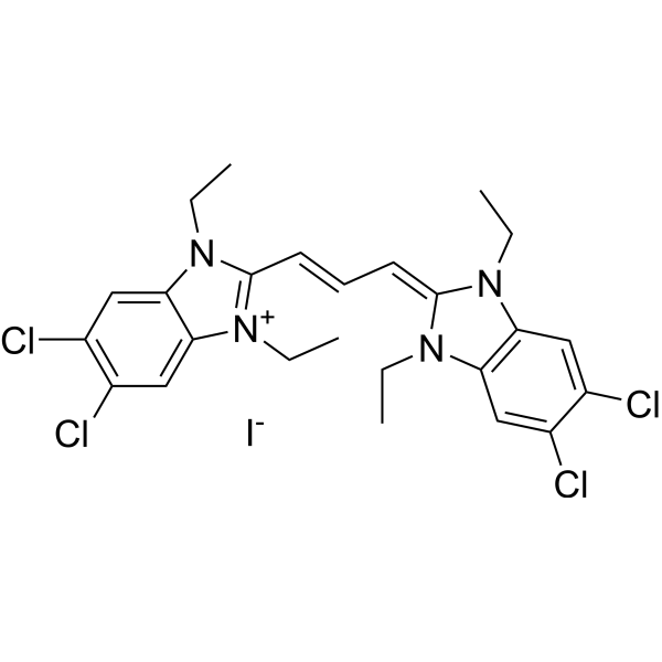
HY-15534
JC-1 -

HY-K1073
Annexin V-FITC/PI Apoptosis Detection Kit -

HY-K0322
Cell Ferrous Iron (Fe2+) Assay Kit (Fluorometric)


Description & Advantages
Reactive oxygen species (ROS) are generated as a result of the reduction of oxygen during aerobic respiration and by various enzymatic systems within the cell. At physiological levels, ROS contribute to cell signaling and host defense. Increased ROS generation, above the detoxification capacity of the biological system, results in oxidative stress and cellular damage. The main damage to cells results from the ROS-induced alteration of macromolecules such as polyunsaturated fatty acids in membrane lipids, essential proteins, and DNA. ROS has been implicated in disease states, such as Alzheimer′s and Parkinson′s disease, cancer, and aging.
MCE ROS Assay Kit utilizes the fluorescent probe DCFH-DA to detect ROS. The principle is that DCFH-DA initially has no fluorescence and can penetrate the cell membrane, be hydrolyzed by intracellular esterases into DCFH which cannot penetrate the cell membrane, and the intracellular ROS can oxidize the non-fluorescent DCFH to generate fluorescent DCF. This fluorescence is then detected using a fluorescence microscope, flow cytometer, or laser confocal microscope to analyze the levels of ROS within the cell.
Features of MCE ROS (Reactive Oxygen Species) Assay Kit
1. Positive control: Provides an ROS positive control reagent (Rosup, 50 mg/mL)
2. High sensitivity: High sensitivity, low background and wide linear range


Protocol
Preparation of DCFH-DA Working Solution
Dilute DCFH-DA (10 mM) 1:1000 with serum-free culture medium to obtain a 10 μM working solution. Prepare immediately before use and protect from light.
Adherent Cells
1. Complete drug or other experimental treatments as designed. Remove the culture medium and gently wash once with serum-free medium.
2. Add sufficient 10 μM DCFH-DA working solution to cover the cells, for example approximately 1 mL per well of a 6-well plate.
3. Incubate at 37°C for 20 min protected from light.
4. Wash the cells three times with serum-free medium to thoroughly remove extracellular DCFH-DA.
5. Analyze by fluorescence microscopy, microplate reader, or flow cytometry. Recommended excitation/emission wavelengths are 488/525 nm.
Suspension Cells
1. Collect cells and adjust the cell density to 1 × 10⁶-2 × 10⁷ cells/mL.
2. Resuspend cells in 10 μM DCFH-DA working solution and incubate at 37°C for 20 min protected from light. Gently invert the tube every 3-5 min to ensure adequate contact between the probe and cells.
3. Centrifuge at 600 × g for 5 min, discard the supernatant, and wash the cells three times with serum-free medium.
4. Resuspend the cells in PBS or serum-free medium and immediately analyze by flow cytometry or microplate reader. Recommended excitation/emission wavelengths are 488/525 nm.
Positive Control
Dilute ROS Positive Control Reagent (Rosup, 50 mg/mL) 1:1000 with serum-free medium to 50 μg/mL. Treat cells with the Rosup working solution at 37°C for 20-30 min, then perform DCFH-DA staining according to the procedure for the corresponding cell type.
Note: Sensitivity to Rosup differs among cell types. Optimize the concentration and treatment time according to cell condition. Protect samples from light throughout the procedure and analyze immediately after staining whenever possible.


Publications
-
Journal Impact Factor
-
Most Recent
-
J Control ReleaseBioswitchable intracellular delivery of small activating RNA by tetrahedral framework nucleic acid: Application to p21-mediated anti-tumor therapy. [Abstract]2026 Jan 10:389:114488. PMID: 41330514
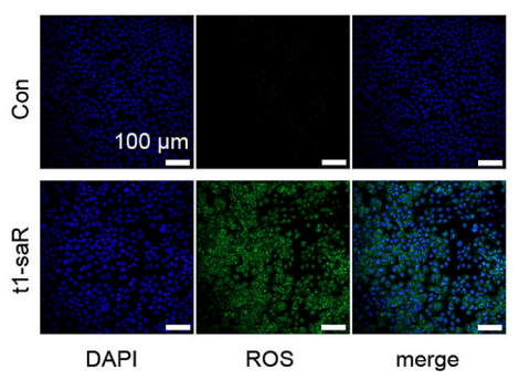
ROS Assay Kit purchased from MedChemExpress. Usage Cited in: J Control Release. 2026 Jan 10:389:114488. [Abstract]
SCC-9 cells stained for intracellular ROS and DAPI according to the instructions of the ROS Assay Kit and observation using LSCM.
-
Phytother ResPaeoniflorin Ameliorates Liver Fibrosis by Inhibiting HIF-1α-Mediated Mitophagy in Hepatic Stellate Cells. [Abstract]2026 Jul;40(7):3990-4004. PMID: 42017683
-
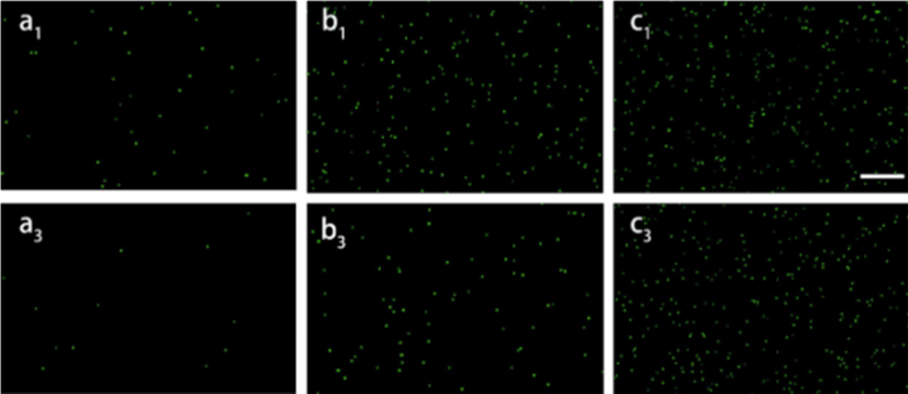
ROS Assay Kit purchased from MedChemExpress. Usage Cited in: Ind Crops Prod. 2024 Dec 15.
The intracellular ROS generation in bacteria detected with a ROS detection kit after 1, 3, 7, 14, and 30 days of incubation in (a) Cu2O, (b) GPC, and (c) BRC, respectively. Note: Scale bar = 100 μm.
-
Pharmaceuticals (Basel)Anti-Inflammatory and Antioxidant Effects of Irigenen Alleviate Osteoarthritis Progression through Nrf2/HO-1 Pathway. [Abstract]2024 Sep 26;17(10):1268. PMID: 39458910
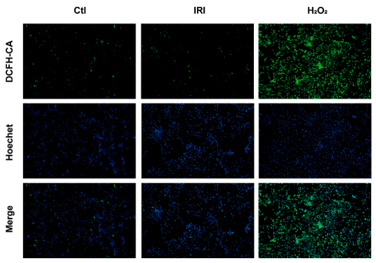
ROS Assay Kit purchased from MedChemExpress. Usage Cited in: Pharmaceuticals (Basel). 2024 Sep 26;17(10):1268. [Abstract]
The representative images of intracellular ROS detected by DCFH-DA fluorescent staining (ROS Assay Kit) in Chondrocytes.
-
Tissue CellPCSK9 exacerbates sevoflurane-induced neuroinflammatory response and apoptosis by up-regulating cGAS-STING signal. [Abstract]2025 Apr:93:102739. PMID: 39818066
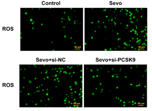
ROS Assay Kit purchased from MedChemExpress. Usage Cited in: Tissue Cell. 2025 Apr:93:102739. [Abstract]
DCFH-DA staining (ROS Assay Kit) was used to detect ROS levels in BV2 cells, Scale bar = 50 μm (magnification × 200);
-
Biomed PharmacotherSulforaphane improves post-resuscitation myocardial dysfunction by inhibiting cardiomyocytes ferroptosis via the Nrf2/IRF1/GPX4 pathway. [Abstract]2024 Oct:179:117408. PMID: 39244999
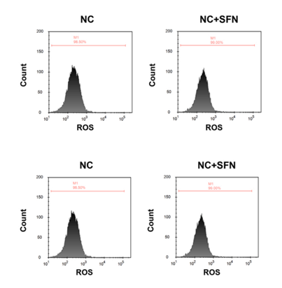
ROS Assay Kit purchased from MedChemExpress. Usage Cited in: Biomed Pharmacother. 2024 Oct:179:117408. [Abstract]
Flow cytometric analysis and quantification of lipid ROS production in AC16 cardiomyocytes using the ROS Assay Kit.
Storage
-20℃, 1 year.
Avoid repeated freeze-thaw cycles.


Components
| Components | HY-K0320-100 T | HY-K0320-500 T |
|---|---|---|
| ROS Positive Control Reagent (Rosup, 50 mg/mL) | 1 mL | 5 mL |
| DCFH-DA (10 mM) | 0.1 mL | 0.5 mL |


 Please enter the correct Lot Number
