



NHS Magnetic Beads (200nm, 10 mg/mL)

Based on 1 Customer Validation
MCE NHS Magnetic Beads (200 nm, 10 mg/mL) contain N-hydroxysuccinimide (NHS) functional groups, which react with primary amines on proteins or other molecules to form stable amide linkages,can covalently immobilize proteins for the affinity purification of antibodies, antigens and other biomolecules.
-
Storage :
4℃, 2 years. Do not freeze.


To place orders, for customer services and technical support, please contact: MedChemExpress USA
Tel: 609-228-6898 E-mail: [email protected] [email protected]



-
HY-K0202
Protein A/G Magnetic Beads -
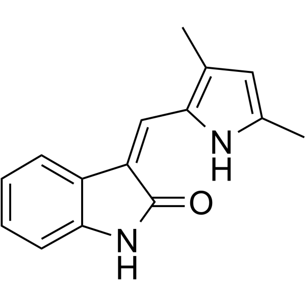
HY-10374
Semaxinib -
HY-K0207
Anti-Flag Magnetic Beads -
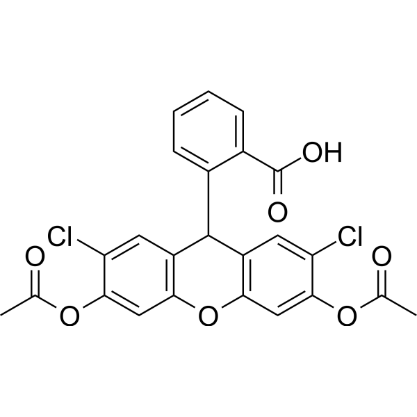
HY-D0940
H2DCFDA -
HY-13629
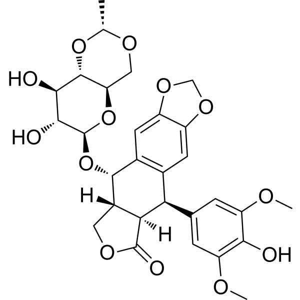
Etoposide -
HY-K0208
Streptavidin Magnetic Beads -

HY-P0319
3X FLAG peptide -
HY-K0210
Ni-NTA His-Tag Purification Agarose -
HY-K0225
Carboxyl Magnetic Beads (200 nm, 10 mg/mL) -

HY-K0224
Amino Magnetic Beads (200 nm, 10 mg/mL)


Description & Advantages
MCE NHS Magnetic Beads (200 nm, 10 mg/mL) contain N-hydroxysuccinimide (NHS) functional groups, which react with primary amines on proteins or other molecules to form stable amide linkages,can covalently immobilize proteins for the affinity purification of antibodies, antigens and other biomolecules. Compared with the traditional carboxyl and amino magnetic beads, NHS magnetic beads do not need to be activated by EDC/NHS or glutaraldehyde. Simply dissolve the primary amine-containing biological ligands in Coupling Buffer and incubate at room temperature to covalently couple the bioconjugate to the beads, which is easy to operate, mild coupling conditions, fast and efficient. The beads coupling process must be carried out in a buffered solvent without any amine, and automated instruments are especially useful for large screening of multiple samples.


Protocol
Recommended Buffers (Not Provided)
| Buffer | Composition |
|---|---|
| Wash Buffer | 1 mM HCl, pre-chilled at 4°C |
| Binding Buffer I | 0.1 M MES, pH 5.0, for biomolecules with pI < 7 |
| Binding Buffer II | 0.1 M PBS, pH 8.0, for biomolecules with pI > 7 |
| Blocking Buffer | 0.5 M ethanolamine, pH 8.3; or 0.1 M Tris-HCl, 150 mM NaCl, pH 8.5 |
| Storage Buffer | PBS containing 0.02% (w/v) NaN3 |
Operating Instructions
Coupling of Magnetic Beads to Biomolecules (For Reference Only)
The following procedure is described using 500 μL of magnetic beads, 500 μL of biomolecule sample, and a 1.5 mL EP tube as an example. The quantities may be adjusted proportionally according to experimental requirements.
1. Protein Solution Preparation
Dissolve an appropriate amount of the protein to be coupled in binding buffer to prepare a protein solution at a concentration of 0.5-2.0 mg/mL. Store the prepared solution at 4°C until use.
Note: The protein solution must not contain substances with amino groups. Previously stored proteins should first be dialyzed or desalted to completely remove substances containing primary amino groups from the original buffer. The protein should then be prepared at a concentration of 0.5-2.0 mg/mL using the binding buffer.
2. Magnetic Bead Pretreatment
Thoroughly mix the magnetic beads and transfer 500 μL into a 1.5 mL EP tube. Perform magnetic separation and discard the supernatant. Add 1 mL of pre-chilled wash buffer and vortex for 15 s. Perform magnetic separation again and discard the supernatant. Repeat the washing procedure twice.
3. Magnetic Bead Coupling
Add 500 μL of protein solution to the magnetic beads and vortex for 30 s. Place the tube on a mixer and incubate for 1-2 h at room temperature or overnight at 4°C.
Note: Keep the magnetic beads evenly suspended during incubation. During the first 30 min of the reaction, remove the tube and vortex for 15 s every 5 min. Thereafter, remove the tube and vortex for 15 s every 15 min.
4. Blocking
Perform magnetic separation and collect the supernatant. Add 1 mL of blocking buffer to resuspend the magnetic beads, vortex for 30 s, and incubate on a mixer at room temperature for 2 h.
5. Storage
Perform magnetic separation and discard the supernatant. Add 1 mL of ultrapure water to resuspend the magnetic beads and wash the coupled product three times. Add 500 μL of storage buffer to resuspend the magnetic beads and store at 4°C.
Storage
4℃, 2 years.
Do not freeze.


Components
| Components | HY-K0227-1 mL | HY-K0227-5 mL | HY-K0227-10 mL |
|---|---|---|---|
| NHS Magnetic Beads (200nm, 10 mg/mL) | 1 mL | 5 mL | 10 mL |


 Please enter the correct Lot Number


