



Cell Senescence β-Galactosidase Staining Kit
Based on 13 publication(s) in Google Scholar
MCE Cell Senescence β-Galactosidase Staining Kit can be used to evaluate the senescence of cells or tissues.
-
Storage :
-20℃, 1 year. X Gal solution needs to be stored away from light.


To place orders, for customer services and technical support, please contact: MedChemExpress USA
Tel: 609-228-6898 E-mail: [email protected] [email protected]











Description & Advantages
Due to limited replicative lifespan, normal cells enter cell cycle arrest, also known as cellular senescence1. While in this senescence phase the cells remain metabolically active without undergoing cell death or division. These senescent cells adopt a specific phenotypic state that includes the appearance of multinucleated cells, increased vacuolization, expression of pH-dependent β-galactosidase, and morphological changes where cells become enlarged and extended2, 3. Senescence, through a variety of mechanisms, can also play a role in tumor suppression, tumor progression, aging, and tissue repair.
β-galactosidase (also known as β-gal) is an essential hydrolase enzyme that catalyzes the hydrolysis of galactose-containing carbohydrates into monosaccharides. Substrates of β-galactosides include lactose, various glycoproteins, ganglioside GM1, and lactosylceramides. β-galactosidase is used widely in molecular biology; for example, isolation of recombinant bacteria during molecular cloning utilizes α-complementation of the bacterial β-galactosidase gene (lacZ) in the presence of a β-gal substrate to identify recombinant clones. In cell biology, Senescence-Associated beta-galactosidase (SA-β-gal), defined as β-gal activity at pH 6.0, is a widely used marker of replicative senescence.
MCE Cell Senescence β-Galactosidase Staining Kit is based on SA-β-gal catalyzing the transformation of X Gal (5-bromo-4-chloro-3-indolyl-β-D-galactopyranoside, 5-bromo-4-chloro-3-indolyl-β-D-galactopyranoside, X-gal) into a blue product at pH 6. This allows for the visualization of cell or tissue senescence under the microscope.


Protocol
1. Preparation of β-Galactosidase staining working solution
Prepare and mix the staining working solution before use. Protect X Gal Solution from light:
| Component | Recommended amount per sample |
|---|---|
| β-Galactosidase Staining Solution C | 1 mL |
| β-Galactosidase Staining Solution A | 15 μL |
| β-Galactosidase Staining Solution B | 15 μL |
| X Gal Solution | 50 μL |
2. Staining of cultured cells
1. Remove culture medium and wash cells once with PBS.
2. Add enough β-Galactosidase Staining Fixative to cover the cells and fix for approximately 15 min at room temperature.
3. Remove the fixative and wash three times with PBS for approximately 3 min each.
4. Add sufficient freshly prepared β-Galactosidase staining working solution.
5. Incubate overnight at 37°C. Avoid a CO2 incubator because CO2 may change the pH of the staining system.
6. Observe under a microscope. Cells containing blue precipitate are scored as SA-β-gal-positive senescent cells.
3. Tissue samples
After appropriate fixation and PBS washing of tissue sections, add staining working solution and incubate at 37°C until a clear blue signal develops, then examine microscopically.


Publications
-
Journal Impact Factor
-
Most Recent
-
Int J Mol SciFN9-10ELP, an ECM-Mimetic Fusion Protein, Protects Human Mesenchymal Stem Cells from Etoposide-Induced Senescence. [Abstract]2025 Sep 21;26(18):9218. PMID: 41009780
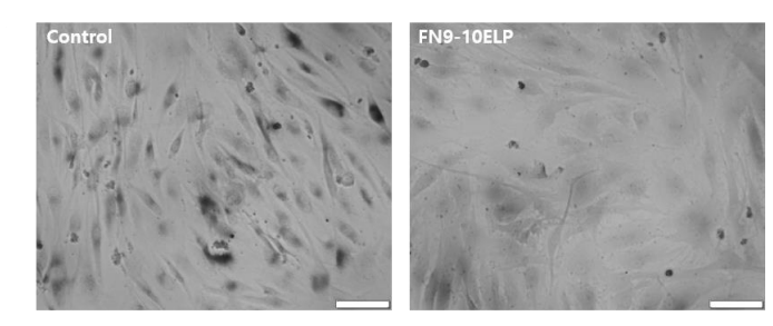
Cell Senescence β-Galactosidase Staining Kit purchased from MedChemExpress. Usage Cited in: Int J Mol Sci. 2025 Sep 21;26(18):9218. [Abstract]
Representative images of SA-β-gal-stained (Cell Senescence β-Galactosidase Staining Kit ) hMSCs treated with 20 µM etoposide for 5 days under control or FN9- 10ELP-coated conditions.
-
Carbohydr ResOat β-Glucooligosaccharides: Preparation, characterization, skin permeability, and photodamage repair for skincare applications. [Abstract]2025 Nov:557:109648. PMID: 40865351
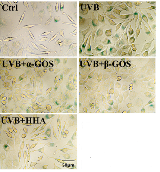
Cell Senescence β-Galactosidase Staining Kit purchased from MedChemExpress. Usage Cited in: Carbohydr Res. 2025 Nov:557:109648. [Abstract]
Cellular senescence induced by UVB damage and the effects of treatment with three oligosaccharides by β-galactosidase stain (Cell Senescence β-Galactosidase Staining Kit ).
-
Histochem Cell Biol2025 Nov 6;163(1):101. PMID: 41196372
-
Oncol Lett2026 Jun 3;32(2):322. PMID: 42290671
Storage
-20℃, 1 year.
X Gal solution needs to be stored away from light.


Components
| Components | HY-K1089-100 T |
|---|---|
| β-Galactosidase Staining Fixative | 100 mL |
| β-Galactosidase Staining Solution A | 1.5 mL |
| β-Galactosidase Staining Solution B | 1.5 mL |
| β-Galactosidase Staining Solution C | 100 mL |
| X Gal Solution | 5 mL |


 Please enter the correct Lot Number
