



Protein L Magnetic Beads
Based on 20 publication(s) in Google Scholar
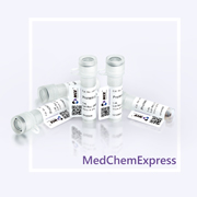
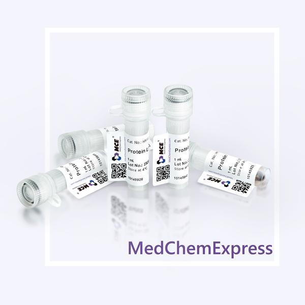
Protein L Magnetic Beads provide a fast and convenient method for Immunuoprecipitaion, Co-Immunoprecipitation and Chromatin Immunoprecipitation. The 1 mL is defined as the base specification. All larger sizes correspond to incremental volumes of this base.
-
Storage :
4°C, 2 years. Do not centrifuge, dry or freeze the magnetic beads.

To place orders, for customer services and technical support, please contact: MedChemExpress USA
Tel: 609-228-6898 E-mail: [email protected] [email protected]



-
HY-K0202
Protein A/G Magnetic Beads -

HY-K0204
Protein G Magnetic Beads -
HY-13948
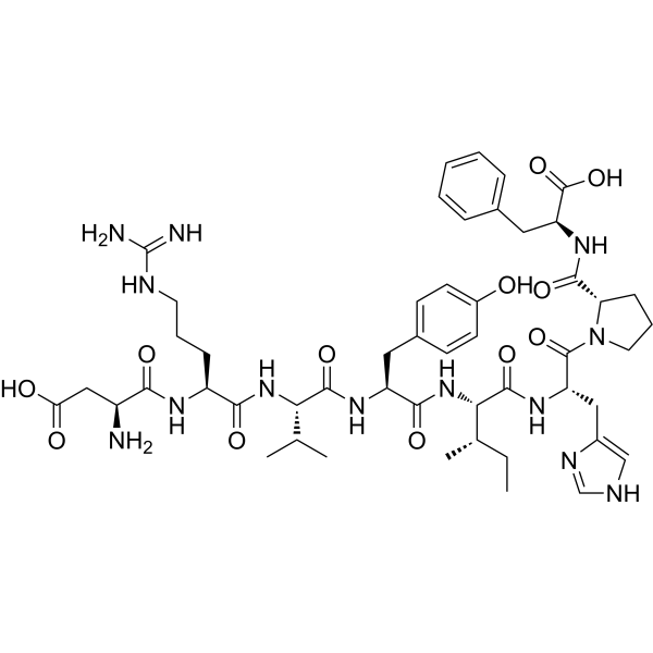
Angiotensin II human -
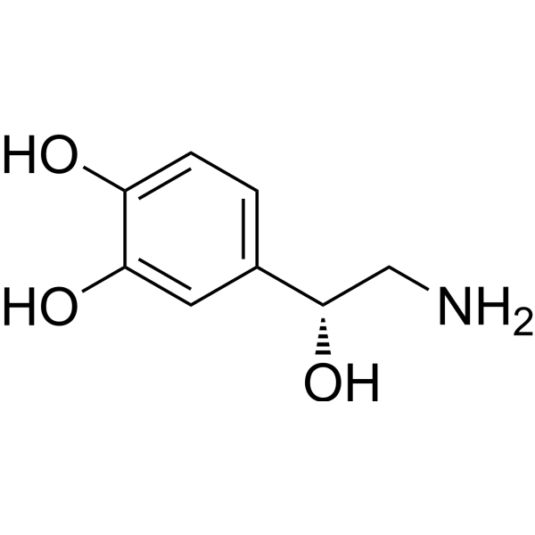
HY-13715
Norepinephrine -

HY-18739
Phorbol 12-myristate 13-acetate -
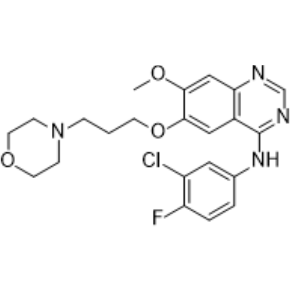
HY-50895
Gefitinib -
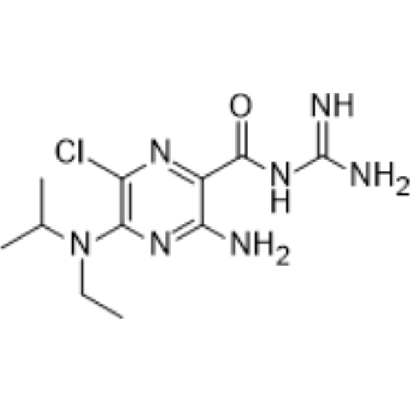
HY-101840
EIPA -

HY-K0215
Protein L Agarose -
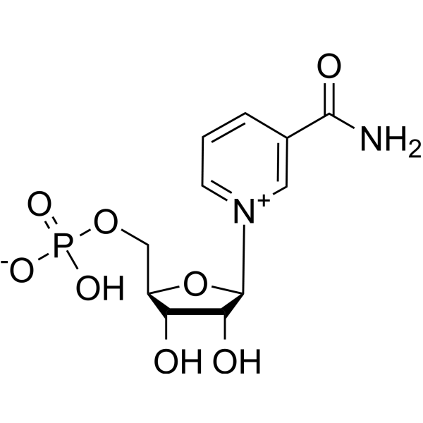
HY-F0004
Fosribnicotinamide -
HY-K0203
Protein A Magnetic Beads -
HY-K0010
Protease Inhibitor Cocktail (EDTA-Free, 100× in DMSO) -
HY-13979
DDR1-IN-1


Description & Advantages
Protein L Magnetic Beads contain a recombinant Protein L that binds to antibody species and subtypes with appropriate Kappa light chains, while Lambda light chains and some Kappa light chains do not bind. During IP, less magnetic beads are needed, non-specific binding is low, enabling Protein L magnetic beads to be used in IP conveniently and efficiently.


Protocol
1. Preparation of Magnetic Beads
1.1 Resuspend the Magnetic Beads in the vial (tilt and rotate for 2 minutes or gently pipette for 10 times).
1.2 Transfer 25-50 μL of Protein L Magnetic Beads into a 1.5 mL tube (Transfer amount may be adjusted as required).
1.3 Add 400 μL of binding/wash buffer to the beads and gently pipette to mix. Place the tube into a magnetic stand to collect the beads against the side of the tube (Hereinafter referred to as magnetic separation). Remove and discard the supernatant. Repeat this step for 2 times.
2. Binding of Antibody
2.1 Dilute antibody (Ab) to the final concentration of 5-50 μg/mL with binding/wash buffer. The optimal amount of Ab may be adjusted as required.
2.2 Add 400 μL of diluted Ab to the Protein L Magnetic Beads. Rotate tube for 30 minutes at room temperature or 2 hours at 4°C.
2.3 Perform magnetic separation. Transfer the supernatant into a new tube for further analysis, if desired. The supernatant is the non-binding fraction.
2.4 Add 400 μL of binding/wash buffer to the beads and gently pipette to mix. Place the tube into a magnetic stand to collect the beads against the side of the tube. Remove and discard the supernatant. Repeat this step for 4 times.
3. Immunoprecipitation of Target Antigen
3.1 Remove the tubes from the magnetic separator and add your sample containing the antigen (Ag) (typically 5-50 μg in 400 μL binding/wash buffer) and gently pipette to resuspend the Protein L Magnetic Beads-Ab complex.
3.2 Incubate with rotation for 30 minutes at room temperature or 2 hours at 4°C to allow Ag to bind to the Protein L Magnetic Beads-Ab complex.
3.3 Perform magnetic separation. Remove and discard the supernatant.
3.4 Wash the Magbeads-Ab-Ag complex 5 times using 400 μL binding/wash buffer for each wash. Perform magnetic separation between each wash, remove supernatant and resuspend by gentle pipetting.
3.5 Resuspend the Protein L Magnetic Beads-Ab-Ag complex in 400 μL binding/wash buffer and transfer the bead suspension into a clean tube. This is recommended to avoid co-elution of the proteins bound to the tube wall.
4. Elution
This is a non-denaturation elution method.
4.1 Perform magnetic separation and remove the supernatant. Add 400 μL of binding/wash buffer into the tube and rotate for 5 minutes. Perform magnetic separation for 1 minute and remove the supernatant. Then add 25-50 μL elution buffer into the tube with magnetic beads-Ab-Ag complex, rotate for 5 minutes.
4.2 Perform magnetic separation, collect the supernatant.
4.3 The final solution can be used as samples for denaturing SDS-PAGE. Or the elution can be adjusted to neutral pH with neutralization buffer immediately and used for further analysis.


Publications
-
Journal Impact Factor
-
Most Recent
-
Cell Metab2026 Jan 23:S1550-4131(25)00584-4. PMID: 41579859
-
AutophagyPTK6 drives HNRNPH1 phase separation to activate autophagy and suppress apoptosis in colorectal cancer. [Abstract]2025 Aug;21(8):1680-1699. PMID: 40103198
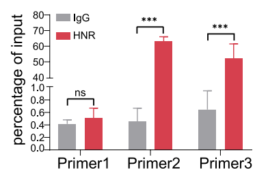
Protein L Magnetic Beads purchased from MedChemExpress. Usage Cited in: Autophagy. 2025 Aug;21(8):1680-1699. [Abstract]
Protein L Magnetic Beads (50 µL; 1 h) were used for clip-qPCR analysis of HNRNPH1 association with NBR1 pre-rRNA at the indicated locations.
-
Adv Sci (Weinh)Inhibition of CARM1-Mediated Methylation of ACSL4 Promotes Ferroptosis in Colorectal Cancer. [Abstract]2023 Dec;10(36):e2303484. PMID: 37946697
-
Mol TherFcεRγI promotes canine CD8 chimeric antigen receptor T cell cytotoxicity through a Syk-NF-κB axis. [Abstract]2025 Dec 10:S1525-0016(25)01033-0. PMID: 41376156
-
Clin Transl MedAlternative splicing of HSPA12A pre-RNA by SRSF11 contributes to metastasis potential of colorectal cancer. [Abstract]2022 Nov;12(11):e1113. PMID: 36394206
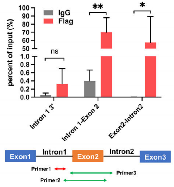
Protein L Magnetic Beads purchased from MedChemExpress. Usage Cited in: Clin Transl Med. 2022 Nov;12(11):e1113. [Abstract]
Protein L Magnetic Beads (50 µL; 1 h) were used for CLIP-qPCR analysis of SRSF11 association with HSPA12A pre-RNA at the indicated locations.
-
J Cell Biol2023 Oct 2;222(10):e202302037. PMID: 37676315
-
Mol Med RepLINC01116 promotes the proliferation and invasion of glioma by regulating the microRNA‑744‑5p‑MDM2‑p53 axis. [Abstract]2021 May;23(5):366. PMID: 33760190
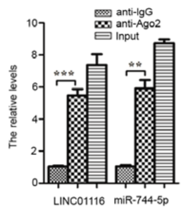
Protein L Magnetic Beads purchased from MedChemExpress. Usage Cited in: Mol Med Rep. 2021 May;23(5):366. [Abstract]
RNA immunoprecipitation assays using Protein L Magnetic Beads (30 µL; 4°C; 6-8 h) showed that both LINC01116 and miR‑744‑5p could bind Ago2.
-
Sci RepMicrofibrillar-associated protein 5 suppresses adipogenesis by inhibiting essential coactivator of PPARγ. [Abstract]2023 Apr 5;13(1):5589. PMID: 37020143
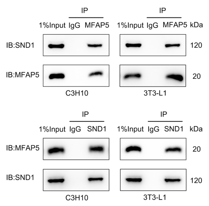
Protein L Magnetic Beads purchased from MedChemExpress. Usage Cited in: Sci Rep. 2023 Apr 5;13(1):5589. [Abstract]
Protein L Magnetic Beads (40 μL; 30 min) were used for co-immunoprecipitation to show the interaction of MFAP5 and SND1 at the protein level in C3H10 and 3T3-L1 cells.
-
Cell SignalMitochondrial outer membrane protein Samm50 protects against hypoxia-induced cardiac injury by interacting with Shmt2. [Abstract]2024 Aug:120:111219. PMID: 38723737
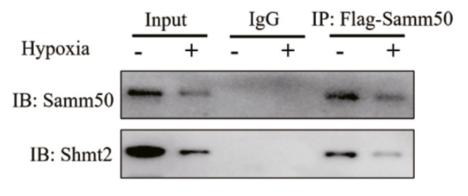
Protein L Magnetic Beads purchased from MedChemExpress. Usage Cited in: Cell Signal. 2024 Aug:120:111219. [Abstract]
Protein L Magnetic Beads (50 μL; 4 h) were used for co-IP analysis of Samm50 and Shmt2 in normoxia and hypoxia-exposed NRCMs.
-
J Biol ChemHexosamine biosynthesis drives hemocyanin O-GlcNAcylation to potentiate antibacterial immunity in shrimp. [Abstract]2026 May 28;302(7):113213. PMID: 42214671
Storage
4°C, 2 years.
Do not centrifuge, dry or freeze the magnetic beads.


Components
| Components | HY-K0205- 1 mL | HY-K0205- 5 mL |
|---|---|---|
| Protein L Magnetic Beads | 1 mL | 1 mL × 5 |


 Please enter the correct Lot Number
