



Protein A/G Agarose
Based on 37 publication(s) in Google Scholar
MCE Protein A/G Agarose is an affinity chromatography medium for separation and purification of immunoglobulins.
-
Storage :
4℃, 2 years. Do not dry or freeze.


To place orders, for customer services and technical support, please contact: MedChemExpress USA
Tel: 609-228-6898 E-mail: [email protected] [email protected]



-
HY-K0202
Protein A/G Magnetic Beads -
HY-K0214
Protein G Agarose -

HY-13259
MG-132Proteasome Inhibitor -
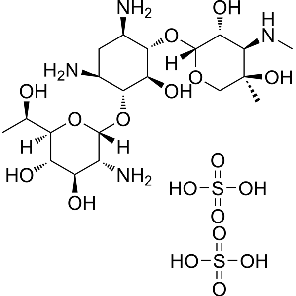
HY-17561
G-418 disulfateAminoglycoside Antibiotic -

HY-10219
RapamycinmTOR Inhibitor -

HY-10182
LaduviglusibGSK-3 Inhibitor -
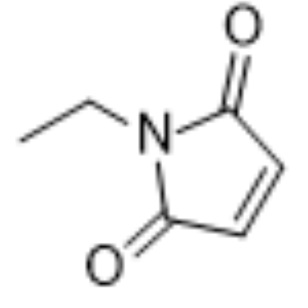
HY-D0843
N-EthylmaleimideCysteine Residues Modifier -

HY-15371
ForskolinAdenylate Cyclase Activator -
HY-13966
2-Deoxy-D-glucoseGlycolysis Inhibitor -
HY-12041
SP600125JNK Inhibitor -

HY-P7109
EGF Protein, Human -

HY-15763
ErastinFerroptosis Inducer


Description & Advantages
MCE Protein A/G Agarose is an affinity chromatography medium for separation and purification of immunoglobulins.
MCE Protein A/G Agarose consists of purified Protein A/G recombinant fusion protein that has been covalently immobilized onto high-quality cross-linked 4% beaded agarose, binds nearly all isotypes and mammalian species of IgG from serum, ascites fluid, cell culture supernatant and other ant.
The product contains 0.5 mL of gel per 1 mL of total volume. Prior to use, ensure the gel is thoroughly resuspended and mixed before aspiration.
The specifications of the product correspond to the actual resin volume, with the resin content of 50%.


Protocols
Buffer Preparation
| Buffer | Composition |
|---|---|
| Binding/Washing Buffer | 0.15 M NaCl, 20 mM Na2HPO4, pH 7.0 |
| Elution Buffer | 0.1 M Glycine, pH 3.0 |
| Neutralization Buffer | 1 M Tris-HCl, pH 8.0 |
| Storage Buffer | 1× PBS, 20% ethanol |
Note: It is recommended to prepare all buffers with ultrapure water. After preparation, filter them through a 0.45 μm or 0.22 μm membrane for sterilization.
Protein Purification
Before sample loading, centrifuge the sample or filter it through a 0.22 μm or 0.45 μm membrane to reduce impurities and improve protein purification efficiency.
Medium-Pressure Chromatography Column Method
1. Column Packing: Pack Protein A/G Agarose into a suitable chromatography column and connect the column to the chromatography system.
2. Column Equilibration: Equilibrate the column with 5 column volumes of Binding/Washing Buffer. Repeat 2-3 times.
3. Sample Loading: Load the sample using a pump or sample loop and collect the flow-through. Repeated sample loading may improve binding efficiency.
Notes: a. Select an appropriate amount of gel according to the amount of protein. Do not exceed the binding capacity of the column;
b. Increased sample viscosity or volume may cause increased back pressure in the chromatography column.
4. Washing: Wash the column with 10-20 column volumes of Binding/Washing Buffer to remove nonspecifically adsorbed contaminating proteins. Collect the wash fractions until the UV absorbance baseline becomes stable.
5. Elution: Elute with 3-5 column volumes of Elution Buffer. Collect the eluate in separate tubes and immediately neutralize the pH with Neutralization Buffer at 1/10 of the total eluate volume. The sample may be used for subsequent functional analysis.
Note: The eluted protein may be stored at 4°C for a short period. For long-term storage, store it at -20°C.
6. Regeneration: Continue washing the column with 5-10 column volumes of Elution Buffer to completely elute the column and regenerate the resin. Then equilibrate the column to neutral pH using Binding/Washing Buffer.
Note: The agarose should be equilibrated with Binding/Washing Buffer immediately after acidic elution, and Protein A/G Agarose should not be kept in the Elution Buffer for more than 20 min.
7. Storage: Equilibrate the column with 5-10 column volumes of Storage Buffer, disconnect the column, and store it at 2-8°C.
Gravity-Flow Column Method
1. Column Packing: According to the amount of sample to be purified, transfer an appropriate volume of Protein A/G Agarose suspension into a gravity-flow chromatography column and remove the storage solution.
2. Column Equilibration: Equilibrate the packed gravity-flow column with 5 column volumes of Binding/Washing Buffer. Repeat 2-3 times.
3. Sample Loading: After adding the sample, allow it to remain in the column for at least 2 min to ensure sufficient contact between the sample and the resin. Collect the flow-through.
Note: Repeated sample loading may improve binding efficiency.
4. Washing: Wash the column with 10-15 column volumes of Binding/Washing Buffer to remove nonspecifically adsorbed contaminating proteins. Collect the wash fractions.
5. Elution: Elute with 3-5 column volumes of Elution Buffer. Collect the eluate in separate tubes and immediately neutralize the pH with Neutralization Buffer at 1/10 of the total eluate volume. The sample may be used for subsequent functional analysis.
Note: The eluted protein may be stored at 4°C for a short period. For long-term storage, store it at -20°C.
6. Regeneration: Continue washing the column with 5-10 column volumes of Elution Buffer to completely elute the column and regenerate the resin. Then equilibrate the column to neutral pH using Binding/Washing Buffer.
7. Storage: Equilibrate the column with 5 column volumes of Storage Buffer and store it at 2-8°C.
Centrifugation Method
1. Agarose Pretreatment: According to the amount of sample to be purified, transfer an appropriate volume of Protein A/G Agarose suspension into a centrifuge tube. Centrifuge at 1,000 rpm for 1 min and discard the supernatant. Add 5 gel volumes of Binding/Washing Buffer, centrifuge at 1,000 rpm for 1 min, and discard the supernatant. Repeat 2-3 times.
2. Binding: Add the sample, close the centrifuge tube, and incubate at 4°C for 2-4 h or at 37°C for 0.5-2 h.
3. Washing: After incubation, centrifuge at 1,000 rpm for 1 min and discard the supernatant. The supernatant may be retained as the flow-through fraction for electrophoretic analysis. Wash with 5 gel volumes of Binding/Washing Buffer, centrifuge at 1,000 rpm for 1 min, and discard the supernatant. Repeat 3-5 times.
4. Elution: Elute with 3-5 gel volumes of Elution Buffer and incubate at room temperature for 5-10 min. Centrifuge at 1,000 rpm for 1 min, collect the supernatant in separate tubes, and immediately neutralize the pH with Neutralization Buffer at 1/10 of the total eluate volume. The sample may be used for subsequent functional analysis. The elution step may be repeated 2-3 times, with each supernatant collected separately.
Note: The agarose should be equilibrated with Binding/Washing Buffer immediately after acidic elution, and Protein A/G Agarose should not be kept in the Elution Buffer for more than 20 min. The eluted protein may be stored at 4°C for a short period. For long-term storage, store it at -20°C.
5. Regeneration and Storage: Wash the resin with 5-10 gel volumes of Binding/Washing Buffer, followed by 5-10 gel volumes of ddH2O. Finally, wash with 2 gel volumes of Storage Buffer and store at 2-8°C.
Immunoprecipitation
Preparation of Agarose
1) Add the required amount of Protein A/G Agarose suspension to a 2 mL tube, centrifuge at 1,000 rpm for 1 min and discard the supernatant.
2) Add 500 μL of Binding Buffer and mix well, centrifuge at 1,000 rpm for 1 min and discard the supernatant. Repeat 3-4 times.
Antibody Binding
1) Introduce an appropriate volume of diluted antibody into the agarose, gently rotate the tube for 30 min at room temperature or 2 h at 4°C. Centrifuge at 1,000 rpm for 1 min and retain the supernatant for future analysis.
2) Add 500 μL Washing Buffer to the agarose and mix well, rotate the tube for 3-5 min at room temperature to eliminate hetero-proteins that have been non-specifically adsorbed. Centrifuge at 1,000 rpm for 1 min and discard the supernatant. Repeat 2-3 times.
Immunoprecipitation of Target Antigen
1) Add the antigen-containing sample to the agarose and mix well, then rotate the tube for 30 min at room temperature or leave overnight at 4°C.
2) Centrifuge at 1,000 rpm for 1 min and retain the supernatant for future analysis.
3) Add 1 mL Washing Buffer to the agarose and mix thoroughly, centrifuge at 1,000 rpm for 1 min and discard the supernatant. Repeat the washing procedure until the OD280 of the supernatant is below 0.05.
Note: If the OD280 of the supernatant is greater than 0.05, increase the number of washing steps as appropriate.
4) Add 1 mL Washing Buffer to the agarose again, mix well and transfer to a new tube.
Antigen Elution
Two elution methods are provided. Select the appropriate method according to the downstream analysis.
1) Acidic Elution: Add 50-100 μL of Elution Buffer, mix thoroughly and incubate at room temperature for 5-10 min. Centrifuge at 1,000 rpm for 1 min, collect the supernatant and immediately neutralize the pH with Neutralization Buffer at 1/10 of the total eluate volume.
2) Denaturing Elution: Add 25 μL of 1× SDS-PAGE Loading Buffer, mix thoroughly and heat at 95°C for 5 min. Centrifuge at 1,000 rpm for 1 min and collect the supernatant for SDS-PAGE analysis.


Publications
-
Journal Impact Factor
-
Most Recent
-
Exp Mol MedCdyl2-60aa encoded by CircCDYL2 accelerates cardiomyocyte death by blocking APAF1 ubiquitination in rats. [Abstract]2023 Apr;55(4):860-869. PMID: 37009805
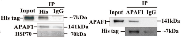
Protein A/G Agarose purchased from MedChemExpress. Usage Cited in: Exp Mol Med. 2023 Apr;55(4):860-869. [Abstract]
IP of His tag (Upper Right) with APAF1 and HSP70; or APAF1 (Upper Left) with His tag in NRCMs transfected with ad-Cdyl2-60aa (containing His tag).The lysates were then centrifuged and cleared via incubation with 25 μl protein A/G agarose for 1.5 h at 4 °C. The precleared supernatant was subjected to IP using the indicated primary antibodies at 4 °C overnight. Then, the protein complexes were collected via incubation with 30 μl protein A/G gel for 2 h at 4 °C.
-
Gut MicrobesSynergism of Fusobacterium periodonticum and N-nitrosamines promote the formation of EMT subtypes in ESCC by modulating Wnt3a palmitoylation. [Abstract]2024 Jan-Dec;16(1):2391521. PMID: 39193618
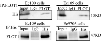
Protein A/G Agarose purchased from MedChemExpress. Usage Cited in: Gut Microbes. 2024 Jan-Dec;16(1):2391521. [Abstract]
The protein interaction between FadAL-His and FLOT1 was verified by CO-IP. The equal lysates (1.5 mg, containing 3 μg recombinant FadAL protein) were then incubated with IP-graded primary antibody (anti-FLOT1, anti-His-Tag or IgG) on a shaking platform at 4°C overnight. Following this, the protein A/G agarose beads were added and maintained for 6 h. Lastly, the protein samples were denatured and analyzed through western blot.
-
J Exp Clin Cancer ResAdipocytes-induced ANGPTL4/KLF4 axis drives glycolysis and metastasis in triple-negative breast cancer. [Abstract]2025 Jul 4;44(1):192. PMID: 40616161
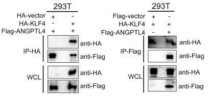
Protein A/G Agarose purchased from MedChemExpress. Usage Cited in: J Exp Clin Cancer Res. 2025 Jul 4;44(1):192. [Abstract]
Co-IP assay showing the exogenous interaction between ANGPTL4 and KLF4 in 293T cells transfected with Flag-ANGPTL4 or HA-KLF4. Cell lysates were incubated with primary antibodies in a rotating incubator overnight at 4 °C, followed by incubation with protein A/G agarose beads for another 6 h. The immunoprecipitates were washed seven times with lysis buffer and analyzed by immunoblots.
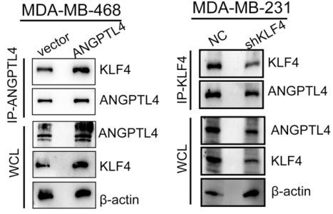
Protein A/G Agarose purchased from MedChemExpress. Usage Cited in: J Exp Clin Cancer Res. 2025 Jul 4;44(1):192. [Abstract]
Co-IP assay showing the endogenous interaction between ANGPTL4 and KLF4 in ANGPTL4 overexpression MDA-MB-468 cells, or KLF4-knockdown MDA-MB-231 cellsCell lysates were incubated with primary antibodies in a rotating incubator overnight at 4 °C, followed by incubation with protein A/G agarose beads for another 6 h. The immunoprecipitates were washed seven times with lysis buffer and analyzed by immunoblots.
-
MedComm (2020)CircMALAT1 promotes cancer stem-like properties and chemoresistance via regulating Musashi-2/c-Myc axis in esophageal squamous cell carcinoma. [Abstract]2024 Jun 14;5(6):e612. PMID: 38881674
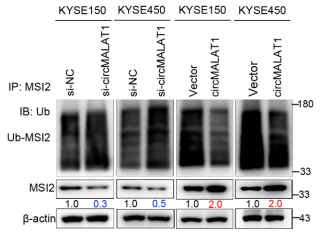
Protein A/G Agarose purchased from MedChemExpress. Usage Cited in: MedComm (2020). 2024 Jun 14;5(6):e612. [Abstract]
KYSE150 cells were overexpressed/knocked down circMALAT1 and treated with MG132 (10 μM) for 12 h before collection. co-IP was performed with MSI2 antibody in the above-mentioned cells. The polyubiquitinated (Poly-Ub) MSI2 protein was analyzed by Western blot with anti-ubiquitin after co-IP.
-
Cell Mol Biol LettThe miR-1269a/PCDHGA9/CXCR4/β-catenin pathway promotes colorectal cancer invasion and metastasis. [Abstract]2024 Nov 26;29(1):144. PMID: 39587482
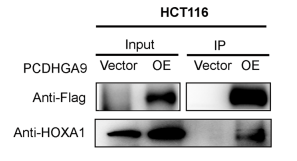
Protein A/G Agarose purchased from MedChemExpress. Usage Cited in: Cell Mol Biol Lett. 2024 Nov 26;29(1):144. [Abstract]
Co-IP assays revealed the presence of the FLAG and HOXA1 proteins in both the input and immunoprecipitated samples. A portion of these protein samples was utilized as input samples, while another portion was mixed with anti-Flag antibody and incubated at 4 °C for 2 h. Then, Protein A/G PLUS-Agarose beads were added and incubated at 4 °C for 12 h.
-
Sci SignalUSP5 deubiquitylates and stabilizes FcεRIγ to enhance IgE-induced mast cell activation and allergic inflammation. [Abstract]2025 Jul 29;18(897):eadr3411. PMID: 40729432
-
PLoS Pathog2025 Oct 24;21(10):e1013615. PMID: 41134845
-
Virol SinASFV pA137R protein triggers inflammatory response by inducing NF-κB signaling pathway and facilitating NLRP3 inflammasome assembly. [Abstract]2026 May 29:S1995-820X(26)00086-6. PMID: 42217584
-
Neurochem ResScavenger Receptor Class B Type I Modulates Epileptic Seizures and Receptor α2δ-1 Expression. [Abstract]2024 Oct;49(10):2842-2853. PMID: 39017956
-
J VirolHSPA5 induces autophagy targeting VP2 through the PERK-eIF2α signaling pathway to inhibit SVA replication. [Abstract]2026 May 19;100(5):e0210325. PMID: 41973539
-
J VirolThe neddylation of the RNA-dependent RNA polymerase 3D of Coxsackievirus B3 promotes viral replication. [Abstract]2025 Nov 25;99(11):e0153525. PMID: 41170971
-
PLoS Genet2025 Jul 10;21(7):e1011794. PMID: 40638681
Storage
4℃, 2 years.
Do not dry or freeze.


Components
| Components | HY-K0230-5 mL | HY-K0230-10 mL | HY-K0230-50 mL |
|---|---|---|---|
| Protein A/G Agarose | 5 mL | 10 mL | 10 mL × 5 |


 Please enter the correct Lot Number
