



Protein G Magnetic Beads
Based on 66 publication(s) in Google Scholar
Protein G Magnetic Beads provide a fast and convenient method for Immunoprecipitation and Co-Immunoprecipitation and Chromatin Immunoprecipitation. The 1 mL is defined as the base specification. All larger sizes correspond to incremental volumes of this base.
-
Storage :
4°C, 2 years. Do not centrifuge, dry or freeze the magnetic beads.



To place orders, for customer services and technical support, please contact: MedChemExpress USA
Tel: 609-228-6898 E-mail: [email protected] [email protected]



-

HY-K0203
Protein A Magnetic Beads -

HY-K0202
Protein A/G Magnetic Beads -

HY-K0207
Anti-Flag Magnetic Beads -

HY-10219
RapamycinmTOR Inhibitor -

HY-K0010
Protease Inhibitor Cocktail (EDTA-Free, 100× in DMSO) -

HY-K0201
Anti-HA Magnetic Beads -

HY-K0206
Anti-c-Myc Magnetic Beads -

HY-K0208
Streptavidin Magnetic Beads -
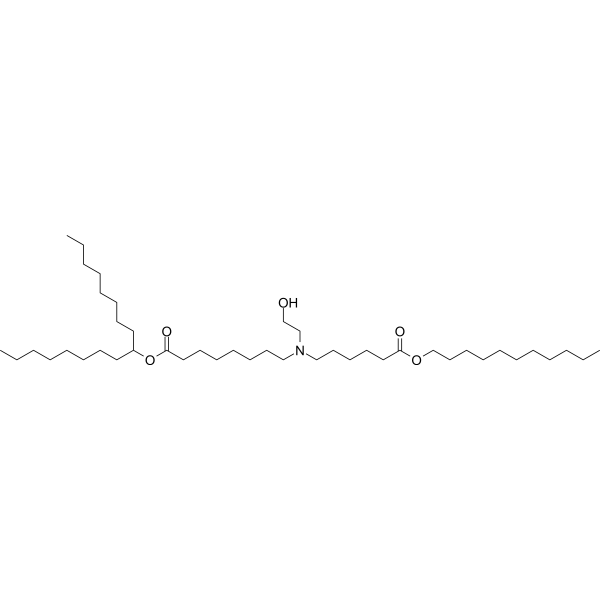
HY-134541
SM-102Ionizable Amino Lipid -
HY-K0209
Anti-His Magnetic Beads -

HY-K0205
Protein L Magnetic Beads -

HY-18739
Phorbol 12-myristate 13-acetatePKC Activator


Description & Advantages
The MCE Protein G Magnetic Beads are typically used for isolating antibodies from serum, cell culture supernatant or ascites and for Immunoprecipitation and Co-immunoprecipitation of antigens from cell or tissue extracts. Protein G Magnetic Beads contain a recombinant Protein G with the binding domains for IgG.
1. During immunoprecipitation, only a small amount of magnetic beads are needed.
2. Convenient and time saving.
3. Low non-specific binding.
4. Minimal sample loss.
5. Antibody binding capacity up to 0.85 mg/mL.
6. Stable, one bottle solution.


Protocol
1. Preparation of Magnetic Beads
1.1 Resuspend the Magnetic Beads in the vial (tilt and rotate for 2 minutes or gently pipette for 10 times).
1.2 Transfer 25-50 μL of Protein G Magnetic Beads into a 1.5 mL tube (Transfer amount may be adjusted as required).
1.3 Add 400 μL of binding/wash buffer to the beads and gently pipette to mix. Place the tube into a magnetic stand to collect the beads against the side of the tube (Hereinafter referred to as magnetic separation). Remove and discard the supernatant. Repeat this step for 2 times.
2. Binding of Antibody
2.1 Dilute antibody (Ab) to the final concentration of 5-50 μg/mL with binding/wash buffer. The optimal amount of Ab may be adjusted as required.
2.2 Add 400 μL of diluted Ab to the Protein G Magnetic Beads. Rotate tube for 30 minutes at room temperature or 2 hours at 4°C.
2.3 Perform magnetic separation. Transfer the supernatant into a new tube for further analysis, if desired. The supernatant is the non-binding fraction.
2.4 Add 400 μL of binding/wash buffer to the beads and gently pipette to mix. Place the tube into a magnetic stand to collect the beads against the side of the tube. Remove and discard the supernatant. Repeat this step for 4 times.
3. Immunoprecipitation of Target Antigen
3.1 Remove the tubes from the magnetic separator and add your sample containing the antigen (Ag) (typically 5-50 μg in 400 μL binding/wash buffer) and gently pipette to resuspend the Protein G Magnetic Beads-Ab complex.
3.2 Incubate with rotation for 30 minutes at room temperature or 2 hours at 4°C to allow Ag to bind to the Protein G Magnetic Beads-Ab complex.
3.3 Perform magnetic separation. Remove and discard the supernatant.
3.4 Wash the Magbeads-Ab-Ag complex 5 times using 400 μL binding/wash buffer for each wash. Perform magnetic separation between each wash, remove supernatant and resuspend by gentle pipetting.
3.5 Resuspend the Protein G Magnetic Beads-Ab-Ag complex in 400 μL binding/wash buffer and transfer the bead suspension into a clean tube. This is recommended to avoid co-elution of the proteins bound to the tube wall.
4. Elution
This is a non-denaturation elution method.
4.1 Perform magnetic separation and remove the supernatant. Add 400 μL of binding/wash buffer into the tube and rotate for 5 minutes. Perform magnetic separation for 1 minute and remove the supernatant. Then add 25-50 μL elution buffer into the tube with magnetic beads-Ab-Ag complex, rotate for 5 minutes.
4.2 Perform magnetic separation, collect the supernatant.
4.3 The final solution can be used as samples for denaturing SDS-PAGE. Or the elution can be adjusted to neutral pH with neutralization buffer immediately and used for further analysis.


Publications
-
Journal Impact Factor
-
Most Recent
-
BloodLoss of sphingosine kinase 2 promotes the expansion of hematopoietic stem cells by improving their metabolic fitness. [Abstract]2022 Oct 13;140(15):1686-1701. PMID: 35881840
-
Cell Stem CellAmino acid catabolism regulates hematopoietic stem cell proteostasis via a GCN2-eIF2α axis. [Abstract]2022 Jul 7;29(7):1119-1134.e7. PMID: 35803229
-
Nat CommunTherapeutic potential of the secreted Kazal-type serine protease inhibitor SPINK4 in colitis. [Abstract]2024 Jul 12;15(1):5874. PMID: 38997284
-
J NanobiotechnologyComparative study of extracellular vesicles derived from mesenchymal stem cells and brain endothelial cells attenuating blood-brain barrier permeability via regulating Caveolin-1-dependent ZO-1 and Claudin-5 endocytosis in acute ischemic stroke. [Abstract]2023 Feb 28;21(1):70. PMID: 36855156
-
Acta Pharm Sin BFunctional aptamer evolution-enabled elucidation of a melanoma migration-related bioactive epitope. [Abstract]2025 Jun;15(6):3196-3209. PMID: 40654334
-
Sci AdvFeaSion decodes the regulatory landscape and functional diversity of RNA polymerase II CTD phosphorylation. [Abstract]2025 Nov 28;11(48):eadz2345. PMID: 41313762
-
Mol MedExtinguishing the flames of inflammation: retardant effect of chlorquinaldol on NLRP3-driven diseases. [Abstract]2024 Dec 19;30(1):245. PMID: 39701924
-
Cancer Immunol ResUBA2-High Osteosarcoma Suppresses Immune Infiltration by Autophagy-Mediated MHC-I Degradation. [Abstract]2026 Jul 2;14(7):1080-1099. PMID: 42085301
-
Int J Mol Sci2025 Mar 22;26(7):2888. PMID: 40243488
-
J Lipid ResE3 ligase SMURF2 promotes adipogenesis and improves obesity complications by suppressing TGF-β signaling. [Abstract]2026 May 18;67(7):101061. PMID: 42155611
-
PLoS Pathog2024 May 30;20(5):e1012279. PMID: 38814988
-
Oncol ResCircTIAM1 overexpression promotes the progression of papillary thyroid cancer by regulating the miR-338-3p/LASP1 axis. [Abstract]2024 Oct 16;32(11):1747-1763. PMID: 39449799
-
Curr Med Sci2019 Jun;39(3):442-448. PMID: 31209817
Storage
4°C, 2 years.
Do not centrifuge, dry or freeze the magnetic beads.


Components
| Components | HY-K0204-1 mL | HY-K0204-5 mL |
|---|---|---|
| Protein G Magnetic Beads | 1 mL | 1 mL×5 |


 Please enter the correct Lot Number
