



Anti-GFP Magnetic Agarose Beads
Based on 6 publication(s) in Google Scholar
MCE Anti-GFP Magnetic Agarose Beads can be used for the detection and purification of GFP, EGFP, and their fusion-expressed proteins and IP assays.
-
Storage :
4℃,2 years. Do not dry or freeze.


To place orders, for customer services and technical support, please contact: MedChemExpress USA
Tel: 609-228-6898 E-mail: [email protected] [email protected]



-
HY-K0207
Anti-Flag Magnetic Beads -
HY-K0201
Anti-HA Magnetic Beads -
HY-K0202
Protein A/G Magnetic Beads -

HY-13259
MG-132 -
HY-K0206
Anti-c-Myc Magnetic Beads -
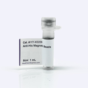
HY-K0209
Anti-His Magnetic Beads -

HY-K0236
Anti-Flag Magnetic Agarose Beads -
HY-K0011
Protease Inhibitor Cocktail, mini-Tablet (EDTA-Free) -
HY-K0010
Protease Inhibitor Cocktail (EDTA-Free, 100× in DMSO) -
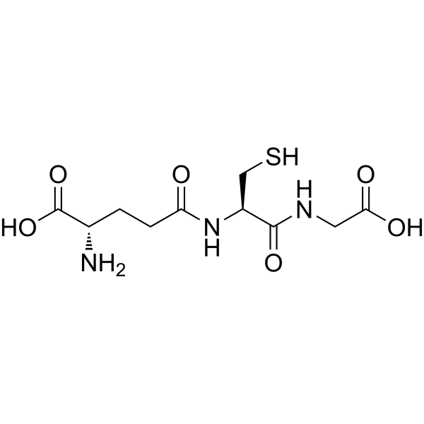
HY-D0187
L-Glutathione reduced -
HY-15921
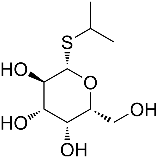
IPTG -
HY-19312
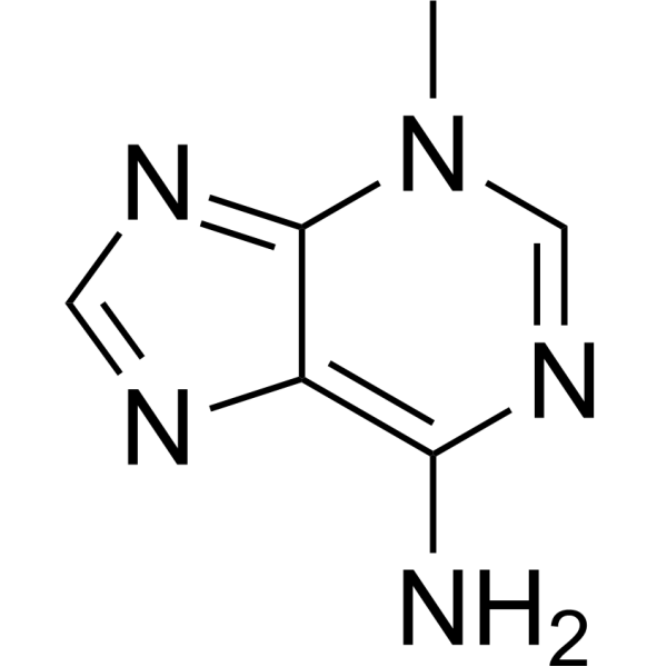
3-Methyladenine


Description & Advantages
Green Fluorescent Protein (GFP), derived from Aequorea Victoria, is comprised of 238 amino acids and has a molecular weight of 26.9 kDa. Its native fluorescent group emits a distinct green fluorescence when excited by ultraviolet or blue light, exhibiting stable fluorescent properties. GFP can be stably expressed in various cellular contexts without species, tissue, or unknown specificity constraints. As it is non-toxic to cells and easy to detect, GFP has been extensively utilized as a reporter gene in the fields of cell biology and molecular biology.
MCE Anti-GFP Magnetic Agarose Beads is produced through the covalent coupling of high-quality GFP antibody with magnetic agarose beads. It has high loading capacity, exceptional specificity, and stability, can be used for the detection and purification of GFP, EGFP, and their fusion-expressed proteins. Moreover, it can be utilized for IP assays without binding to BFP-tagged proteins.
The specifications of the product correspond to the actual beads volume, with the beads content of 20%.


Protocol
Recommended Buffers (Not Provided)
| Buffer | Composition |
|---|---|
| Binding/Wash Buffer | 50 mM Tris, 0.15 M NaCl, pH 7.4 |
| Acidic Elution Buffer | 0.1 M glycine, pH 2.0-3.0 |
| Neutralization Buffer | 1 M Tris-HCl, pH 8.0 |
| Storage Buffer | PBS containing 0.02% (w/v) NaN3 |
Note: Prepare all buffers with ultrapure water and sterilize them by filtration through a 0.45 μm or 0.22 μm membrane.
Protein Purification/Immunoprecipitation
1. Bead Pretreatment: Thoroughly resuspend the Anti-GFP Magnetic Agarose Beads. Transfer the required volume into a 1.5 mL tube, add 500 μL of binding/wash buffer, mix thoroughly, perform magnetic separation, and discard the supernatant. Repeat 2-3 times.
2. Sample Binding: Add 200-1,000 μL of clarified sample and incubate on a rotator at 4°C for 2 h or overnight. Perform magnetic separation and transfer the supernatant to a new tube for analysis if required.
3. Washing: Add 500 μL-1 mL of binding/wash buffer, resuspend thoroughly, perform magnetic separation, and discard the supernatant. Repeat at least three times, or until the OD280 of the wash supernatant is below 0.05.
4. Elution: Select acidic or denaturing elution according to downstream requirements. For acidic elution, add 50-100 μL of elution buffer, incubate at room temperature for 5-10 min, perform magnetic separation, and immediately add 1/10 volume of neutralization buffer. For denaturing elution, add 20-50 μL of 2× SDS-PAGE Loading Buffer, heat at 95°C for 5 min, perform magnetic separation, and collect the supernatant.
Note: Acid-eluted proteins may be stored at 4°C for a short period or at -20°C for long-term storage. After denaturing elution with SDS, β-mercaptoethanol, or DTT, the immobilized antibody or ligand may be inactivated and the beads should not be reused.
5. Storage: If the beads will be reused, wash thoroughly with binding/wash buffer, resuspend in storage buffer, and store at 2-8°C. Do not freeze, dry, or expose the beads to a magnetic field for an extended period.


Publications
-
Journal Impact Factor
-
Most Recent
-
JCI InsightTargeting KIFC1 to disrupt centrosome clustering and trigger anaphase catastrophe in small-cell lung cancer. [Abstract]2026 Apr 8;11(7):e199352. PMID: 41948931
-
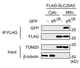
Anti-GFP Magnetic Agarose Beads purchased from MedChemExpress. Usage Cited in: bioRxiv. 2025 Nov 11.
Coimmunoprecipitation and immunoblot analysis of cytosolic (Cyto) or mitochondrial (Mito) fractions isolated from HEK293T cells transfected with FLAG-SLC25A5 and GFP-PE18. The GFP vector (-) was used as a control. TOMM20 and β-tubulin were used as reference proteins for lysates of either mitochondria or the cytosol.
Storage
4℃,2 years.
Do not dry or freeze.


Components
| Components | HY-K0246-1 mL | HY-K0246-5 mL |
|---|---|---|
| Anti-GFP Magnetic Agarose Beads | 1 mL | 5 mL |


 Please enter the correct Lot Number
