



One Step TUNEL Apoptosis Detection Kit (FITC)
Based on 43 publication(s) in Google Scholar
MCE One Step TUNEL Apoptosis Detection Kit (FITC) provides a rapid and convenient method to detect cell apoptosis. After staining cells with this kit, live cells have no fluorescence, apoptosis cells show green fluorescence.
-
Storage :
-20°C, 1 year.Protect from light.



To place orders, for customer services and technical support, please contact: MedChemExpress USA
Tel: 609-228-6898 E-mail: [email protected] [email protected]



-

HY-108717
Proteinase K -

HY-15141
Staurosporine -
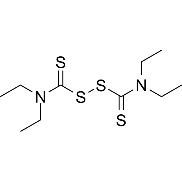
HY-B0240
Disulfiram -
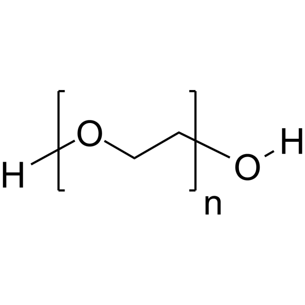
HY-Y0873
PEG300 -
HY-K1042
AntiFade Mounting Medium -

HY-P7118
TGF beta 1/TGFB1 Protein, Human (CHO) -

HY-10219
Rapamycin -

HY-K0301
Cell Counting Kit-8 -

HY-K1073
Annexin V-FITC/PI Apoptosis Detection Kit -

HY-128868A
FITC-Dextran (MW 4000) -
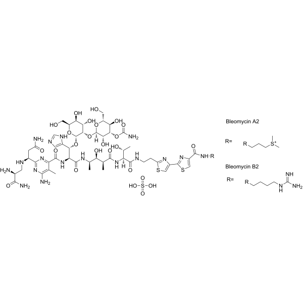
HY-17565
Bleomycin sulfate -
HY-K0302
Cell-ATP Viability Detection Kit


Description & Advantages
MCE One Step TUNEL Apoptosis Detection Kit (FITC) provides a rapid and convenient method to detect cell apoptosis. When cells undergo apoptosis, specific DNA endonucleases will be activated, cutting the genomic DNA between the nucleosomes. The DNA of apoptotic cells is cleaved into multimers of 180-200 bp fragments. The exposed 3'-OH of the broken DNA can be catalyzed by Terminal Deoxynucleotidyl Transferase (TdT) with fluorescein labeled dUTP, which can be detected with fluorescence microscope or flow cytometer. The maximum excitation wavelength and emission wavelength of FITC were 488 nm and 525 nm respectively.


Protocol
1. Preparation of TUNEL working solution
Prepare the TUNEL working solution on ice and protected from light immediately before use:
| Component | 1 reaction (50 μL) |
|---|---|
| TdT Enzyme | 5 μL |
| FITC-12-dUTP Labeling Mix | 45 μL |
2. Adherent cells
1. Wash cells twice with PBS for 5 min each.
2. Fix with 4% paraformaldehyde in PBS for 30 min at 4°C.
3. Wash twice with PBS for 5 min each.
4. Permeabilize with PBS containing 0.3% Triton X-100 for 5 min at room temperature.
5. Wash twice with PBS for 5 min each.
6. Add 50 μL TUNEL working solution per sample and incubate at 37°C for 60 min protected from light.
7. Wash three times with PBS. Counterstain/mount with DAPI-containing antifade mounting medium if desired and observe FITC green fluorescence by fluorescence microscopy.
3. Suspension cells
1. Collect approximately 2 × 10⁶ cells by centrifugation and wash twice with PBS for 5 min each.
2. Fix for 30 min at 4°C. Gentle shaking during fixation is recommended to reduce extensive cell clumping.
3. Wash twice with PBS for 5 min each.
4. Resuspend cells in permeabilization solution and incubate for 5 min at room temperature.
5. Wash twice with PBS for 5 min each.
6. Add 50 μL TUNEL working solution and incubate at 37°C for 60 min protected from light.
7. Wash with PBS and resuspend in an appropriate volume of PBS for FITC detection by flow cytometry, or prepare slides for fluorescence microscopy.
Note: For a negative control, omit TdT Enzyme. FITC-positive cells indicate apoptotic cells with DNA strand breaks.


Publications
-
Journal Impact Factor
-
Most Recent
-
J Adv ResParvimonas micra-derived SirTM: An ADP-ribosyltransferase as a novel driver in colorectal cancer progression. [Abstract]2026 Mar 17:S2090-1232(26)00256-0. PMID: 41850426
-
Gut MicrobesProgranulin protects against Clostridioides difficile infection by enhancing IL-22 production. [Abstract]2024 Jan-Dec;16(1):2409220. PMID: 39349385
-
Nat Plants2024 Jun;10(6):1027-1038. PMID: 38831045
-
ApoptosisTNF-α and RPLP0 drive the apoptosis of endothelial cells and increase susceptibility to high-altitude pulmonary edema. [Abstract]2024 Oct;29(9-10):1600-1618. PMID: 39110356
-
CNS Neurosci TherRFWD2 Mitigates AD-Like Cognitive Impairments via the JNK-SGK1 Signaling Pathway in Mice. [Abstract]2026 Apr;32(4):e70860. PMID: 41954517
-
Ecotoxicol Environ Saf2025 Jan 15:290:117747. PMID: 39823667
-
Ren FailH-151 attenuates lipopolysaccharide-induced acute kidney injury by inhibiting the STING-TBK1 pathway. [Abstract]2024 Dec;46(2):2363591. PMID: 38856314
-
StressRole of ATF4 in fibroblast proliferation and apoptosis induced by mechanical trauma through the miR-93-3p/Smad7 axis. [Abstract]2025 Dec 31;28(1):2579503. PMID: 41312780
Storage
-20°C, 1 year.
Protect from light.


Components
| Components | HY-K1078-20T | HY-K1078-50T |
|---|---|---|
| TdT Enzyme | 100 μL | 250 μL |
| FITC-12-dUTP Labeling Mix | 900 μL | 2×1.2 mL |
| TdT Dilution Buffer | 300 μL | 1 mL |


 Please enter the correct Lot Number
