



Hematoxylin-Eosin Staining Kit
Based on 15 publication(s) in Google Scholar
MCE Hematoxylin-Eosin Staining Kit can be used for cell staining, the combination of hematoxylin and eosin results in a staining pattern where the cytoplasm appears red or pink and the nucleus takes on a blue or blue-violet.
-
Storage :
Room temperature, 1 year.


To place orders, for customer services and technical support, please contact: MedChemExpress USA
Tel: 609-228-6898 E-mail: [email protected] [email protected]



-

HY-10583
Y-27632 dihydrochloride -
HY-K0301
Cell Counting Kit-8 -
HY-B0673
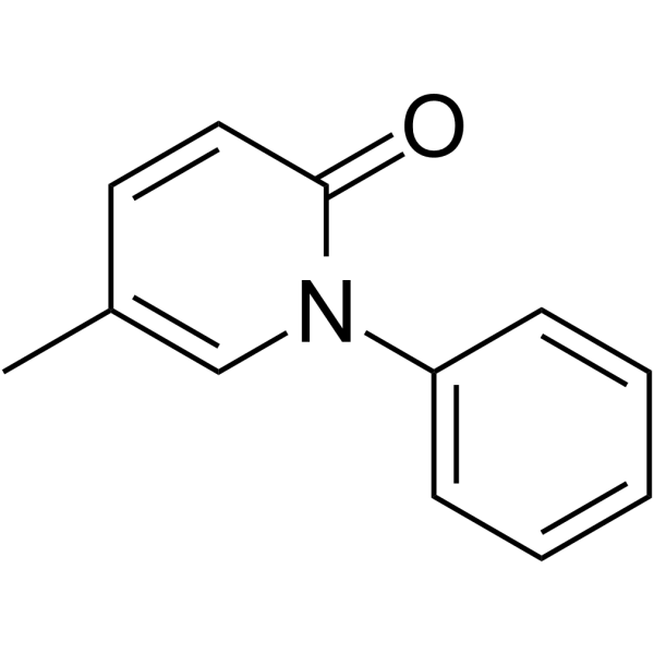
Pirfenidone -
HY-K1073
Annexin V-FITC/PI Apoptosis Detection Kit -
HY-K1001
RIPA Lysis Buffer (Strong) -

HY-100579
Ferrostatin-1 -
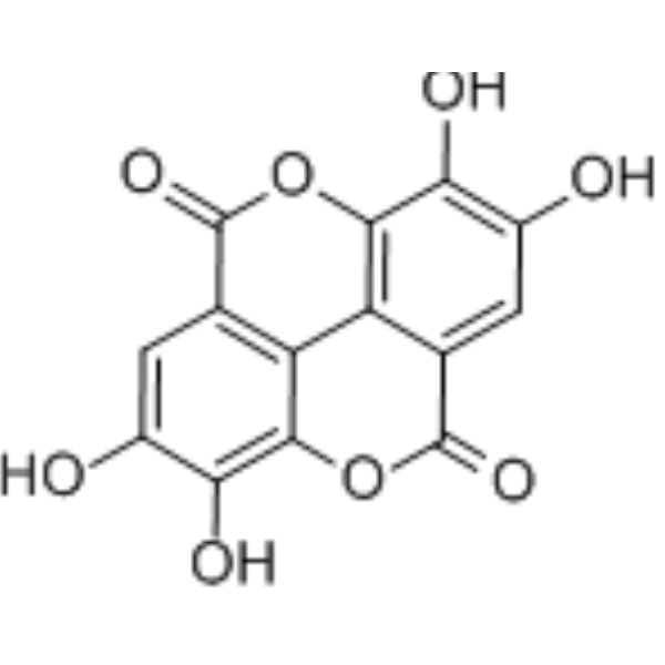
HY-B0183
Ellagic acid -
HY-K1059
BM-Cyclin -

HY-K0320
ROS Assay Kit -
HY-K1071
Cell Cycle and Apoptosis Analysis Kit (PI staining)


Description & Advantages
Hematoxylin is a naturally sourced basic dye derived from the Hematoxylin tree, it binds to negatively charged acids (such as DNA), staining the chromatin in the nucleus and cytoplasmic ribosomes a blue-violet color. Eosin is a chemically synthesized acidic dye that binds to positively charged amino cations in proteins, staining the cytoplasm and extracellular matrix red. The combination of hematoxylin and eosin results in a staining pattern where the cytoplasm appears red or pink and the nucleus takes on a blue or blue-violet.
MCE Hematoxylin-Eosin Staining Kit streamlines experimental procedures, reduces operation time, and eliminates the use of toxic reagents. It can be directly employed for staining tissue sections or cultured cells in immunohistochemistry.


Protocol
Paraffin Section Staining
1. Deparaffinize sections in xylene I and xylene II for 5-10 min each.
2. Rehydrate in absolute ethanol I and II for 3 min each, followed by 95%, 85%, and 75% ethanol for 2 min each, and finally rinse with distilled water.
3. Stain the sections with Hematoxylin Staining Solution for 3-10 min.
4. Rinse under running tap water for 5-10 min to allow complete nuclear bluing. If necessary, briefly differentiate with a routine laboratory acid-alcohol solution and blue again.
5. Stain with Eosin Staining Solution for 1-3 min.
6. Rapidly dehydrate through 75%, 85%, and 95% ethanol, then absolute ethanol I and II for 2-3 min each, and clear in xylene I and II for 3-5 min each.
7. Mount with neutral mounting medium or another suitable mounting reagent and observe under a microscope.
Frozen Section Staining
1. Equilibrate frozen sections to room temperature, fix with 4% paraformaldehyde for 10-20 min, and wash 2-3 times with PBS.
2. Stain with Hematoxylin Staining Solution for 3-10 min and rinse under running tap water for 5-10 min.
3. Stain with Eosin Staining Solution for 1-3 min and briefly rinse with distilled water.
4. Dehydrate, clear, and mount as required.
Cultured Cell Staining
1. Remove the culture medium, wash the cells twice with PBS, and fix with 4% paraformaldehyde for 10-20 min.
2. Wash 2-3 times with PBS, add sufficient Hematoxylin Staining Solution to cover the cells, and stain for 3-10 min.
3. Rinse gently under running tap water until the background is clear and the nuclei are fully blued.
4. Add Eosin Staining Solution and stain for 30 s-3 min, then briefly rinse with distilled water.
5. Select an appropriate dehydration, clearing, and mounting method according to the slide or culture vessel used, and observe the staining.


Publications
-
Journal Impact Factor
-
Most Recent
-
J Adv ResRutin triggers IRE1-mediated GSDMD-dependent pyroptosis in macrophages to suppress systemic Salmonella infection. [Abstract]2026 Apr 1:S2090-1232(26)00273-0. PMID: 41932494
-
FASEB JSENP3 Exacerbates Spinal Cord Injury by Increasing H6PD deSUMOylation to Promote Glycolysis. [Abstract]2026 Jun 15;40(11):e71995. PMID: 42247250
-
Tissue CellComprehensive amelioration of glucose and lipid metabolism dysregulation in T2DM by a Lycium blend. [Abstract]2026 Jun 10:103:103685. PMID: 42275865
Storage
Room temperature, 1 year.


Components
| Components | HY-K0315-100 mL |
|---|---|
| Hematoxylin Staining Solution | 50 mL |
| Eosin Staining Solution | 50 mL |


 Please enter the correct Lot Number
