



Streptavidin Magnetic Beads
Based on 226 publication(s) in Google Scholar
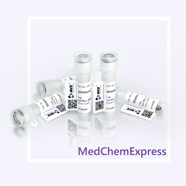
MCE Streptavidin Magnetic Beads provide a fast and convenient method for numerous applications, including purification of proteins and nucleic acids, protein interaction studies, immunoprecipitation, immunoassays, pull-down and cell isolation. The 1 mL is defined as the base specification. All larger sizes correspond to incremental volumes of this base.
-
Storage :
4°C, 2 years. Do not centrifuge, dry or freeze the magnetic beads.


To place orders, for customer services and technical support, please contact: MedChemExpress USA
Tel: 609-228-6898 E-mail: [email protected] [email protected]



-
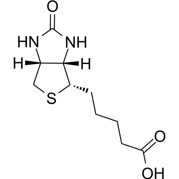
HY-B0511
Biotin -

HY-K0202
Protein A/G Magnetic Beads -

HY-K0207
Anti-Flag Magnetic Beads -

HY-K0010
Protease Inhibitor Cocktail (EDTA-Free, 100× in DMSO) -

HY-10071
Y-27632 -

HY-13259
MG-132 -
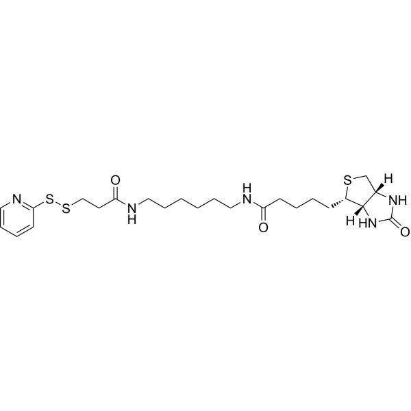
HY-136769
Biotin-HPDP -

HY-K0201
Anti-HA Magnetic Beads -
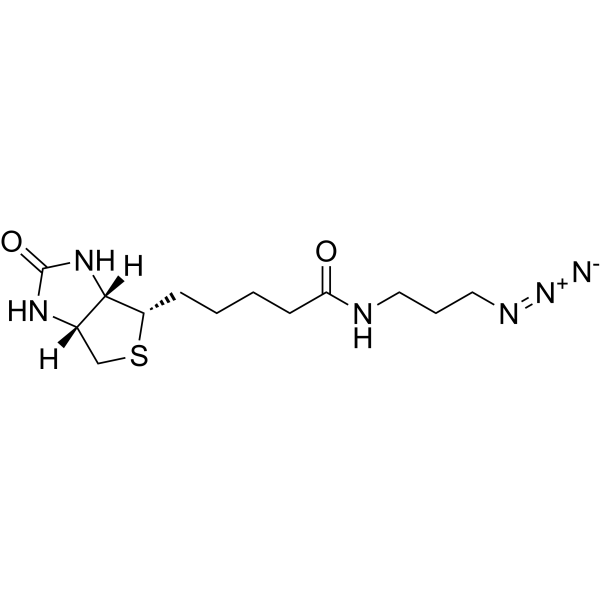
HY-129832
Biotin-azide -

HY-10219
Rapamycin -

HY-100558
Bafilomycin A1 -

HY-Y0320C
Dimethyl sulfoxide


Description & Advantages
MCE Streptavidin Magnetic Beads use recombinant streptavidin covalently coupling to the surface of the paramagnetic beads. Streptavidin, with no carbohydrate group, is different from avidin ensuring low nonspecific binding. The biotinylated molecules (e.g. peptides, proteins, antibodies, sugars, lectins, oligonucleotides, DNA/RNA) bind to the beads due to the high affinity between streptavidin and biotin. MCE Streptavidin Magnetic Beads are removed from the solution manually by using a magnetic stand or automatically by using an instrument.
1. High binding capacity.
2. Low non-specific binding.
3. Minimal sample loss.
4. Ready to use.


Protocol
1. Immobilization Nucleic Acids
1) Resuspend the magnetic beads in the vial (or vortex for 20 seconds), transfer 100 μL of Streptavidin Magnetic Beads into a 1.5 mL tube (transfer amount may be adjusted as required). Place the tube into a magnetic stand to collect the beads against the side of the tube (Hereinafter referred to as magnetic separation). Remove and discard the supernatant.
Note: The amount of beads is sufficient for one reaction. Scale up the reaction as needed.
2) Add 1 mL Wash Buffer I to the beads, invert the tube several times or vortex gently for 15 seconds to mix. Remove and discard the supernatant from magnetic separation. Repeat this step for 2 times.
3) Add 500 μL of biotinylated nucleic acids diluted with Wash Buffer I, makes the beads at a final concentration of 2 mg/mL. Rotate the tube for 30 minutes at room temperature or 2 hours at 4ºC.
4) Separate the biotinylated nucleic acids coated beads with a magnetic stand.
5) Add 1 mL Wash Buffer I to the beads, invert the tube several times or vortex gently for 15 seconds to mix. Remove and discard the supernatant from magnetic separation. Repeat this step for 2 times.
Note: The amount of biotinylated nucleic acids can be calculated by measuring absorbance of the flow through along with absorbance of starting material.
2. Immobilization Antibodies/Proteins
1) Resuspend the magnetic beads in the vial (or vortex for 20 seconds), transfer 100 μL of Streptavidin Magnetic Beads into a 1.5 mL tube (transfer amount may be adjusted as required). Place the tube into a magnetic stand to collect the beads against the side of the tube (Hereinafter referred to as magnetic separation). Remove and discard the supernatant.
Note: The amount of beads is sufficient for one reaction. Scale up the reaction as needed.
2) Add 1 mL Wash Buffer II to the beads, invert the tube several times or vortex gently for 15 seconds to mix. Remove and discard the supernatant from magnetic separation. Repeat this step for 2 times.
3) Add 1 mL of biotinylated antibodies/proteins with Wash Buffer II, makes the beads at a final concentration of 1 mg/mL. Rotate the tube for 60 minutes at room temperature or 2 hours at 4ºC.
4) Separate the biotinylated antibodies/proteins coated beads with a magnetic stand.
5) Add 1 mL Wash Buffer II to the beads, invert the tube several times or vortex gently for 15 seconds to mix. Remove and discard the supernatant from magnetic separation. Repeat this step for 5 times.
6) Binding is now complete. Resuspend the beads in Buffer II or a buffer suitable for downstream applications to a desired concentration. Use the beads immediately, or store at 4ºC for later use.
3. Elution
3.1 Nucleic acid Elution
Step: Add 50–100 μL of Elution Buffer I to the magnetic beads, incubate for 5 minutes at 65°C or 2 minutes at 90°C, place on a magnetic rack, perform magnetic separation and collect the supernatant.
3.2 Antibody/Protein Elution
a. Denaturation elution: The samples eluted by this method are suitable for SDS–PAGE. Step: Add 50–100 μL of 1 × SDS-PAGE Loading Buffer to the magnetic beads, mix well and heat at 95°C for 5 minutes. Place on a magnetic rack, perform magnetic separation and collect the supernatant.
Note: If you choose denaturation elution, the eluent will contain streptavidin monomers and polymers, biotin-labeled antibody or protein.
b. Non-denaturation elution: The sample eluted by this method retains its original biological activity and can be used for later functional analysis. Step: Add 50–100 μL Elution Buffer II to the magnetic beads and incubate at room temperature for 5-10 minutes. Place on a magnetic rack, perform magnetic separation and collect the supernatant, immediately add 1/10 volume of the total volume of Neutralization Buffer (0.1 M NaOH), adjust the pH of the eluted product to neutral, and the samples is used for later functional analysis.
Note: If you choose non-denaturation elution method, the streptavidin may be dislodged under acidic conditions, and the incubation time should not exceed 10 minutes. Acidic eluents can destroy most of the antibody and antigen interactions, you can wash the beads once with 1mL of 0.1% Tween-20 before elution.


Publications
-
Journal Impact Factor
-
Most Recent
-
Signal Transduct Target TherNuclear mitochondrial acetyl-CoA acetyltransferase 1 orchestrates natural killer cell-dependent antitumor immunity in colorectal cancer. [Abstract]2025 Apr 28;10(1):138. PMID: 40289129
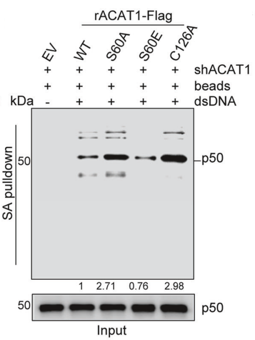
Streptavidin Magnetic Beads purchased from MedChemExpress. Usage Cited in: Signal Transduct Target Ther. 2025 Apr 28;10(1):138. [Abstract]
ACAT1-depleted HCT116 cells were rescued with ACAT1-Flag WT, S60A, S60E, C126A or EV (as a control). Subcellular fractionation assay and DNA pulldown assay were performed to detect nuclear p50 bound to biotinylated dsDNA (5 µg) conjugated to Streptavidin magnetic beads.
-
Cancer CellLymphatic-localized Treg-mregDC crosstalk limits antigen trafficking and restrains anti-tumor immunity. [Abstract]2024 Aug 12;42(8):1415-1433.e12. PMID: 39029466
-
CellStreptococcus anginosus promotes gastric inflammation, atrophy, and tumorigenesis in mice. [Abstract]2024 Feb 15;187(4):882-896.e17 PMID: 38295787
-
Cell ResA thermosensor FUST1 primes heat-induced stress granule formation via biomolecular condensation in Arabidopsis. [Abstract]2025 Jul;35(7):483-496. PMID: 40360668
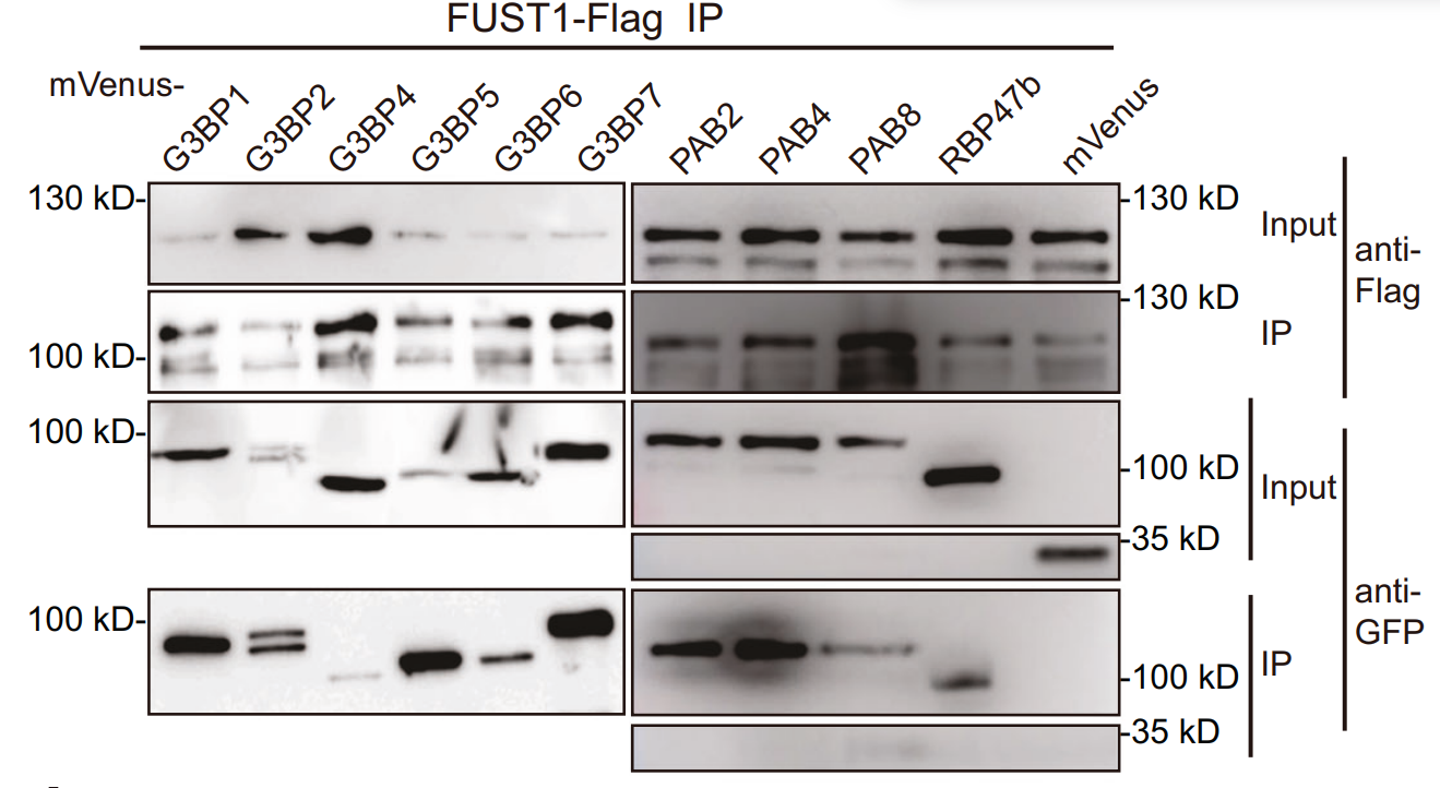
Streptavidin Magnetic Beads purchased from MedChemExpress. Usage Cited in: Cell Res. 2025 Jul;35(7):483-496. [Abstract]
Streptavidin Magnetic Beads (4 mg/ml). Co-immunoprecipitation of FUST1 with indicated proteins expressed in tobacco epidermal cells. Cells were treated at 37 °C for 1 h and crosslinked with UV. Empty vector was included as a negative control.
-
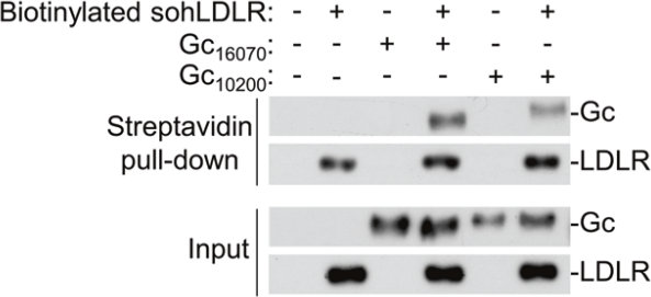
Streptavidin Magnetic Beads purchased from MedChemExpress. Usage Cited in: Cell Res. 2024 Feb;34(2):140-150. [Abstract]
Recombinant Gc of CCHFV YL16070 and IbAr 10200 strains and biotinylated sohLDLR were co-incubated as indicated. Pull-down assay was performed with magnetic streptavidin beads and the pellets were subjected to immunoblots with the indicated antibodies.
-
Adv MaterCell Surface-Confined DNAzyme Coordination-Facilitated Logical Engineering for Precise Cell Identification. [Abstract]2025 Aug;37(34):e2504363. PMID: 40489061
-
Cell Host MicrobeImmune activation reprograms growth mRNA stability to shape plant growth-defense trade-offs. [Abstract]2026 Mar 11;34(3):479-493.e8. PMID: 41763212
-
J Extracell VesiclesIdentification of a Biomarker Panel in Extracellular Vesicles Derived From Non-Small Cell Lung Cancer (NSCLC) Through Proteomic Analysis and Machine Learning. [Abstract]2025 May;14(5):e70078. PMID: 40366616
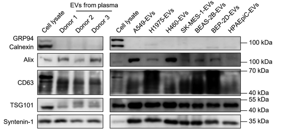
Streptavidin Magnetic Beads purchased from MedChemExpress. Usage Cited in: J Extracell Vesicles. 2025 May;14(5):e70078. [Abstract]
Streptavidin Magnetic Beads (4 ℃; 6 h). EV biomarker detection using western blotting
-
AutophagyPhase separation of OPTN initiates mitophagy to orchestrate craniofacial bone mineralization. [Abstract]2026 May;22(5):1021-1043. PMID: 41692957
-
AutophagyMyotubularin 2 interacts with SEC23A and negatively regulates autophagy at ER exit sites in Arabidopsis. [Abstract]2025 Jan;21(1):141-159. PMID: 39177202
-
Nat CommunTimosaponin AIII enhances CAR-T cell potency and prevents relapse through impairing CAR-Tregs. [Abstract]2026 Mar 31;17(1):3045. PMID: 41916982
-
Nat Commun2025 Dec 12;16(1):11092. PMID: 41387436
-
Nat Commun2025 Nov 26;16(1):10514. PMID: 41298370
-
Nat CommunA cohort of mRNAs undergo high-stoichiometry NSUN6-mediated site-specific m5C modification. [Abstract]2025 Jul 4;16(1):6119. PMID: 40615396
-
Nat Commun2024 Aug 24;15(1):7287. PMID: 39179589
-
Nat CommunMycobacterium tuberculosis suppresses host antimicrobial peptides by dehydrogenating L-alanine. [Abstract]2024 May 17;15(1):4216. PMID: 38760394
-
Nat Commun2023 Nov 21;14(1):7597. PMID: 37989750
-
Nat Commun2023 Mar 30;14(1):1731. PMID: 36997512
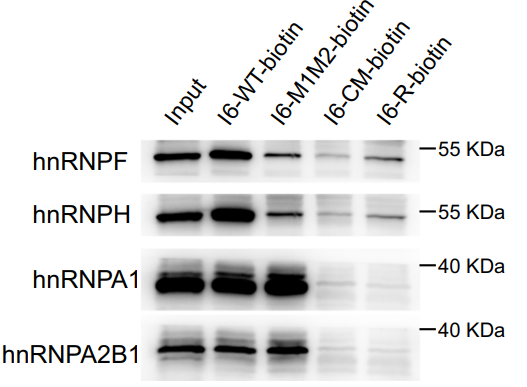
Streptavidin Magnetic Beads purchased from MedChemExpress. Usage Cited in: Nat Commun. 2023 Mar 30;14(1):1731. [Abstract]
The RNA pull-down assays showed that both hnRNP F/H and A1/A2B1 could bind to I6-WT RNA but not the negative control I6-R biotin (lanes 2 and 5).
-
Nat CommunCryptococcal Hsf3 controls intramitochondrial ROS homeostasis by regulating the respiratory process. [Abstract]2022 Sep 15;13(1):5407. PMID: 36109512
-
J Adv ResLong noncoding RNA Nron and its functional motif modulate macrophage-mediated inflammatory response. [Abstract]2025 Aug 22:S2090-1232(25)00647-2. PMID: 40850682
-
Mol CellHSPA1A and DNAJB1 regulate NELF condensate dynamics to safeguard transcriptional recovery under heat stress. [Abstract]2026 Feb 19;86(4):674-692.e10. PMID: 41653920
-
Mol CellCPT1A induction following epigenetic perturbation promotes MAVS palmitoylation and activation to potentiate antitumor immunity. [Abstract]2023 Dec 7;83(23):4370-4385.e9. PMID: 38016475
-
Nucleic Acids Res2025 Aug 27;53(16):gkaf820. PMID: 40902005
-
Nucleic Acids ResSelective recognition of RNA G-quadruplex in vitro and in cells by L-aptamer-D-oligonucleotide conjugate. [Abstract]2024 Dec 11;52(22):13544-13560. PMID: 39558155
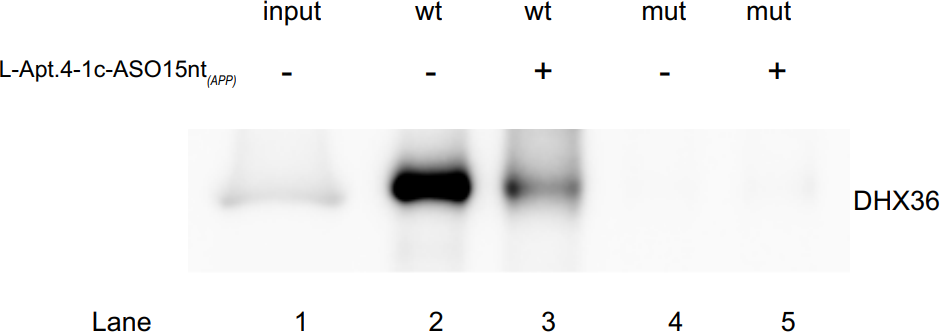
Streptavidin Magnetic Beads purchased from MedChemExpress. Usage Cited in: Nucleic Acids Res. 2024 Dec 11;52(22):13544-13560. [Abstract]
L-Apt.4–1c-ASO15nt(APP) conjugate can disrupt APP rG4–DHX36 interactions according to RIP assay (100 nM L-Apt.4–1c-ASO15nt(APP).
-
Nucleic Acids Res2023 Jan 25;51(2):e12. PMID: 36477375
-
TheranosticsHilnc- mediated UCP1 translation repression contributes to thermogenesis and energy expenditure. [Abstract]2026 Jan 1;16(7):3263-3285. PMID: 41608567
-
TheranosticsTherapeutic potential of targeting MKK3-p38 axis with Capsaicin for Nasopharyngeal Carcinoma. [Abstract]2020 Jun 24;10(17):7906-7920. PMID: 32685028
-
Acta Pharm Sin B2025 Aug;15(8):4078-4095. PMID: 40893681
-
Adv Sci (Weinh)Targeting Histone Deacetylase 11 with a Highly Selective Inhibitor for the Treatment of MASLD. [Abstract]2025 Apr;12(15):e2412903. PMID: 39976110
-
Cancer Lett2025 Apr 28:616:217532. PMID: 40021040
-
Mol TherFOXO3A-induced LINC00926 suppresses breast tumor growth and metastasis through inhibition of PGK1-mediated Warburg effect. [Abstract]2021 Sep 1;29(9):2737-2753. PMID: 33940159
-
Mol TherMicroglial lnc-U90926 facilitates neutrophil infiltration in ischemic stroke via MDH2/CXCL2 axis. [Abstract]2021 Sep 1;29(9):2873-2885. PMID: 33895326
-
Phytomedicine2025 Mar:138:156434. PMID: 39884078
-
J Transl Med2025 Nov 7;23(1):1243. PMID: 41204251
-
Proc Natl Acad Sci U S AA brain-enriched lncRNA shields cancer cells from immune-mediated killing for metastatic colonization in the brain. [Abstract]2022 May 31;119(22):e2200230119. PMID: 35617432
-
Dev CellHematopoietic stem cells activate a latent differentiation pathway to facilitate recovery after 5-fluorouracil-induced myeloablation. [Abstract]2026 Mar 2:S1534-5807(26)00043-2. PMID: 41775265
-
OncogeneZDHHC9-mediated KLF5 palmitoylation enhances the cAMP/PKA/CREB axis to promote colorectal cancer progression. [Abstract]2026 Apr;45(15):1370-1385. PMID: 41882103
-
Mol MedAcid ceramidase targeting pyruvate kinase affected trypsinogen activation in acute pancreatitis. [Abstract]2022 Sep 6;28(1):106. PMID: 36068514
-
Cell RepInterleukin-1β modulates lymphoid differentiation of Flt3-positive multipotent progenitors after transplantation. [Abstract]2024 Oct 18;43(11):114890. PMID: 39425929
-
Cell RepCholesterol suppresses GOLM1-dependent selective autophagy of RTKs in hepatocellular carcinoma. [Abstract]2022 Apr 19;39(3):110712. PMID: 35443161
-
Anal ChemUniversal Enrichment and Sequencing of DNA Modifications Using a Click-Coupled IgG-Protein A/G System. [Abstract]2026 Mar 3;98(8):5964-5975. PMID: 41693615
-
Anal ChemProximity-Based Phospho-Interactome (Prob-PhI) Characterization Reveals Distinct Signaling Activities of MEK1 and MEK2. [Abstract]2026 Feb 10;98(5):3688-3698. PMID: 41611650
-
Anal ChemSelf-Priming DNA Polymerization-Propelled Stochastic Walkers on Magnetic Microbeads for Amplified Detection of miRNA. [Abstract]2023 Mar 7;95(9):4529-4535. PMID: 36814089
-
Anal ChemMultivalent Duplexed-Aptamer Networks Regulated a CRISPR-Cas12a System for Circulating Tumor Cell Detection. [Abstract]2021 Sep 28;93(38):12921-12929. PMID: 34533940
-
Emerg Microbes InfectInhibition of RAN attenuates influenza a virus replication and nucleoprotein nuclear export. [Abstract]2024 Dec;13(1):2387910. PMID: 39087696
-
Anal Chim ActaAn aptamer-mediated EXPAR fluorescence biosensor for early and sensitive detection of Aβ42 and Aβ40. [Abstract]2026 Jul 8:1406:345513. PMID: 42067300
-
EMBO RepEhbp1 orchestrates orderly sorting of Wnt/Wingless to the basolateral and apical cell membranes. [Abstract]2024 Nov;25(11):5053-5079. PMID: 39402333
-
Pharmaceuticals (Basel)2026 Jun 22;19(6):967. PMID: 42356584
-
Cell Signal2024 Dec:124:111427. PMID: 39304099
-
iScienceEpitranscriptome analysis of NAD-capped RNA by spike-in-based normalization and prediction of chronological age. [Abstract]2023 Nov 22;26(12):108558. PMID: 38094247
-
FASEB JCircTLK1: A novel regulator involved in VSMC phenotypic switching and the developmental process of atherosclerosis. [Abstract]2024 Mar 31;38(6):e23557. PMID: 38498343
-
J VirolYBX1 is required for assembly of viral replication complexes of chikungunya virus and replication of multiple alphaviruses. [Abstract]2025 Feb 25;99(2):e0201524. PMID: 39745458
-
J Biol ChemSplicing factor hnRNPA1 regulates alternative splicing of LOXL2 to enhance the production of LOXL2Δ13. [Abstract]2024 Jul;300(7):107414. PMID: 38810697
-
J Med Virol2020 Dec;92(12):3057-3066. PMID: 32776519
-
AnalystA highly-efficient isothermal nano-detection platform coupling CRISPR/Cas technology for detection of circRNA. [Abstract]2026 Mar 2;151(5):1304-1309. PMID: 41705504
-
PLoS OneProteomic screening of TMEM43 binding partners identifies VDAC leading to mitochondrial dysfunction. [Abstract]2025 Dec 18;20(12):e0339129. PMID: 41411330
-
Chembiochem2024 Sep 2;25(17):e202400269. PMID: 38923255
-
bioRxivPhosphorylation-induced flexibility of proto-oncogenic Bcl3 regulates transcriptional activation by NF-κB p52 homodimer. [Abstract]2024 Jul 17:2024.06.30.601400. PMID: 38979190
Storage
4°C, 2 years.
Do not centrifuge, dry or freeze the magnetic beads.


Components
| Components | HY-K0208-1 mL | HY-K0208-5 mL | HY-K0208-10 mL |
|---|---|---|---|
| Streptavidin Magnetic Beads | 1 mL | 1 mL×5 | 1 mL×10 |


 Please enter the correct Lot Number
