


Bafilomycin A1
Based on 815 publication(s) in Google Scholar
Bafilomycin A1 (BafA1) is a specific and reversible inhibitor of vacuolar H+-ATPase (V-ATPase) with IC50 values of 4-400 nmol/mg. Bafilomycin A1, a macrolide antibiotic, is also used as an autophagy inhibitor at the late stage. Bafilomycin A1 blocks autophagosome-lysosome fusion and inhibits acidification and protein degradation in lysosomes of cultured cells. Bafilomycin A1 induces apoptosis.
For research use only. We do not sell to patients.
- Purity: 99.95%
- CAS No.: 88899-55-2
- Formula: C35H58O9
- Molecular Weight:622.83
-
Storage:
-20°C, protect from light
* In solvent : -80°C, 6 months; -20°C, 1 month (protect from light)


-
 Biological Activity
-
Chemical Information
-
Solvent & Solubility
- Protocol
- Purity & Documentation
- References
-
 Help & FAQs


Publications Citing Use of MedChemExpress (MCE) Bafilomycin A1
More- Signal Transduct Target Ther. 2025 Dec 15;10(1):406. [Abstract]
- Signal Transduct Target Ther. 2024 Dec 30;9(1):367. [Abstract]
- Signal Transduct Target Ther. 2023 Jan 20;8(1):22. [Abstract]
- Signal Transduct Target Ther. 2021 Feb 17;6(1):67. [Abstract]
- Signal Transduct Target Ther. 2021 Jan 25;6(1):29. [Abstract]
- Nature. 2022 Aug;608(7922):413-420. [Abstract]
- Cancer Cell. 2023 Jul 10;41(7):1276-1293.e11. [Abstract]
- Cancer Cell. 2021 Mar 8;39(3):423-437.e7. [Abstract]
- Cell. 2026 Apr 2;189(7):1923-1941.e26. [Abstract]
- Cell. 2025 Jun 26;188(13):3441-3458.e25. [Abstract]
- Cell. 2023 Aug 31;186(18):3903-3920.e21. [Abstract]
- Nat Biotechnol. 2022 Dec;40(12):1834-1844. [Abstract]
- Nat Nanotechnol. 2025 Feb;20(2):296-302. [Abstract]
- Nat Nanotechnol. 2022 Sep;17(9):993-1003. [Abstract]
- Mol Cancer. 2024 Sep 2;23(1):184. [Abstract]
- Cell Metab. 2025 Oct 7:S1550-4131(25)00389-4. [Abstract]
- Cell Metab. 2021 May 4;33(5):971-987.e6. [Abstract]
- Nat Immunol. 2025 Apr 11. [Abstract]
- Adv Mater. 2025 May 22:e2504050. [Abstract]
- Immunity. 2025 Nov 11;58(11):2670-2684.e10. [Abstract]
- Immunity. 2025 Apr 8:S1074-7613(25)00128-1. [Abstract]
- Gut. 2025 Apr 23:gutjnl-2025-335220. [Abstract]
- Cancer Commun (Lond). 2025 Aug;45(8):1010-1037. [Abstract]
- Cancer Commun (Lond). 2025 Mar;45(3):218-244. [Abstract]
- Mil Med Res. 2022 Feb 14;9(1):9. [Abstract]
- Exploration. 2025 Dec 12;5(6):20240069. [Abstract]
- Drug Resist Updat. 2023 Nov:71:101005. [Abstract]
- Nat Metab. 2022 Sep;4(9):1202-1213. [Abstract]
- Cell Stem Cell. 2021 Jun 3;28(6):1074-1089.e7. [Abstract]
- Cell Mol Immunol. 2025 May;22(5):512-526. [Abstract]
- Cell Mol Immunol. 2022 Jan;19(1):67-78. [Abstract]
- Nat Microbiol. 2025 Nov;10(11):2949-2965. [Abstract]
- Nat Aging. 2024 May;4(5):647-663. [Abstract]
- Nat Cell Biol. 2026 Apr;28(4):797-811. [Abstract]
- Nat Cell Biol. 2026 Apr;28(4):812-827. [Abstract]
- Adv Funct Mater. 2023 Apr 28.
- Adv Funct Mater. 2023 Mar.
- Adv Funct Mater. 2019, 1808556.
- Cell Host Microbe. 2026 Apr 8;34(4):657-671.e7. [Abstract]
- Cell Host Microbe. 2023 Nov 8;31(11):1792-1803.e7. [Abstract]
- Cell Host Microbe. 2023 Nov 8;31(11):1820-1836.e10. [Abstract]
- Mol Cell. 2026 Feb 25:S1097-2765(26)00097-3. [Abstract]
- Cancer Res. 2025 Mar 14;85(6):1113-1129. [Abstract]
- Mol Cell. 2025 Feb 6;85(3):620-637.e6. [Abstract]
- ACS Nano. 2024 Jul 2;18(26):16790-16807. [Abstract]
- ACS Nano. 2024 Jan 23;18(3):2162-2183. [Abstract]
- Nat Commun. 2026 Feb 9;17(1):2544. [Abstract]
- Nat Commun. 2026 Feb 6;17(1):2434. [Abstract]
- Nat Commun. 2025 Nov 19;16(1):10181. [Abstract]
- Nat Commun. 2025 Jul 24;16(1):6777. [Abstract]
- Nat Commun. 2025 Feb 24;16(1):1917. [Abstract]
- Nat Commun. 2025 Jan 2;16(1):50. [Abstract]
- Nat Commun. 2024 Sep 4;15(1):7711. [Abstract]
- Nat Commun. 2024 Jul 26;15(1):6311. [Abstract]
- Nat Commun. 2023 May 5;14(1):2602. [Abstract]
- Nat Commun. 2023 Jan 17;14(1):265. [Abstract]
- Nat Commun. 2020 Sep 9;11(1):4510. [Abstract]
- J Am Chem Soc. 2026 Mar 11;148(9):9478-9493. [Abstract]
- Cell Death Differ. 2026 Jan 28. [Abstract]
- Cell Death Differ. 2025 Nov 10. [Abstract]
- Cell Death Differ. 2025 Jul;32(7):1317-1335. [Abstract]
- Cell Death Differ. 2025 Jun;32(6):1041-1057. [Abstract]
- Cell Death Differ. 2024 Nov;31(11):1519-1533. [Abstract]
- Cell Death Differ. 2021 Apr;28(4):1222-1236. [Abstract]
- Bone Res. 2025 Mar 3;13(1):30. [Abstract]
- Bone Res. 2024 Aug 28;12(1):49. [Abstract]
- Acta Pharm Sin B. 2025 May;15(5):2545-2558. [Abstract]
- Acta Pharm Sin B. 2025 Mar;15(3):1643-1658. [Abstract]
- Acta Pharm Sin B. 2024 Oct;14(10):4312-4328. [Abstract]
- Acta Pharm Sin B. 2023 Sep;13(9):3744-3755. [Abstract]
- Acta Pharm Sin B. 2022 Jul;12(7):3085-3102. [Abstract]
- Acta Pharm Sin B. 2022 May;12(5):2506-2521. [Abstract]
- J Extracell Vesicles. 2026 Feb;15(2):e70238. [Abstract]
- J Extracell Vesicles. 2025 Aug;14(8):e70146. [Abstract]
- J Extracell Vesicles. 2021 Oct;10(12):e12153. [Abstract]
- Autophagy. 2026 May;22(5):1021-1043. [Abstract]
- Autophagy. 2025 Dec 8. [Abstract]
- Autophagy. 2025 Oct 25. [Abstract]
- Autophagy. 2025 Oct 14:1-23. [Abstract]
- Autophagy. 2025 Sep 29:1-19. [Abstract]
- Autophagy. 2025 Aug 6:1-17. [Abstract]
- Autophagy. 2025 Aug 3:1-20. [Abstract]
- Autophagy. 2025 Jul 27:1-23. [Abstract]
- Autophagy. 2025 Jun 7:1-19. [Abstract]
- Autophagy. 2025 Apr 9. [Abstract]
- Autophagy. 2025 Apr 13. [Abstract]
- Autophagy. 2025 Aug;21(8):1824-1842. [Abstract]
- Autophagy. 2025 Aug;21(8):1644-1661. [Abstract]
- Autophagy. 2025 Jul;21(7):1473-1491. [Abstract]
- Autophagy. 2025 Jul;21(7):1591-1607. [Abstract]
- Autophagy. 2025 Jun;21(6):1228-1244. [Abstract]
- Autophagy. 2024 Dec 11:1-18. [Abstract]
- Autophagy. 2025 Apr;21(4):807-826. [Abstract]
- Autophagy. 2025 Apr;21(4):840-859. [Abstract]
- Autophagy. 2024 Dec;20(12):2697-2718. [Abstract]
- Autophagy. 2024 Aug;20(8):1798-1814. [Abstract]
- Autophagy. 2023 Oct;19(10):2639-2656. [Abstract]
- Autophagy. 2023 Mar;19(3):839-857. [Abstract]
- Autophagy. 2022 Apr 26:1-17. [Abstract]
- Autophagy. 2022 Dec;18(12):2781-2798. [Abstract]
- Autophagy. 2022 Oct;18(10):2350-2367. [Abstract]
- Autophagy. 2022 Aug;18(8):1969-1981. [Abstract]
- Autophagy. 2022 Sep;18(9):2178-2197. [Abstract]
- Autophagy. 2022 Aug;18(8):1932-1954. [Abstract]
- Autophagy. 2022 Jul;18(7):1583-1598. [Abstract]
- Autophagy. 2022 Apr;18(4):726-744. [Abstract]
- Autophagy. 2021 Dec;17(12):4401-4422. [Abstract]
- Autophagy. 2021 Nov;17(11):3475-3490. [Abstract]
- Autophagy. 2021 Sep;17(9):2475-2493. [Abstract]
- Autophagy. 2020 Jan;16(1):106-122. [Abstract]
- Autophagy. 2021 Feb;17(2):457-475. [Abstract]
- Autophagy. 2019 Jun;15(6):1017-1030. [Abstract]
- Adv Sci (Weinh). 2026 May;13(25):e15933. [Abstract]
- Adv Sci (Weinh). 2026 Jan 20:e16355. [Abstract]
- Adv Sci (Weinh). 2025 Dec 17:e09927. [Abstract]
- Adv Sci (Weinh). 2025 Dec 2:e04665. [Abstract]
- Adv Sci (Weinh). 2025 Oct 14:e11734. [Abstract]
- Adv Sci (Weinh). 2025 Sep 11:e05635. [Abstract]
- Adv Sci (Weinh). 2025 Aug;12(31):e02931. [Abstract]
- Adv Sci (Weinh). 2025 Jul 11:e05479. [Abstract]
- Adv Sci (Weinh). 2025 Jul 25:e15313. [Abstract]
- Adv Sci (Weinh). 2025 Jun 29:e09533. [Abstract]
- Adv Sci (Weinh). 2025 Jun 23:e05070. [Abstract]
- Adv Sci (Weinh). 2024 Nov 5:e2410285. [Abstract]
- Adv Sci (Weinh). 2024 Dec 25:e2413122. [Abstract]
- Adv Sci (Weinh). 2024 Oct 23:e2405153. [Abstract]
- Adv Sci (Weinh). 2024 Aug;11(31):e2308307. [Abstract]
- Adv Sci (Weinh). 2024 Jun;11(21):e2309010. [Abstract]
- Adv Sci (Weinh). 2024 Jan;11(3):e2306535. [Abstract]
- Adv Sci (Weinh). 2023 Mar;10(7):e2203869. [Abstract]
- Adv Sci (Weinh). 2023 Feb;10(4):e2204018. [Abstract]
- Nat Chem Biol. 2025 Apr;21(4):490-500. [Abstract]
- J Clin Invest. 2025 Dec 1;135(23):e195279. [Abstract]
- J Clin Invest. 2020 Jun 1;130(6):3253-3269. [Abstract]
- Leukemia. 2025 Sep;39(9):2181-2195. [Abstract]
- Neuro Oncol. 2023 Jan 5;25(1):82-96. [Abstract]
- Theranostics. 2024 Aug 26;14(14):5443-5460. [Abstract]
- Theranostics. 2019 Jul 9;9(17):4878-4892. [Abstract]
- Theranostics 2018; 8(19):5434-5451. [Abstract]
- Chem Eng J. 2026 Feb 12.
- J Adv Res. 2026 Feb 21:S2090-1232(26)00175-X. [Abstract]
- J Adv Res. 2026 Jan 2:S2090-1232(26)00003-2. [Abstract]
- J Adv Res. 2025 Nov 7:S2090-1232(25)00877-X. [Abstract]
- Biomaterials. 2025 Dec 18:329:123925. [Abstract]
- Exp Mol Med. 2025 Aug;57(8):1847-1860. [Abstract]
- Biomaterials. 2026 Jan:324:123541. [Abstract]
- Exp Mol Med. 2023 Sep;55(9):2025-2038. [Abstract]
- Biomaterials. 2022 Sep;288:121743. [Abstract]
- Biomaterials. 2022 Aug:287:121645. [Abstract]
- J Exp Clin Cancer Res. 2024 Sep 6;43(1):255. [Abstract]
- J Exp Clin Cancer Res. 2024 Jun 20;43(1):173. [Abstract]
- J Nanobiotechnology. 2025 Mar 22;23(1):239. [Abstract]
- J Nanobiotechnology. 2024 Mar 1;22(1):85. [Abstract]
- J Nanobiotechnology. 2022 Mar 9;20(1):124. [Abstract]
- Sci Adv. 2026 Mar 6;12(10):eadw0891. [Abstract]
- Cell Discov. 2026 Feb 24;12(1):13. [Abstract]
- Carbohydr Polym. 2026 Feb 15:374:124706. [Abstract]
- Sci Adv. 2025 Aug 29;11(35):eady0240. [Abstract]
- Carbohydr Polym. 2023 Oct 1:317:121108. [Abstract]
- Cell Discov. 2022 May 3;8(1):40. [Abstract]
- Carbohydr Polym. 2020 Jun 1;237:116113. [Abstract]
- Int J Oral Sci. 2025 Sep 12;17(1):64. [Abstract]
- Small. 2026 Mar;22(18):e13394. [Abstract]
- Redox Biol. 2025 Aug 27:86:103847. [Abstract]
- Redox Biol. 2025 Jul 31:86:103793. [Abstract]
- Redox Biol. 2025 Sep:85:103751. [Abstract]
- Redox Biol. 2024 Dec 5:79:103460. [Abstract]
- Redox Biol. 2024 Dec 5:79:103456. [Abstract]
- Metabolism. 2023 Aug:145:155579. [Abstract]
- Redox Biol. 2023 Jul:63:102751. [Abstract]
- Redox Biol. 2023 Jun:62:102663. [Abstract]
- Redox Biol. 2022 Oct:56:102456. [Abstract]
- Redox Biol. 2022 Feb:49:102217. [Abstract]
- J Control Release. 2026 May 10:393:114828. [Abstract]
- J Control Release. 2025 Dec 30:114591. [Abstract]
- Biomark Res. 2025 Jan 23;13(1):17. [Abstract]
- J Hazard Mater. 2025 Dec 2:501:140687. [Abstract]
- J Hazard Mater. 2024 Dec 5:480:135952. [Abstract]
- J Hazard Mater. 2024 Jun 13:475:134855. [Abstract]
- J Hazard Mater. 2024 May 8:473:134560. [Abstract]
- J Hazard Mater. 2023 Jul 5:453:131354. [Abstract]
- J Hazard Mater. 15 October 2022, 129849.
- J Hazard Mater. 2021 Sep 5;417:126092. [Abstract]
- EBioMedicine. 2025 Apr:114:105646. [Abstract]
- EBioMedicine. 2023 Feb:88:104444. [Abstract]
- EBioMedicine. 2022 Apr;78:103980. [Abstract]
- MedComm (2020). 2024 Jun 27;5(7):e611. [Abstract]
- Cell Rep Med. 2026 Apr 21;7(4):102724. [Abstract]
- Cell Rep Med. 2025 Dec 16;6(12):102478. [Abstract]
- J Immunother Cancer. 2025 Nov 28;13(11):e011976. [Abstract]
- J Immunother Cancer. 2025 Oct 5;13(10):e012182. [Abstract]
- Cell Rep Med. 2025 Mar 19:102039. [Abstract]
- Cell Rep Med. 2024 Feb 20;5(2):101357. [Abstract]
- Pharmacol Res. 2025 Jan 30:107632. [Abstract]
- Pharmacol Res. 2024 Jun 28:107252. [Abstract]
- Pharmacol Res. 2021 Jul:169:105642. [Abstract]
- Clin Cancer Res. 2025 Jul 8. [Abstract]
- Mater Today Bio. 2025 Jun 21:33:102009. [Abstract]
- Cell Mol Biol Lett. 2025 Jan 29;30(1):14. [Abstract]
- J Neuroinflammation. 2025 Oct 31;22(1):253. [Abstract]
- Mol Biomed. 2025 Nov 21;6(1):113. [Abstract]
- Cancer Lett. 2025 Apr 16:217726. [Abstract]
- Cancer Lett. 2025 Apr 10:217715. [Abstract]
- J Neuroinflammation. 2025 Mar 7;22(1):68. [Abstract]
- Cancer Lett. 2024 May 1:589:216811. [Abstract]
- J Neuroinflammation. 2023 Oct 20;20(1):240. [Abstract]
- Cancer Lett. 2023 Nov 28:577:216435. [Abstract]
- Cancer Lett. 2022 Oct 28:547:215862. [Abstract]
- J Neuroinflammation. 2022 Apr 12;19(1):87. [Abstract]
- J Neuroinflammation. 2022 Jan 29;19(1):25. [Abstract]
- J Neuroinflammation. 2021 Dec 3;18(1):281. [Abstract]
- Cancer Lett. 2020 Jul 1;481:15-23. [Abstract]
- Cancer Lett. 2019 Sep 10:459:135-144. [Abstract]
- Int J Biol Sci. 2025 Jan 1;21(2):842-859. [Abstract]
- Int J Biol Sci. 2023 Jul 9;19(11):3576-3594. [Abstract]
- Int J Biol Sci. 2023 May 8;19(8):2531-2550. [Abstract]
- Cell Death Dis. 2026 Mar 2;17(1):288. [Abstract]
- Cell Death Dis. 2025 Aug 28;16(1):654. [Abstract]
- Cell Death Dis. 2025 Mar 25;16(1):201. [Abstract]
- Cell Death Dis. 2024 Nov 1;15(11):786. [Abstract]
- Cell Death Dis. 2024 Sep 11;15(9):665. [Abstract]
- Cell Death Dis. 2024 Jan 5;15(1):14. [Abstract]
- Cell Death Dis. 2023 Jun 14;14(6):361. [Abstract]
- Cell Death Dis. 2022 Nov 28;13(11):1007. [Abstract]
- Cell Death Dis. 2022 Jul 28;13(7):653. [Abstract]
- Cell Death Dis. 2022 May 20;13(5):479. [Abstract]
- Cell Death Dis. 2022 May 12;13(5):451. [Abstract]
- Cell Death Dis. 2022 Apr 23;13(4):400. [Abstract]
- Cell Death Dis. 2021 Oct 16;12(11):955. [Abstract]
- Cell Death Dis. 2021 Jul 3;12(7):673. [Abstract]
- Cell Death Dis. 2020 Apr 27;11(4):288. [Abstract]
- Cell Death Dis. 2020 Jan 13;11(1):23. [Abstract]
- Cell Death Dis. 2018 Dec 13;9(12):1195. [Abstract]
- Cell Death Dis. 2018 Oct 9;9(10):1032. [Abstract]
- Cell Death Dis. 2018 Oct 3;9(10):1015. [Abstract]
- Sci China Life Sci. 2026 May;69(5):1604-1619. [Abstract]
- Sci China Life Sci. 2025 Oct 24. [Abstract]
- Genes Dis. 2026 Mar 17.
- Genes Dis. 2025 Sep 23.
- Proc Natl Acad Sci U S A. 2025 Oct 7;122(40):e2502841122. [Abstract]
- Proc Natl Acad Sci U S A. 2025 Jul 15;122(28):e2501347122. [Abstract]
- Proc Natl Acad Sci U S A. 2023 Feb 21;120(8):e2207425120. [Abstract]
- Proc Natl Acad Sci U S A. 2022 Oct 25;119(43):e2207280119. [Abstract]
- Proc Natl Acad Sci U S A. 2022 Sep 6;119(36):e2117396119. [Abstract]
- Nano Res. 2025 Oct.
- Cell Commun Signal. 2026 Mar 24;24(1):262. [Abstract]
- J Pharm Anal. 2025 Dec 31.
- Cell Commun Signal. 2025 Nov 12;23(1):492. [Abstract]
- Cell Commun Signal. 2025 Feb 19;23(1):99. [Abstract]
- Cell Commun Signal. 2025 Jan 7;23(1):8. [Abstract]
- JACS Au. 2025 May 24;5(6):2567-2579. [Abstract]
- Dev Cell. 2024 Aug 2:S1534-5807(24)00447-7. [Abstract]
- Int J Biol Macromol. 2026 Apr:353:151239. [Abstract]
- Int J Biol Macromol. 2025 Sep 25;330(Pt 1):147928. [Abstract]
- Int J Biol Macromol. 2025 May 19:144398. [Abstract]
- Int J Biol Macromol. 2025 Jan 27:140362. [Abstract]
- Int J Biol Macromol. 2024 Dec 29:139289. [Abstract]
- Int J Biol Macromol. 2025 Feb 3:140631. [Abstract]
- Int J Biol Macromol. 2023 Jun 16;244:125453. [Abstract]
- Int J Biol Macromol. 2022 Aug 31:215:23-35. [Abstract]
- Int J Biol Macromol. 2021 Jun 30:181:1081-1091. [Abstract]
- Acta Pharmacol Sin. 2025 Jun 13. [Abstract]
- Acta Pharmacol Sin. 2025 May;46(5):1205-1220. [Abstract]
- Acta Pharmacol Sin. 2023 Nov;44(11):2253-2264. [Abstract]
- Acta Pharmacol Sin. 2023 Apr;44(4):865-876. [Abstract]
- Acta Pharmacol Sin. 2023 Mar;44(3):561-572. [Abstract]
- Acta Pharmacol Sin. 2021 Nov;42(11):1875-1887. [Abstract]
- Phytomedicine. 2026 May:154:158028. [Abstract]
- Phytomedicine. 2026 Apr:153:157916. [Abstract]
- Phytomedicine. 2026 Jan 28:153:157894. [Abstract]
- Phytomedicine. 2025 Dec 28.
- Small Sci. 2025 Dec 15.
- Phytomedicine. 2025 Nov 29:150:157633. [Abstract]
- Phytomedicine. 2025 Oct 3:148:157358. [Abstract]
- Phytomedicine. 2025 Nov:147:157220. [Abstract]
- Phytomedicine. 2025 Sep:145:157034. [Abstract]
- Phytomedicine. 2025 May 8:143:156835. [Abstract]
- EMBO J. 2025 Apr 7. [Abstract]
- EMBO J. 2025 Feb;44(4):1249-1273. [Abstract]
- Phytomedicine. 2024 Aug 22:134:155958. [Abstract]
- Free Radic Biol Med. 2026 Mar 25:250:153-167. [Abstract]
- Free Radic Biol Med. 2026 May:248:162-176. [Abstract]
- Free Radic Biol Med. 2026 Mar 16:246:614-626. [Abstract]
- Free Radic Biol Med. 2026 Mar 16:246:35-50. [Abstract]
- Free Radic Biol Med. 2025 Dec 20:245:237-249. [Abstract]
- Free Radic Biol Med. 2025 Oct 3:241:526-542. [Abstract]
- Free Radic Biol Med. 2025 Sep 19:S0891-5849(25)00942-6. [Abstract]
- Free Radic Biol Med. 2025 Sep 24:241:367-383. [Abstract]
- Free Radic Biol Med. 2025 Aug 1:235:231-247. [Abstract]
- Free Radic Biol Med. 2025 Jun:233:148-161. [Abstract]
- Free Radic Biol Med. 2025 Feb 16:228:173-182. [Abstract]
- Free Radic Biol Med. 2025 Feb 16:228:150-162. [Abstract]
- Free Radic Biol Med. 2024 Aug 1:220:15-27. [Abstract]
- Free Radic Biol Med. 2023 Nov 1:208:402-417. [Abstract]
- Free Radic Biol Med. 2023 Jul:203:114-128. [Abstract]
- Free Radic Biol Med. 2023 May 20:201:111-125. [Abstract]
- Cancer Immunol Res. 2023 May 3;11(5):583-599. [Abstract]
- Free Radic Biol Med. 2021 Jun:169:271-282. [Abstract]
- Free Radic Biol Med. 2021 Jun:169:122-136. [Abstract]
- Free Radic Biol Med. 2021 Feb 20:164:1-12. [Abstract]
- Drug Deliv. 2025 Dec 31;32(1):2585612. [Abstract]
- Chemosphere. 2023 Jun:327:138463. [Abstract]
- Sci Total Environ. 2024 Dec 24:959:178167. [Abstract]
- Sci Total Environ. 2023 Oct 1:893:164787. [Abstract]
- J Eur Acad Dermatol Venereol. 2022 Nov;36(11):2224-2234. [Abstract]
- Sci Total Environ. 2021 Jul 20:779:146523. [Abstract]
- Br J Pharmacol. 2025 May 13. [Abstract]
- Br J Pharmacol. 2024 May 7. [Abstract]
- Environ Res. 2024 Feb 1:242:117733. [Abstract]
- Br J Pharmacol. 2023 Aug;180(15):1930-1948. [Abstract]
- Genes Dev. 2021 Oct 1;35(19-20):1327-1332. [Abstract]
- Neoplasia. 2021 May;23(5):515-528. [Abstract]
- Br J Pharmacol. 2020 Sep;177(18):4147-4165. [Abstract]
- Brain Behav Immun. 2024 Mar:117:20-35. [Abstract]
- Biomed Pharmacother. 2024 Nov:180:117529. [Abstract]
- Biomed Pharmacother. 2024 Aug 7:178:117260. [Abstract]
- Emerg Microbes Infect. 2024 Aug 1:2387910. [Abstract]
- Biomed Pharmacother. 2024 Jun 14:177:116918. [Abstract]
- J Transl Med. 2024 May 14;22(1):457. [Abstract]
- Biomed Pharmacother. 2023 Nov:167:115567. [Abstract]
- Biomed Pharmacother. 2023 Aug:164:114980. [Abstract]
- Emerg Microbes Infect. 2023 Dec;12(1):2207688. [Abstract]
- Biomed Pharmacother. 2023 Jun:162:114555. [Abstract]
- Diabetes. 2023 May 1;72(5):611-626. [Abstract]
- J Transl Med. 2023 Jan 13;21(1):24. [Abstract]
- Biomed Pharmacother. 2022 Jun:150:112966. [Abstract]
- Emerg Microbes Infect. 2022 Dec;11(1):483-497. [Abstract]
- Diabetes. 2021 Oct 21:db210240. [Abstract]
- Biomed Pharmacother. 2021 Dec:144:112297. [Abstract]
- Biomed Pharmacother. 2020 Aug;128:110245. [Abstract]
- Biomed Pharmacother. 2019 Dec;120:109490. [Abstract]
- Oncogene. 2026 Feb;45(6):703-714. [Abstract]
- Stem Cell Res Ther. 2024 May 1;15(1):127. [Abstract]
- Environ Pollut. 2023 Jan 1;316(Pt 2):120627. [Abstract]
- Stem Cell Res Ther. 2022 Jul 15;13(1):294. [Abstract]
- Oncogene. 2021 Nov;40(44):6273-6283. [Abstract]
- Stem Cell Res Ther. 2021 Apr 29;12(1):254. [Abstract]
- Environ Pollut. 2021 Mar 1:272:116413. [Abstract]
- Cell Chem Biol. 2025 Jun 19;32(6):872-884.e7. [Abstract]
- Cell Chem Biol. 2023 Mar 16;30(3):248-260.e4. [Abstract]
- Journal of Future Foods. 7 July 2022.
- Aging Cell. 2026 Feb;25(2):e70373. [Abstract]
- Aging Cell. 2025 Jul 26:e70184. [Abstract]
- J Genet Genomics. 2024 Oct;51(10):1020-1029. [Abstract]
- Aging Cell. 2020 Jul;19(7):e13171. [Abstract]
- Cell Death Discov. 2025 Oct 27;11(1):492. [Abstract]
- Cell Death Discov. 2024 Jul 24;10(1):337. [Abstract]
- Cell Death Discov. 2024 Aug 13;10(1):363. [Abstract]
- Cell Death Discov. 2023 Nov 18;9(1):419. [Abstract]
- Cell Death Discov. 2023 Sep 30;9(1):362. [Abstract]
- Cell Death Discov. 2023 Aug 19;9(1):303. [Abstract]
- Curr Res Food Sci. 2022 May 29:5:904-917. [Abstract]
- Cell Death Discov. 2022 Feb 2;8(1):44. [Abstract]
- Cell Rep. 2026 Jan 22;45(2):116859. [Abstract]
- Cell Rep. 2025 Dec 24;45(1):116797.
- Cell Rep. 2025 Nov 19;44(12):116594. [Abstract]
- Cell Rep. 2025 Nov 25;44(11):116485. [Abstract]
- Cell Rep. 2025 Oct 15;44(10):116433. [Abstract]
- Cell Rep. 2025 Aug 21;44(9):116186. [Abstract]
- Cell Rep. 2025 Jul 26;44(8):116076. [Abstract]
- Cell Rep. 2024 Dec 21;44(1):115094. [Abstract]
- Cell Rep. 2024 Oct 24;43(11):114885. [Abstract]
- Cell Rep. 2024 Oct 8;43(10):114812. [Abstract]
- Plant Physiol. 2024 Jun 27:kiae350. [Abstract]
- Cell Rep. 2024 Mar 27;43(4):114002. [Abstract]
- Cell Rep. 2023 Aug 29;42(8):112851. [Abstract]
- Cell Rep. 2024 Jan 23;43(1):113575. [Abstract]
- Cell Rep. 2023 Nov 10;42(11):113417. [Abstract]
- Cell Rep. 2023 Aug 11;42(8):112975. [Abstract]
- Cell Rep. 2023 Jan 31;42(2):112065. [Abstract]
- Sci Data. 2022 Oct 8;9(1):610. [Abstract]
- Cell Rep. 2022 Aug 16;40(7):111194. [Abstract]
- Neurotherapeutics. 2020 Oct;17(4):1796-1812. [Abstract]
- J Med Chem. 2026 Apr 9;69(7):8417-8432. [Abstract]
- J Med Chem. 2025 Nov 27;68(22):24546-24559. [Abstract]
- J Med Chem. 2025 Jul 24;68(14):14333-14356. [Abstract]
- Clin Transl Med. 2025 Jan;15(1):e70191. [Abstract]
- J Med Chem. 2024 Oct 24;67(20):18247-18264. [Abstract]
- J Med Chem. 2023 Sep 14;66(17):12614-12628. [Abstract]
- Clin Transl Med. 2023 Jun;13(6):e1300. [Abstract]
- Clin Transl Med. 2023 Mar;13(3):e1229. [Abstract]
- J Med Chem. 2022 Jul 19. [Abstract]
- Neural Regen Res. 2025 Sep 3. [Abstract]
- Neural Regen Res. 2025 Sep 1;20(9):2633-2644. [Abstract]
- Sci Signal. 2022 Dec 20;15(765):eabo4356. [Abstract]
- Sci Signal. 2022 Apr 12;15(729):eabg8744. [Abstract]
- J Anim Sci Biotechnol. 2026 Mar 14;17(1):49. [Abstract]
- J Cell Biol. 2025 May 5;224(5):e202405060. [Abstract]
- Mol Med. 2024 Dec 6;30(1):242. [Abstract]
- Mol Med. 2024 Oct 18;30(1):180. [Abstract]
- Mol Med. 2023 Jan 16;29(1):6. [Abstract]
- Elife. 2022 Oct 7;11:e80760. [Abstract]
- Elife. 2022 Jan 13;11:e71256. [Abstract]
- Phytother Res. 2026 May;40(5):2632-2650. [Abstract]
- Phytother Res. 2026 May;40(5):2364-2379. [Abstract]
- J Pineal Res. 2025 Mar;77(2):e70039. [Abstract]
- Phytother Res. 2024 Jun;38(6):2800-2817. [Abstract]
- Phytother Res. 2023 Jul;37(7):3025-3041. [Abstract]
- Phytother Res. 2022 Apr;36(4):1692-1707. [Abstract]
- Cell Mol Life Sci. 2025 Nov 25;82(1):421. [Abstract]
- EMBO Rep. 2025 Apr;26(8):2166-2191. [Abstract]
- EMBO Rep. 2024 Oct;25(10):4594-4624. [Abstract]
- EMBO Rep. 2024 Oct;25(10):4488-4514. [Abstract]
- Cell Biosci. 2024 Aug 2;14(1):101. [Abstract]
- Cell Mol Life Sci. 2024 Aug 13;81(1):349. [Abstract]
- Cell Biosci. 2023 Feb 21;13(1):37. [Abstract]
- EMBO Rep. 2022 Feb 3;23(3):e53373. [Abstract]
- J Agric Food Chem. 2021 Jan 20;69(2):686-697. [Abstract]
- Ecotoxicol Environ Saf. 2025 Aug 25:303:118932. [Abstract]
- JCI Insight. 2025 Apr 29:e190838. [Abstract]
- Ecotoxicol Environ Saf. 2024 Jun 1:277:116392. [Abstract]
- JCI Insight. 2023 Oct 23;8(20):e173144. [Abstract]
- Prog Neurobiol. 2023 Sep:228:102489. [Abstract]
- Ecotoxicol Environ Saf. 2023 Jun 1:257:114947. [Abstract]
- Ecotoxicol Environ Saf. 2022 Nov:246:114172. [Abstract]
- JCI Insight. 2022 May 9; 7(9): e152481. [Abstract]
- Environ Sci Eur. 2025 Mar 08.
- NPJ Aging. 2025 Feb 4;11(1):6. [Abstract]
- Cancer Cell Int. 2025 Jan 13;25(1):11. [Abstract]
- Cancer Cell Int. 2023 Apr 16;23(1):68. [Abstract]
- J Mol Cell Biol. 2025 Dec 18:mjaf049. [Abstract]
- Eur J Med Chem. 2025 Jun 27:297:117918. [Abstract]
- Cell Biol Toxicol. 2025 Mar 20;41(1):61. [Abstract]
- Cell Biol Toxicol. 2025 Jan 28;41(1):35. [Abstract]
- Front Immunol. 2022 Feb 3;13:746890. [Abstract]
- J Invest Dermatol. 2021 Jan;141(1):48-58.e3. [Abstract]
- Biochem Pharmacol. 2026 Apr:246:117721. [Abstract]
- Biochem Pharmacol. 2025 Jul 16:241:117176. [Abstract]
- Breast Cancer Res. 2025 Jul 9;27(1):128. [Abstract]
- Biochem Pharmacol. 2025 Jun 23:117073. [Abstract]
- Biochem Pharmacol. 2025 Apr:234:116822. [Abstract]
- Biochem Pharmacol. 2025 Jan 29:116788. [Abstract]
- Cell Prolif. 2024 Jul 2:e13702. [Abstract]
- Biochem Pharmacol. 2024 Jun 15:116373. [Abstract]
- Biochem Pharmacol. 2024 May 25:116297. [Abstract]
- Biochem Pharmacol. 2020 May;175:113856. [Abstract]
- Am J Chin Med. 2026;54(1):329-348. [Abstract]
- Pharmaceutics. 2022 Mar 17;14(3):663. [Abstract]
- Pharmaceutics. 2021 Nov 19;13(11):1964. [Abstract]
- Pharmaceutics. 2020 Feb 17;12(2):167. [Abstract]
- Food Funct. 2026 Jun 23. [Abstract]
- Geroscience. 2026 Apr 30. [Abstract]
- Geroscience. 2026 Mar 5. [Abstract]
- J Enzyme Inhib Med Chem. 2026 Dec;41(1):2638027. [Abstract]
- World J Gastroenterol. 2026 Feb 28;32(8):113299. [Abstract]
- Chem Biol Interact. 2026 Mar 25:427:111940. [Abstract]
- Chem Biol Interact. 2026 Mar 25:427:111932. [Abstract]
- J Ethnopharmacol. 2026 Apr 24:361:121146. [Abstract]
- Chem Biol Interact. 2025 May 17:111559. [Abstract]
- J Ethnopharmacol. 2024 Aug 3:118658. [Abstract]
- Chem Biol Interact. 2024 Jan 5:387:110794. [Abstract]
- World J Gastroenterol. 2023 Sep 14;29(34):5038-5053. [Abstract]
- Chem Biol Interact. 2023 Sep 1:382:110643. [Abstract]
- Food Funct. 2021 Nov 1;12(21):10524-10537. [Abstract]
- Chem Biol Interact. 2021 Feb 25;336:109319. [Abstract]
- Mar Life Sci Technol. 2026 Feb 2;8(1):144-163. [Abstract]
- Cells. 2021 Sep 6;10(9):2330. [Abstract]
- Cells. 2020 Jan 4;9(1):122. [Abstract]
- Stem Cell Reports. 2026 Jun 25:102977. [Abstract]
- Commun Biol. 2025 Apr 10;8(1):596. [Abstract]
- Drug Des Devel Ther. 2025 Jan 7:19:67-82. [Abstract]
- Commun Biol. 2024 Oct 8;7(1):1279. [Abstract]
- Life Sci. 2020 Sep 15:257:118043. [Abstract]
- Life Sci. 2021 Jan 15;265:118848. [Abstract]
- Life Sci. 2021 Feb 1:266:118884. [Abstract]
- Life Sci. 2020 Apr 15;247:117425. [Abstract]
- Life Sci. 2020 Apr 1;246:117419. [Abstract]
- CNS Neurosci Ther. 2026 Feb;32(2):e70756. [Abstract]
- Respir Res. 2025 Apr 9;26(1):128. [Abstract]
- PLoS Pathog. 2026 Mar 16;22(3):e1014042. [Abstract]
- Int J Mol Sci. 2026 Jan 22;27(2):1119. [Abstract]
- PLoS Pathog. 2026 Jan 8;22(1):e1013854. [Abstract]
- Int J Mol Sci. 2025 Aug 2;26(15):7487. [Abstract]
- Int J Mol Sci. 2025 May 12;26(10):4630. [Abstract]
- PLoS Pathog. 2025 May 9;21(5):e1013163. [Abstract]
- Int J Mol Sci. 2025 Feb 5;26(3):1352. [Abstract]
- PLoS Pathog. 2024 Nov 21;20(11):e1012681. [Abstract]
- PLoS Pathog. 2024 Oct 28;20(10):e1012670. [Abstract]
- PLoS Pathog. 2024 Feb 14;20(2):e1011981. [Abstract]
- PLoS Pathog. 2023 Nov 10;19(11):e1011789. [Abstract]
- Int J Mol Sci. 2023 May 5;24(9):8328. [Abstract]
- Int J Oncol. 2023 Mar;62(3):44. [Abstract]
- PLoS Pathog. 2022 Oct 12;18(10):e1010907. [Abstract]
- Int J Mol Sci. 2022 Apr 1;23(7):3940. [Abstract]
- Int J Mol Sci. 2022 Mar 30;23(7):3788. [Abstract]
- PLoS Pathog. 2022 Jan 14;18(1):e1010204. [Abstract]
- PLoS Pathog. 2020 Oct 27;16(10):e1009035. [Abstract]
- Pharm Biol. 2026 Dec;64(1):514-535. [Abstract]
- Biomolecules. 2025 Dec 18;15(12):1749. [Abstract]
- Pharmaceuticals (Basel). 2024 Feb 2;17(2):197. [Abstract]
- Cell Oncol (Dordr). 2023 Apr;46(2):315-330. [Abstract]
- Pharmaceuticals (Basel). 2023 Jan 12;16(1):113. [Abstract]
- Pharmaceuticals (Basel). 2023 Jan 1;16(1):70. [Abstract]
- Pharmaceuticals (Basel). 2022 Dec 3;15(12):1509. [Abstract]
- Biomolecules. 2022 Jul 31;12(8):1063. [Abstract]
- Front Pharmacol. 2021 Oct 18;12:709526. [Abstract]
- Front Cell Infect Microbiol. 2021 Oct 4;11:731372. [Abstract]
- Front Pharmacol. 2021 Jul 16:12:713491. [Abstract]
- Front Bioeng Biotechnol. 2020 May 8:8:378. [Abstract]
- Biomolecules. 2020 Jan 3;10(1):77. [Abstract]
- Int Immunopharmacol. 2026 Apr 15:175:116405. [Abstract]
- Int Immunopharmacol. 2026 Mar 15:173:116271. [Abstract]
- J Mol Cell Cardiol. 2026 Jan 19:213:14-30. [Abstract]
- Int Immunopharmacol. 2026 Jan 1;168(Pt 1):115747. [Abstract]
- Eur J Pharmacol. 2025 Dec 5:1008:178303. [Abstract]
- Int Immunopharmacol. 2025 Sep 23:166:115590. [Abstract]
- Bioorg Chem. 2025 Aug 9:164:108864. [Abstract]
- Eur J Pharmacol. 2025 Sep 15:1003:177942. [Abstract]
- Eur J Pharmacol. 2025 Jun 13:177837. [Abstract]
- Bioorg Chem. 2025 Apr 8:160:108453. [Abstract]
- Spine J. 2025 Mar;25(3):597-608. [Abstract]
- Bioorg Chem. 2024 Nov 3:153:107937. [Abstract]
- Invest Ophthalmol Vis Sci. 2024 Dec 2;65(14):12. [Abstract]
- mBio. 2024 Oct 16;15(10):e0075624. [Abstract]
- Eur J Pharmacol. 2024 May 15:971:176552. [Abstract]
- Am J Physiol Cell Physiol. 2024 Jun 1;326(6):C1590-C1603. [Abstract]
- Int Immunopharmacol. 2024 Jan 5:126:111188. [Abstract]
- Int Immunopharmacol. 2022 Oct:111:108806. [Abstract]
- Int Immunopharmacol. 2021 Aug:97:107688. [Abstract]
- J Med Virol. 2025 May;97(5):e70393. [Abstract]
- Hepatol Commun. 2025 May 6;9(5):e0680. [Abstract]
- J Med Virol. 2024 Sep;96(9):e29891. [Abstract]
- Neuropharmacology. 2024 Apr 1:247:109812. [Abstract]
- Neuropharmacology. 2024 Mar 1:245:109814. [Abstract]
- Molecules. 2022 Dec 2;27(23):8475. [Abstract]
- J Med Virol. 2023 Jan;95(1):e28175. [Abstract]
- Nanomedicine. 2023 Jan:47:102623. [Abstract]
- Molecules. 2022 Apr 9;27(8):2431. [Abstract]
- Mol Oncol. 2020 Nov 23;15(2):725-738. [Abstract]
- Cancers (Basel). 2026 Jun 2;18(11):1827. [Abstract]
- Cancers (Basel). 2026 Mar 11;18(6):902. [Abstract]
- Cancers (Basel). 2022 Sep 25;14(19):4671. [Abstract]
- Cancers (Basel). 2021 Jul 11;13(14):3467. [Abstract]
- Mol Neurobiol. 2025 Jun;62(6):7557-7575. [Abstract]
- Mol Neurobiol. 2024 Oct;61(10):8438-8453. [Abstract]
- Mol Neurobiol. 2024 Jun;61(6):3678-3696. [Abstract]
- ACS Omega. 2023 May 29;8(23):20739-20754. [Abstract]
- Mol Neurobiol. 2023 May;60(5):2749-2766. [Abstract]
- Front Cell Dev Biol. 2022 Feb 10:10:810327. [Abstract]
- Front Cell Dev Biol. 2021 Aug 26:9:680600. [Abstract]
- Front Cell Dev Biol. 2021 Feb 5:8:621509. [Abstract]
- Front Cell Dev Biol. 2021 Mar 18:9:640667. [Abstract]
- Eur J Nutr. 2021 Aug;60(5):2781-2793. [Abstract]
- Poult Sci. 2026 Mar 17;105(6):106808. [Abstract]
- Biochim Biophys Acta Mol Basis Dis. 2026 Jan 27;1872(4):168170. [Abstract]
- Lipids Health Dis. 2025 Oct 2;24(1):307. [Abstract]
- Poult Sci. 2025 Aug 26;104(11):105730. [Abstract]
- FASEB J. 2025 Jul 31;39(14):e70880. [Abstract]
- Biochim Biophys Acta Mol Basis Dis. 2025 May 28:167930. [Abstract]
- Lipids Health Dis. 2025 Apr 12;24(1):138. [Abstract]
- FEBS J. 2025 Mar;292(5):1052-1069. [Abstract]
- Poult Sci. 2024 Dec;103(12):104238. [Abstract]
- Biochim Biophys Acta Mol Basis Dis. 2024 Aug 10:167459. [Abstract]
- FASEB J. 2024 Jun 30;38(12):e23723. [Abstract]
- FASEB J. 2024 May 31;38(10):e23671. [Abstract]
- FASEB J. 2024 Feb 29;38(4):e23488. [Abstract]
- Biochim Biophys Acta Mol Basis Dis. 2024 Mar;1870(3):167011. [Abstract]
- FEBS J. 2024 Mar;291(6):1199-1219. [Abstract]
- Exp Neurol. 2023 Dec:370:114573. [Abstract]
- J Cell Mol Med. 2024 Jan;28(1):e18007. [Abstract]
- J Cell Mol Med. 2024 Mar;28(5):e17856. [Abstract]
- FASEB J. 2023 May;37(5):e22933. [Abstract]
- FASEB J. 2023 Feb;37(2):e22783. [Abstract]
- FASEB J. 2023 Feb;37(2):e22749. [Abstract]
- Mediators Inflamm. 2022 Sep 13;2022:9603989. [Abstract]
- J Cell Mol Med. 2022 Aug;26(16):4492-4505. [Abstract]
- Bioengineered. 2022 Mar;13(3):7328-7339. [Abstract]
- Mol Nutr Food Res. 2021 Oct;65(19):e2100367. [Abstract]
- J Cell Mol Med. 2021 Jun;25(12):5769-5781. [Abstract]
- J Cell Mol Med. 2020 Jan;24(1):328-341. [Abstract]
- iScience. 2026 Mar 13.
- iScience. 2026 Mar 10;29(4).
- iScience. 2026 Jan 29;29(3):114840. [Abstract]
- iScience. 2025 Oct 27;28(11):113853. [Abstract]
- Biogerontology. 2025 Jul 21;26(4):147. [Abstract]
- iScience. 2024 Oct 4;27(11):111024. [Abstract]
- iScience. 2023 Oct 27;26(12):108330. [Abstract]
- iScience. 2022 Jun 30;25(8):104690. [Abstract]
- Virol Sin. 2025 Aug 5:S1995-820X(25)00107-5. [Abstract]
- Antiviral Res. 2025 Sep:241:106242. [Abstract]
- J Neurochem. 2025 Mar;169(3):e70026. [Abstract]
- J Integr Med. 2024 Nov;22(6):696-708. [Abstract]
- Virol Sin. 2024 Apr;39(2):264-276. [Abstract]
- Virol Sin. 2024 Feb;39(1):113-122. [Abstract]
- J Funct Foods. 2023 Sep, 108, 105742.
- Virol Sin. 2023 Oct;38(5):709-722. [Abstract]
- Virol Sin. 2023 Aug;38(4):520-530. [Abstract]
- Antiviral Res. 2023 Apr:212:105558. [Abstract]
- AAPS PharmSciTech. 2023 Feb 15;24(2):69. [Abstract]
- Virol Sin. 2022 May 3;S1995-820X(22)00077-3. [Abstract]
- Front Cell Neurosci. 2020 Sep 2;14:555591. [Abstract]
- J Biol Chem. 2026 Jan 20:111182. [Abstract]
- Sci Rep. 2025 Oct 22;15(1):36952. [Abstract]
- Sci Rep. 2025 May 28;15(1):18625. [Abstract]
- Sci Rep. 2024 Dec 4;14(1):30163. [Abstract]
- Sci Rep. 2024 Sep 13;14(1):21383. [Abstract]
- Sci Rep. 2024 Aug 30;14(1):20231. [Abstract]
- Sci Rep. 2024 Aug 16;14(1):18974. [Abstract]
- J Biol Chem. 2024 Jul 4:107530. [Abstract]
- Sci Rep. 2024 Mar 18;14(1):6504. [Abstract]
- Sci Rep. 2024 Feb 16;14(1):3925. [Abstract]
- J Biol Chem. 2023 Jun;299(6):104814. [Abstract]
- Biomedicines. 2023 Mar 21;11(3):973. [Abstract]
- Sci Rep. 2022 Dec 24;12(1):22282. [Abstract]
- J Biol Chem. 2022 Nov;298(11):102599. [Abstract]
- J Biol Chem. 2022 Apr;298(4):101753. [Abstract]
- Aging. 2020 Nov 29;12(23):24318-24332. [Abstract]
- Aging. 2020 Nov 16;12(22):23017-23028. [Abstract]
- J Biol Chem. 2020 Apr 3;295(14):4451-4463. [Abstract]
- J Virol. 2025 Dec 23;99(12):e0161625. [Abstract]
- J Virol. 2025 Nov 25:e0164825. [Abstract]
- Microbiol Spectr. 2025 Oct 7;13(10):e0136225. [Abstract]
- J Virol. 2025 Aug 11:e0077125. [Abstract]
- J Virol. 2025 Aug 19;99(8):e0049825. [Abstract]
- Virol J. 2025 Jun 4;22(1):181. [Abstract]
- J Virol. 2025 Jun 18:e0074125. [Abstract]
- Virol J. 2025 Apr 17;22(1):103. [Abstract]
- J Virol. 2025 Mar 18;99(3):e0198024. [Abstract]
- J Virol. 2025 Feb 12:e0191224. [Abstract]
- J Virol. 2024 Aug 30:e0133524. [Abstract]
- ACS Infect Dis. 2024 Aug 9;10(8):2741-2754. [Abstract]
- Microbiol Spectr. 2024 Jan 11;12(1):e0275823. [Abstract]
- J Virol. 2023 Jul 27;97(7):e0051223. [Abstract]
- J Virol. 2023 Apr 27;97(4):e0030223. [Abstract]
- Virol J. 2023 Jan 31;20(1):18. [Abstract]
- J Virol. 2022 May 11;96(9):e0195721. [Abstract]
- Virol J. 2021 Feb 27;18(1):46. [Abstract]
- J Virol. 2020 Nov 23;94(24):e01570-20. [Abstract]
- J Virol. 2020 Nov 23;94(24):e01350-20. [Abstract]
- Cell Signal. 2026 Feb:138:112271. [Abstract]
- Cell Signal. 2025 Sep 8:136:112121. [Abstract]
- Cell Signal. 2025 Nov:135:112024. [Abstract]
- PLoS Genet. 2025 Jul 10;21(7):e1011794. [Abstract]
- Cell Signal. 2025 Apr 23:132:111834. [Abstract]
- Cell Signal. 2025 Jan 3:111583. [Abstract]
- Mol Cell Biochem. 2025 May;480(5):2881-2892. [Abstract]
- Biochim Biophys Acta Mol Cell Res. 2024 Dec;1871(8):119853. [Abstract]
- ACS Pharmacol Transl Sci. 2024 Jun 24;7(7):1951-1970. [Abstract]
- Biochim Biophys Acta Mol Cell Res. 2023 Oct 6;1871(1):119604. [Abstract]
- Mol Cell Biochem. 2024 Sep;479(9):2365-2379. [Abstract]
- Cell Signal. 2021 Jun:82:109956. [Abstract]
- Cell Signal. 2021 Apr:80:109911. [Abstract]
- Mol Cell Biochem. 2020 Sep;472(1-2):219-230. [Abstract]
- Cell Signal. 2019 Jun:58:119-130. [Abstract]
- J Proteome Res. 2025 Jul 4;24(7):3389-3398. [Abstract]
- Heliyon. 2024 Jun 21;10(12):e33371. [Abstract]
- Heliyon. 2024 Feb 23;10(5):e26904. [Abstract]
- Bone. 2023 Feb:167:116643. [Abstract]
- Stem Cells. 2023 Jan 30;41(1):77-92. [Abstract]
- Viruses. 2026 Feb 12;18(2):229. [Abstract]
- Exp Cell Res. 2025 Aug 15;451(2):114713. [Abstract]
- Viruses. 2025 Jul 22;17(8):1025. [Abstract]
- Vet Res. 2025 Apr 1;56(1):71. [Abstract]
- Vet Res. 2025 Jan 31;56(1):23. [Abstract]
- Microb Pathog. 2024 Dec:197:107046. [Abstract]
- Food Chem Toxicol. 2024 Mar:185:114431. [Abstract]
- Exp Cell Res. 2024 Jan 1;434(1):113871. [Abstract]
- Front Med. 2021 Sep 30;8:692781. [Abstract]
- Microb Pathog. 2021 Aug:157:105012. [Abstract]
- Exp Cell Res. 2020 Feb 1;387(1):111772. [Abstract]
- Cardiovasc Ther. 2025 Jun 18:2025:9910333. [Abstract]
- J Immunol. 2025 May 20:vkaf089. [Abstract]
- J Immunol. 2024 Jun 15;212(12):1932-1944. [Abstract]
- J Neurosci Res. 2024 Feb;102(2):e25301. [Abstract]
- Toxicol Appl Pharmacol. 2024 Feb:483:116836. [Abstract]
- J Immunol. 2023 Mar 15;210(6):786-794. [Abstract]
- Am J Physiol Renal Physiol. 2021 May 1;320(5):F838-F858. [Abstract]
- Toxicol Appl Pharmacol. 2020 Aug 15;401:115090. [Abstract]
- Biochim Biophys Acta Mol Cell Biol Lipids. 2026 Jan;1871(1):159705. [Abstract]
- Appl Biochem Biotechnol. 2025 Sep 10. [Abstract]
- Chem Asian J. 2024 Sep 2:e202400824. [Abstract]
- Psychopharmacology (Berl). 2022 Oct;239(10):3201-3212. [Abstract]
- Front Oncol. 2022 May 18:12:874900. [Abstract]
- Psychopharmacology (Berl). 2022 Mar;239(3):951-964. [Abstract]
- J Pharm Pharmacol. 2024 May 3;76(5):567-578. [Abstract]
- IUBMB Life. 2024 Mar;76(3):161-178. [Abstract]
- J Cancer. 2021 Jun 11;12(16):4819-4829. [Abstract]
- Adipocyte. 2025 Dec;14(1):2588888. [Abstract]
- Hum Cell. 2025 Sep 17;38(6):163. [Abstract]
- Cancer Med. 2025 Jun;14(11):e70916. [Abstract]
- mSphere. 2025 Apr 10:e0006125. [Abstract]
- Cell Biol Int. 2024 Oct;48(10):1559-1572. [Abstract]
- Funct Integr Genomics. 2023 May 5;23(2):147. [Abstract]
- J Tradit Complement Med. 2025 Feb 7;15(4):434-445. [Abstract]
- Curr Issues Mol Biol. 2024 Oct 7;46(10):11236-11254. [Abstract]
- Ren Fail. 2024 Dec;46(1):2338933. [Abstract]
- Genomics. 2023 Oct 31;115(6):110739. [Abstract]
- Mol Immunol. 2020 Oct;126:101-109. [Abstract]
- Free Radic Res. 2025 Jun 3:1-18. [Abstract]
- Free Radic Res. 2022 Sep-Oct;56(9-10):651-665. [Abstract]
- Toxicol Lett. 2023 Jan 15:373:41-52. [Abstract]
- Mol Brain. 2022 Jul 18;15(1):61. [Abstract]
- Am J Cancer Res. 2022 Jan 15;12(1):108-122. [Abstract]
- Front Cardiovasc Med. 2021 Apr 13:8:654254. [Abstract]
- Oral Dis. 2021 Oct;27(7):1775-1788. [Abstract]
- Am J Cancer Res. 2021 Jul 15;11(7):3496-3514. [Abstract]
- J Cell Biochem. 2025 Apr;126(4):e70031. [Abstract]
- J Appl Toxicol. 2024 Aug;44(8):1257-1268. [Abstract]
- Mol Biol Rep. 2023 Aug;50(8):6457-6468. [Abstract]
- J Biochem Mol Toxicol. 2023 Mar;37(3):e23280. [Abstract]
- Int J Biochem Cell Biol. 2022 May;146:106208. [Abstract]
- Innate Immun. 2022 Jan;28(1):11-18. [Abstract]
- Nanotechnology. 2020 Nov 13;31(46):465102. [Abstract]
- Int J Biochem Cell Biol. 2020 Jan;118:105644. [Abstract]
- Vet Microbiol. 2026 May:316:111004. [Abstract]
- Vet Microbiol. 2026 May:316:110993. [Abstract]
- Exp Eye Res. 2026 Feb:263:110777. [Abstract]
- Vet Microbiol. 2025 Dec 31:313:110865. [Abstract]
- Vet Microbiol. 2025 Oct 20:311:110776. [Abstract]
- Vet Microbiol. 2025 Sep 6:310:110721. [Abstract]
- Vet Microbiol. 2025 Sep:308:110658. [Abstract]
- Vet Microbiol. 2025 May:304:110456. [Abstract]
- Vet Microbiol. 2024 Nov:298:110290. [Abstract]
- Toxicol In Vitro. 2024 Aug 16:105919. [Abstract]
- J Assist Reprod Genet. 2023 Mar;40(3):491-508. [Abstract]
- Vet Microbiol. 2021 Jul:258:109103. [Abstract]
- PLoS One. 2020 Nov 2;15(11):e0239119. [Abstract]
- Clin Exp Pharmacol Physiol. 2024 Apr;51(4):e13846. [Abstract]
- J Cardiovasc Transl Res. 2023 Oct;16(5):999-1009. [Abstract]
- Traffic. 2023 Jan;24(1):20-33. [Abstract]
- Tissue Cell. 2019 Dec;61:98-108. [Abstract]
- Gene. 2026 Sep 20:1005:150258. [Abstract]
- Amino Acids. 2024 Sep 30;56(1):57. [Abstract]
- BMC Nephrol. 2023 Jul 22;24(1):217. [Abstract]
- Exp Ther Med. 2024 May 22;28(1):293. [Abstract]
- Thorac Cancer. 2024 May 8. [Abstract]
- Immunobiology. 8 June 2022, 152235.
- Biochem Biophys Res Commun. 2026 Jul 2:820:153847. [Abstract]
- Biochem Biophys Res Commun. 2025 Jun 10:776:152184. [Abstract]
- Biochem Biophys Res Commun. 2025 Apr 21:766:151870. [Abstract]
- Biochem Biophys Res Commun. 2024 Jun 3:724:150198. [Abstract]
- Biochim Biophys Acta Gen Subj. 2023 Mar;1867(3):130300. [Abstract]
- Biochem Biophys Res Commun. 2022 May 7:603:130-137. [Abstract]
- Biochem Biophys Res Commun. 2021 Jan 1;534:1083-1090. [Abstract]
- Biochem Biophys Res Commun. 2020 Dec 10;533(3):474-480. [Abstract]
- Biochem Bioph Res Co. 2020 Sep 3;529(4):1045-1052. [Abstract]
- MethodsX. 2024 Jul 11:13:102860. [Abstract]
- Biol Pharm Bull. 2021 Dec 1;44(12):1843-1850. [Abstract]
- Am J Transl Res. 2020 Sep 15;12(9):5252-5268. [Abstract]
- Nat Prod Commun. June 22, 2022.
- Biosci Biotechnol Biochem. 2023 Nov 21;87(12):1485-1494. [Abstract]
- bioRxiv. 2026 Jun 23.
- bioRxiv. 2026 Jun 9:2026.06.05.730503. [Abstract]
- bioRxiv. 2026 Jun 19.
- bioRxiv. 2026 May 29:2026.05.26.727520. [Abstract]
- bioRxiv. 2026 Apr 11.
- bioRxiv. 2026 Apr 15:2026.04.13.718209. [Abstract]
- bioRxiv. 2026 Mar 13.
- bioRxiv. 2026 Mar 17:2026.03.13.711603. [Abstract]
- Res Sq. 2026 Mar 4:rs.3.rs-8437418. [Abstract]
- bioRxiv. 2026 Feb 12.
- Res Sq. 2026 Jan 20.
- Res Sq. 2026 Jan 9.
- bioRxiv. 2025 Dec 20.
- bioRxiv. 2025 Dec 25.
- bioRxiv. 2025 Nov 12.
- Seoul National University. 2025.
- SSRN. 2025 Sep 30.
- bioRxiv. 2025 Oct 24:2025.10.24.684008. [Abstract]
- bioRxiv. 2025 Sep 25:2025.09.23.678070. [Abstract]
- bioRxiv. 2025 Sep 30.
- Res Sq. 2025 Aug 18.
- SSRN. 2025 Aug 7.
- Authorea. 2025 Aug 1.
- SSRN. 2025 Aug 13.
- University of Zurich. 2025.
- bioRxiv. 2025 May 05.
- bioRxiv. 2025 April 01.
- SSRN. 2025 Feb 19.
- bioRxiv. 2025 Feb 1:2025.01.30.635746. [Abstract]
- bioRxiv. 2024 August 26.
- bioRxiv. 2024 November 05.
- bioRxiv. 2024 Aug 19:2024.08.18.608451. [Abstract]
- SSRN. 2024 Jul 11.
- Res Sq. 2024 Jun 28.
- SSRN. 2024 May 22.
- bioRxiv. 2024 May 8.
- bioRxiv. 2024 May 10.
- bioRxiv. 2024 May 22.
- bioRxiv. 2024 Apr 4.
- Research Square Preprint. 2024 Mar 20.
- SSRN. 2023 Nov 28.
- Research Square Preprint. 2023 Sep 15.
- Research Square Preprint. 2023 Sep 26.
- SSRN. 2023 Aug 1.
- Research Square Preprint. 2023 Jul 10.
- SSRN. 2023 Jun 30.
- bioRxiv. 2023 Jun 1:2023.06.01.541136. [Abstract]
- bioRxiv. 2023 Jun 15:2023.06.15.545113. [Abstract]
- Research Square Preprint. 2023 May 30.
- Research Square Preprint. 2023 May 24.
- Research Square Print. 2023 Mar 17.
- Research Square Print. 2023 Mar 24.
- SSRN. 2023 Mar 20.
- SSRN. 2023 Mar 9.
- bioRxiv. 2023 Feb 23.
- SSRN. 2023 Feb 14.
- SSRN. 2023 Jan 28.
- bioRxiv. 2023 Jan 17.
- SSRN. 15 Oct 2022.
- Research Square Print. October 11th, 2022.
- Research Square Print. September 9th, 2022.
- Research Square Print. 2022 Jun.
- J Oncol. 2022 Jun 24:2022:7104592. [Abstract]
- Oxid Med Cell Longev. 2022 Apr 5:2022:1898844. [Abstract]
- Oxid Med Cell Longev. 2022 Mar 10:2022:1863098. [Abstract]
- Research Square Preprint. 2022 Feb.
- Evid Based Complement Alternat Med. 2021 Oct 15:2021:3372403. [Abstract]
- Research Square Preprint. 2021 Jul.
- Research Square Preprint. 2021 Jul.
- Oxid Med Cell Longev. 2021 Jun 9:2021:6694964. [Abstract]
- Research Square Preprint. 2020 Dec.
- Oxid Med Cell Longev. 2020 Aug 6;2020:3186306. [Abstract]
- bioRxiv. 2020 Jan.
- Oxid Med Cell Longev. 2019 Oct 15;2019:8783197. [Abstract]
 Customer Validation & Images

-
WB
-
WB
-
Cell Proliferation/Viability Assay
-
WB
-
WB
All Antibiotic Isoforms
More

Biological Activity
|
Macrolide |
|
Cell Line
|
Type | Value | Description | References |
|---|---|---|---|---|
| A549 | EC50 |
0.6 nM
Compound: Baf A1
|
Cytoprotection activity against diphtheria toxin-induced cytotoxicity against human A549 cells assessed as effect on protein synthesis incubated for 20 hrs followed by replacement of [14c]-Leucine containing medium and measured after 4 hrs by liquid scint
Cytoprotection activity against diphtheria toxin-induced cytotoxicity against human A549 cells assessed as effect on protein synthesis incubated for 20 hrs followed by replacement of [14c]-Leucine containing medium and measured after 4 hrs by liquid scint
|
[PMID: 31413797] |
| BGC-823 | IC50 |
6.47 μM
Compound: 4
|
Cytotoxicity against human BGC823 cells assessed as reduction in cell viability by MTT assay
Cytotoxicity against human BGC823 cells assessed as reduction in cell viability by MTT assay
|
[PMID: 26933756] |
| Caco-2 | IC50 |
5.49 μM
Compound: 4
|
Cytotoxicity against human Caco2 cells assessed as reduction in cell viability by MTT assay
Cytotoxicity against human Caco2 cells assessed as reduction in cell viability by MTT assay
|
[PMID: 26933756] |
| MDCK | CC50 |
1.41 μM
Compound: 3
|
Cytotoxicity against dog MDCK cells incubated for 48 hrs by MTT assay
Cytotoxicity against dog MDCK cells incubated for 48 hrs by MTT assay
|
[PMID: 33631936] |
| NCI-H460 | IC50 |
12.41 μM
Compound: 4
|
Cytotoxicity against human NCI-H460 cells assessed as reduction in cell viability by MTT assay
Cytotoxicity against human NCI-H460 cells assessed as reduction in cell viability by MTT assay
|
[PMID: 26933756] |
| SMMC-7721 | IC50 |
2.03 μM
Compound: 4
|
Cytotoxicity against human SMMC7721 cells assessed as reduction in cell viability by MTT assay
Cytotoxicity against human SMMC7721 cells assessed as reduction in cell viability by MTT assay
|
[PMID: 26933756] |
Bafilomycin A1 is treated to different types of membrane ATPases with the I50 of 400 nmol/mg, 4 nmol/mg and 50 nmol/mg for the vacuolar ATPases of a fungus (N. crassa), a plant (Z. mays), and an animal (bovine abrenal medulla). The I50 values refer as μmol of Bafilomycin A1 per mg of protein giving 50% inhibition of ATPase activity[1].
Bafilomycin A1 ((-)-Bafilomycin A1) disrupts autophagic flux by inhibiting both V-ATPase-dependent acidification and Ca-P60A/SERCA-dependent autophagosome-lysosome fusion[2].
Bafilomycin A1 at a low concentration (1 nM) effectively and specifically inhibits and kills pediatric B-cell acute lymphoblastic leukemia cells. It targets both early and late stages of the autophagy pathway, mitochondria and induces caspase-independent apoptosis. Bafilomycin A1 induces the binding of Beclin 1 to Bcl-2, which further inhibits autophagy and promotes apoptotic cell death[5].
The growth of the BEL-7402 hepatocellular carcinoma and HO-8910 ovarian cancer cell lines are retarded and the metastatic potential is inhibited by Bafilomycin A1. Transmission electron microscopy and assays of capsase-3 and -9 suggest that Bafilomycin A1 induces apoptosis[6].
Bafilomycin A1 inhibits the growth of a variety of cultured cells dose-dependently, including golden hamster embryo and NIH-3T3 fibroblasts, whether or not they are transformed, and PC12 and HeLa cells. The IC50 of Bafilomycin A1 for inhibition of cell growth ranges from 10 to 50 nM[7].
MedChemExpress (MCE) has not independently confirmed the accuracy of these methods. They are for reference only.
Bafilomycin A1 (0.1 mg/kg or 1 mg/kg; i.p. daily for 3 days) extends the survival of B-cell acute lymphoblastic leukemia (B-ALL) xenograft mice with advanced disease[9].
MedChemExpress (MCE) has not independently confirmed the accuracy of these methods. They are for reference only.


Chemical Information
-
CAS No. 88899-55-2
-
Appearance Solid
-
Molecular Weight 622.83
-
Formula C35H58O9
-
Color White to light yellow
-
SMILES
C[C@H]([C@](O[C@@H]1C(C)C)(C[C@@H](O)[C@@H]1C)O)[C@H](O)[C@@H]([C@](OC(/C(OC)=C/C(C)=C\[C@@H](C)[C@@H](O)[C@@H](C)C2)=O)([H])[C@H](/C=C/C=C2\C)OC)C
-
Synonyms
BafA1
-
Structure Classification
-
Initial Source
Streptomyces griseus strains
-
Shipping
Room temperature in continental US; may vary elsewhere.
-
Storage
-20°C, protect from light
* In solvent : -80°C, 6 months; -20°C, 1 month (protect from light)


Publications (815)
-
Journal Impact Factor
-
Most Recent
-
Signal Transduct Target Ther
Selective depletion of tumor-associated SAMHD1 enhances chemotherapeutic efficacy and antitumor immune responses. [Abstract]2025 Dec 15;10(1):406. PMID: 41392286 -
Signal Transduct Target Ther
2024 Dec 30;9(1):367. PMID: 39737965 -
Signal Transduct Target Ther
Chemo-drugs in cell microparticles reset antitumor activity of macrophages by activating lysosomal P450 and nuclear hnRNPA2B1. [Abstract]2023 Jan 20;8(1):22. PMID: 36658134 -
Signal Transduct Target Ther
Targeting autophagy as a therapeutic strategy for identification of liganans from Peristrophe japonica in Parkinson's disease. [Abstract]2021 Feb 17;6(1):67. PMID: 33594053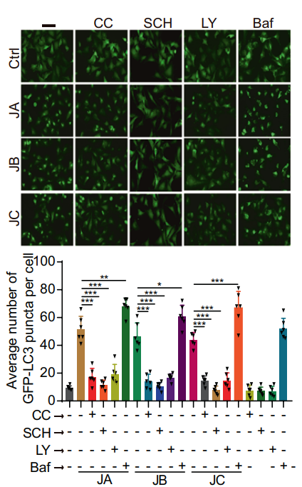
Bafilomycin A1 purchased from MedChemExpress. Usage Cited in: Signal Transduct Target Ther. 2021 Feb 17;6(1):67. [Abstract]
Representative images of cells with GFP or GFP-LC3 puncta, and the average number of GFP-LC3 puncta per cell in stable RFP-GFP-LC3 U87 cells treated with Justicidin A (JA) (0.13 μM), Justicidin B (JB) (0.13 μM), or Justicidin C (JC) (4 μM) in the presence or absence of Compound C (2.5 μM), SCH772984 (10 μM), LY294002 (25 μM), or Bafilomycin A1 (1 nM) for 24 h.
Bafilomycin A1 purchased from MedChemExpress. Usage Cited in: Signal Transduct Target Ther. 2021 Feb 17;6(1):67. [Abstract]
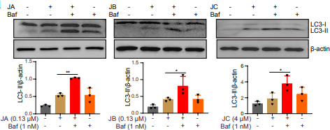
SHSY5Y cells were treated with JA, JB or JC in the presence or absence of Baf (Bafilomycin A1) (1 nM) at the indicated concentrations for 24 h.
-
Signal Transduct Target Ther
2021 Jan 25;6(1):29. PMID: 33487631 -
Nature
2022 Aug;608(7922):413-420. PMID: 35922515
Bafilomycin A1 purchased from MedChemExpress. Usage Cited in: Nature. 2022 Aug;608(7922):413-420. [Abstract]
Huh7 cells and ASGR1 KO cells are treated with 20 nM Bafilomycin A1 for 2 h.
-
Cancer Cell
Exhaustion-associated cholesterol deficiency dampens the cytotoxic arm of antitumor immunity. [Abstract]2023 Jul 10;41(7):1276-1293.e11. PMID: 37244259 -
Cancer Cell
Cathepsin C promotes breast cancer lung metastasis by modulating neutrophil infiltration and neutrophil extracellular trap formation. [Abstract]2021 Mar 8;39(3):423-437.e7. PMID: 33450198 -
Cell
2026 Apr 2;189(7):1923-1941.e26. PMID: 41785850 -
Cell
2025 Jun 26;188(13):3441-3458.e25. PMID: 40280132 -
Cell
2023 Aug 31;186(18):3903-3920.e21. PMID: 37557169 -
Nat Biotechnol
2022 Dec;40(12):1834-1844. PMID: 35879364
Bafilomycin A1 purchased from MedChemExpress. Usage Cited in: Nat Biotechnol. 2022 Dec;40(12):1834-1844. [Abstract]
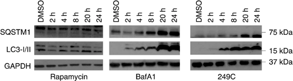
Treatment with 249C or the autophagy inhibitor Bafilomycin A1 (BafA1; 30 nM), but not with the autophagy inducer rapamycin, resulted in upregulation of autophagy markers SQSTM1/p62 and LC3-I/II over time.
Bafilomycin A1 purchased from MedChemExpress. Usage Cited in: Nat Biotechnol. 2022 Dec;40(12):1834-1844. [Abstract]
Treatment with 249C or the autophagy inhibitor Bafilomycin A1 (BafA1; 30 nM), but not with the autophagy inducer rapamycin, resulted in upregulation of autophagy markers SQSTM1/p62 and LC3-I/II over time.
-
Nat Nanotechnol
2025 Feb;20(2):296-302. PMID: 39468359
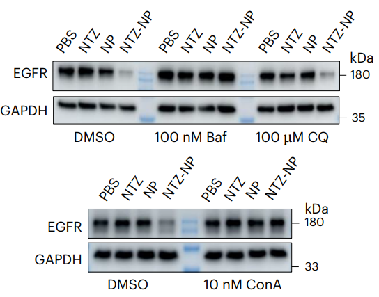
Bafilomycin A1 purchased from MedChemExpress. Usage Cited in: Nat Nanotechnol. 2025 Feb;20(2):296-302. [Abstract]
Bafilomycin A1 (100 nM; 24 h) significantly reduces TPD-NP-based EGFR degradation in MDA-MB-231 cells.
-
Nat Nanotechnol
A nanomaterial targeting the spike protein captures SARS-CoV-2 variants and promotes viral elimination. [Abstract]2022 Sep;17(9):993-1003. PMID: 35995853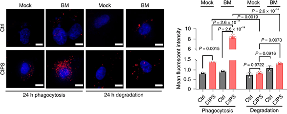
Bafilomycin A1 purchased from MedChemExpress. Usage Cited in: Nat Nanotechnol. 2022 Sep;17(9):993-1003. [Abstract]
Immunofluorescence (IF) images of phagocytosis and elimination of SC2-P (red) in the absence or presence of CIPS in THP-1 differentiated macrophages. The lysosomal inhibitor Bafilomycin A1 (BM; 100 nM; 6 h) is added to macrophages 6 h before the end of culture.
-
Mol Cancer
LAMTOR1 decreased exosomal PD-L1 to enhance immunotherapy efficacy in non-small cell lung cancer. [Abstract]2024 Sep 2;23(1):184. PMID: 39223601 -
Cell Metab
2025 Oct 7:S1550-4131(25)00389-4. PMID: 41061696 -
Cell Metab
Serine metabolism antagonizes antiviral innate immunity by preventing ATP6V0d2-mediated YAP lysosomal degradation. [Abstract]2021 May 4;33(5):971-987.e6. PMID: 33798471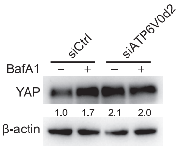
Bafilomycin A1 purchased from MedChemExpress. Usage Cited in: Cell Metab. 2021 May 4;33(5):971-987.e6. [Abstract]
Western blot detection of YAP expression in HEK293T cells transfected with siControl or siATP6V0d2, followed by treatment with 100 nM bafilomycin A1 (BafA1) for 6 h.
-
Nat Immunol
2025 Apr 11. PMID: 40217111 -
Adv Mater
Highly Specific Cytokine Receptor-Targeting Chimeras for Targeted Membrane Protein Degradation and Sensitization of Osimertinib in EGFR-Mutated Non-Small-Cell Lung Cancer. [Abstract]2025 May 22:e2504050. PMID: 40401615 -
Immunity
Membrane integrity changes upon viral infection activate sphingomyelinase SMPDL3B to restrict cGAS-STING signaling via cGAMP degradation. [Abstract]2025 Nov 11;58(11):2670-2684.e10. PMID: 41175872 -
Immunity
Alcaligenes faecalis induces intestinal T helper-17 cells by enhancing Rorc transcription through E3 ligase Trim21-mediated Fbxw7 degradation. [Abstract]2025 Apr 8:S1074-7613(25)00128-1. PMID: 40215968 -
Gut
Long-chain sulfatide enrichment is an actionable metabolic vulnerability in intraductal papillary mucinous neoplasm (IPMN)-associated pancreatic cancers. [Abstract]2025 Apr 23:gutjnl-2025-335220. PMID: 40268349 -
Cancer Commun (Lond)
Lipid metabolism reprograming by SREBP1-PCSK9 targeting sensitizes pancreatic cancer to immunochemotherapy. [Abstract]2025 Aug;45(8):1010-1037. PMID: 40439109 -
Cancer Commun (Lond)
Radiotherapy-resistant prostate cancer cells escape immune checkpoint blockade through the senescence-related ataxia telangiectasia and Rad3-related protein. [Abstract]2025 Mar;45(3):218-244. PMID: 39698847 -
Mil Med Res
Ultrasound-triggered microbubble destruction enhances the radiosensitivity of glioblastoma by inhibiting PGRMC1-mediated autophagy in vitro and in vivo. [Abstract]2022 Feb 14;9(1):9. PMID: 35152910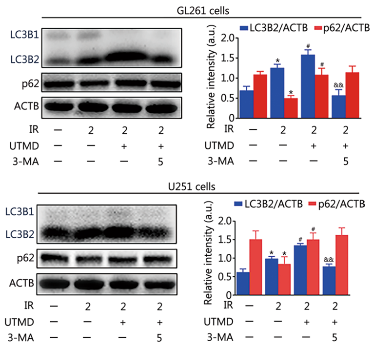
Bafilomycin A1 purchased from MedChemExpress. Usage Cited in: Mil Med Res. 2022 Feb 14;9(1):9. [Abstract]
GL261 and U251 cells are treated with 3-MA (5 mM), BafA1 (10 nM), AG-205 (10 µM), or RAPA (20 nM) for 1 h followed by IR or IR plus UTMD treatment for another 24 h.
-
Exploration
Caffeic Acid Acts as a Potent Senomorphic and Alleviates Inflammation and Lung Fibrosis by Covalently Targeting Annexin A5 Protein in Mice. [Abstract]2025 Dec 12;5(6):20240069. PMID: 41476649 -
Drug Resist Updat
Cell membrane-camouflaged bufalin targets NOD2 and overcomes multidrug resistance in pancreatic cancer. [Abstract]2023 Nov:71:101005. PMID: 37647746 -
Nat Metab
2022 Sep;4(9):1202-1213. PMID: 36131205 -
Cell Stem Cell
Sequential CRISPR gene editing in human iPSCs charts the clonal evolution of myeloid leukemia and identifies early disease targets. [Abstract]2021 Jun 3;28(6):1074-1089.e7. PMID: 33571445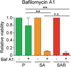
Bafilomycin A1 purchased from MedChemExpress. Usage Cited in: Cell Stem Cell. 2021 Jun 3;28(6):1074-1089.e7. [Abstract]
Viability determined by cell counts of P, SA, and SAR iPSC-HSPCs treated with the V-ATPase inhibitor Bafilomycin A1 (Baf A1; 10 nM; 48 hours) compared with untreated cells.
-
Cell Mol Immunol
GPNMB disrupts SNARE complex assembly to maintain bacterial proliferation within macrophages. [Abstract]2025 May;22(5):512-526. PMID: 40038549 -
Cell Mol Immunol
SARS-CoV-2 ORF10 suppresses the antiviral innate immune response by degrading MAVS through mitophagy. [Abstract]2022 Jan;19(1):67-78. PMID: 34845370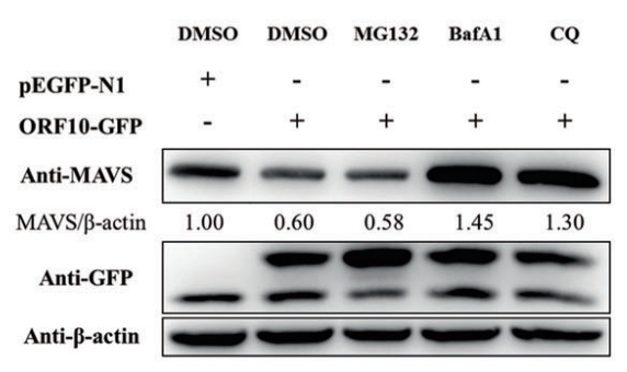
Bafilomycin A1 purchased from MedChemExpress. Usage Cited in: Cell Mol Immunol. 2022 Jan;19(1):67-78. [Abstract]
HeLa cells transfected with the ORF10-GFP plasmid and pEGFP-N1 are stimulated by poly(I:C) transfection and are then treated with MG132 (5 μM), Bafilomycin A1 (Baf A1; 5 nM), or Chloroquine (CQ; 40 μM) for 24 h.
-
Nat Microbiol
Mycobacterium tuberculosis-derived linoleic acid increases regulatory T cell function to promote bacterial survival within macrophages. [Abstract]2025 Nov;10(11):2949-2965. PMID: 41073667 -
Nat Aging
Targeting dysregulated phago-/auto-lysosomes in Sertoli cells to ameliorate late-onset hypogonadism. [Abstract]2024 May;4(5):647-663. PMID: 38649614 -
Nat Cell Biol
The lncRNA DAMER selectively guides m6A-dependent regulation of ATF4 and asparagine metabolism under nutrient stress in cancer. [Abstract]2026 Apr;28(4):797-811. PMID: 41792266 -
Nat Cell Biol
TOLLIP targets GSDME-NT-carrying endocytic vesicles for autophagy to regulate pyroptosis and chemotherapy efficacy. [Abstract]2026 Apr;28(4):812-827. PMID: 41803502 -
-
-
-
Cell Host Microbe
2026 Apr 8;34(4):657-671.e7. PMID: 41794037 -
Cell Host Microbe
2023 Nov 8;31(11):1792-1803.e7. PMID: 37944492 -
Cell Host Microbe
Mycobacterium tuberculosis suppresses host DNA repair to boost its intracellular survival. [Abstract]2023 Nov 8;31(11):1820-1836.e10. PMID: 37848028 -
Mol Cell
Functional nutrient-genetic profiling reveals biotin and FBXW7 are essential to bypass glutamine addiction. [Abstract]2026 Feb 25:S1097-2765(26)00097-3. PMID: 41747732 -
Cancer Res
MTHFD2 Enhances cMYC O-GlcNAcylation to Promote Sunitinib Resistance in Renal Cell Carcinoma. [Abstract]2025 Mar 14;85(6):1113-1129. PMID: 39804969 -
Mol Cell
Ca2+/calmodulin-dependent protein kinase II β decodes ER Ca2+ transients to trigger autophagosome formation. [Abstract]2025 Feb 6;85(3):620-637.e6. PMID: 39742665 -
ACS Nano
Realistic Nanoplastics Induced Pulmonary Damage via the Crosstalk of Ferritinophagy and Mitochondrial Dysfunction. [Abstract]2024 Jul 2;18(26):16790-16807. PMID: 38869479 -
ACS Nano
Ligand Phase Separation-Promoted, "Squeezing-Out" Mode Explaining the Mechanism and Implications of Neutral Nanoparticles That Escaped from Lysosomes. [Abstract]2024 Jan 23;18(3):2162-2183. PMID: 38198577 -
Nat Commun
CD38 degrades MAVS through mitophagy to inhibit type I interferon secretion in nasopharyngeal carcinoma cells and impairs CD8+T cell-mediated anti-tumor immunity. [Abstract]2026 Feb 9;17(1):2544. PMID: 41663422 -
Nat Commun
Mycobacterium tuberculosis modulates phosphorylation of host ATP6V1E1 to promote intracellular survival. [Abstract]2026 Feb 6;17(1):2434. PMID: 41651829 -
Nat Commun
PRDX1 promotes testosterone synthesis and attenuates aging via redox regulation of ATG4B to modulate lipophagy. [Abstract]2025 Nov 19;16(1):10181. PMID: 41261096 -
Nat Commun
PEAK1 maintains tight junctions in intestinal epithelial cells and resists colitis by inhibiting autophagy-mediated ZO-1 degradation. [Abstract]2025 Jul 24;16(1):6777. PMID: 40707483 -
Nat Commun
Endosomal trafficking participates in lipid droplet catabolism to maintain lipid homeostasis. [Abstract]2025 Feb 24;16(1):1917. PMID: 39994216 -
Nat Commun
Liver-specific gene PGRMC1 blocks c-Myc-induced hepatocarcinogenesis through ER stress-independent PERK activation. [Abstract]2025 Jan 2;16(1):50. PMID: 39747098 -
Nat Commun
EXTL3 and NPC1 are mammalian host factors for Autographa californica multiple nucleopolyhedrovirus infection. [Abstract]2024 Sep 4;15(1):7711. PMID: 39231976 -
Nat Commun
Cholesterol-rich lysosomes induced by respiratory syncytial virus promote viral replication by blocking autophagy flux. [Abstract]2024 Jul 26;15(1):6311. PMID: 39060258 -
Nat Commun
KK-LC-1 as a therapeutic target to eliminate ALDH+ stem cells in triple negative breast cancer. [Abstract]2023 May 5;14(1):2602. PMID: 37147285 -
Nat Commun
Tumor-intrinsic YTHDF1 drives immune evasion and resistance to immune checkpoint inhibitors via promoting MHC-I degradation. [Abstract]2023 Jan 17;14(1):265. PMID: 36650153 -
Nat Commun
2020 Sep 9;11(1):4510. PMID: 32908143 -
J Am Chem Soc
2026 Mar 11;148(9):9478-9493. PMID: 41739600 -
Cell Death Differ
TUFT1 stabilizes TGF-β receptor II protein and facilitates activation of hepatic stellate cells into metastasis-promoting myofibroblasts. [Abstract]2026 Jan 28. PMID: 41593321 -
Cell Death Differ
OR2T6 modulates autophagy through the PPP3CA-mediated pathways to suppress gastric cancer. [Abstract]2025 Nov 10. PMID: 41214150 -
Cell Death Differ
TRIM21-driven K63-linked ubiquitination of RBM38c, as a novel interactor of BECN1, contributes to DNA damage-induced autophagy. [Abstract]2025 Jul;32(7):1317-1335. PMID: 40133668 -
Cell Death Differ
Redox regulation of TRIM28 facilitates neuronal ferroptosis by promoting SUMOylation and inhibiting OPTN-selective autophagic degradation of ACSL4. [Abstract]2025 Jun;32(6):1041-1057. PMID: 39875520 -
Cell Death Differ
Vps34 sustains Treg cell survival and function via regulating intracellular redox homeostasis. [Abstract]2024 Nov;31(11):1519-1533. PMID: 39117783 -
Cell Death Differ
Branched-chain amino acid aminotransferase 2 regulates ferroptotic cell death in cancer cells. [Abstract]2021 Apr;28(4):1222-1236. PMID: 33097833 -
Bone Res
Enhanced SIRT3 expression restores mitochondrial quality control mechanism to reverse osteogenic impairment in type 2 diabetes mellitus. [Abstract]2025 Mar 3;13(1):30. PMID: 40025004 -
Bone Res
2024 Aug 28;12(1):49. PMID: 39198395 -
Acta Pharm Sin B
Deubiquitinase USP13 alleviates doxorubicin-induced cardiotoxicity through promoting the autophagy-mediated degradation of STING. [Abstract]2025 May;15(5):2545-2558. PMID: 40487656 -
Acta Pharm Sin B
USP25 ameliorates vascular remodeling by deubiquitinating FOXO3 and promoting autophagic degradation of FOXO3. [Abstract]2025 Mar;15(3):1643-1658. PMID: 40370563 -
Acta Pharm Sin B
Demethylzeylasteral induces PD-L1 ubiquitin-proteasome degradation and promotes antitumor immunity via targeting USP22. [Abstract]2024 Oct;14(10):4312-4328. PMID: 39525573 -
Acta Pharm Sin B
Translocation of IGF-1R in endoplasmic reticulum enhances SERCA2 activity to trigger Ca2+ER perturbation in hepatocellular carcinoma. [Abstract]2023 Sep;13(9):3744-3755. PMID: 37719369 -
Acta Pharm Sin B
Repurposing econazole as a pharmacological autophagy inhibitor to treat pancreatic ductal adenocarcinoma. [Abstract]2022 Jul;12(7):3085-3102. PMID: 35865101 -
Acta Pharm Sin B
Intravenous route to choroidal neovascularization by macrophage-disguised nanocarriers for mTOR modulation. [Abstract]2022 May;12(5):2506-2521. PMID: 35646523 -
J Extracell Vesicles
Extracellular Vesicles Coordinate Bacterial Cloaking in Lung Epithelial Cells to Alleviate Acute Inflammatory Injury. [Abstract]2026 Feb;15(2):e70238. PMID: 41656978 -
J Extracell Vesicles
Exomeres From Adventitial Fibroblasts of Spontaneously Hypertensive Rats Promote Vascular Remodelling via Transferring Osteopontin. [Abstract]2025 Aug;14(8):e70146. PMID: 40767027 -
J Extracell Vesicles
Neddylation of Coro1a determines the fate of multivesicular bodies and biogenesis of extracellular vesicles. [Abstract]2021 Oct;10(12):e12153. PMID: 34623756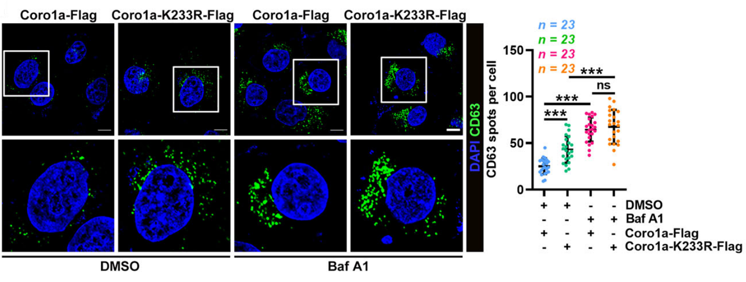
Bafilomycin A1 purchased from MedChemExpress. Usage Cited in: J Extracell Vesicles. 2021 Oct;10(12):e12153. [Abstract]
Confocal microscopy analysis of the MVB marker CD63 in Coro1a‐Flag or Coro1a‐K233R‐Flag overexpressing HeLa cells treated with DMSO or 20 nM Bafilomycin A1 (Baf A1) for 12 h.
-
Autophagy
Phase separation of OPTN initiates mitophagy to orchestrate craniofacial bone mineralization. [Abstract]2026 May;22(5):1021-1043. PMID: 41692957 -
Autophagy
Trans-Golgi network-associated noncanonical autophagy depends on the V-ATPase-ATG16L1 axis and mediates IL1B secretion. [Abstract]2025 Dec 8. PMID: 41361996 -
Autophagy
XIAP-ULK1-Mediated mitophagy modulates carnitine metabolism to mitigate diabetic kidney disease. [Abstract]2025 Oct 25. PMID: 41139215 -
Autophagy
2025 Oct 14:1-23. PMID: 41037659 -
Autophagy
2025 Sep 29:1-19. PMID: 40963239 -
Autophagy
Dysfunctional autophagy triggers STING1 activation to exacerbate cartilage degeneration in obesity-associated osteoarthritis. [Abstract]2025 Aug 6:1-17. PMID: 40728163 -
Autophagy
Autophagy suppression via SRC induction represents a therapeutic vulnerability for BAP1-mutant cancers. [Abstract]2025 Aug 3:1-20. PMID: 40754831 -
Autophagy
Restricting intracellular Salmonella proliferation by coordinating p-TBK1 mediated mitophagy and xenophagy. [Abstract]2025 Jul 27:1-23. PMID: 40660474 -
Autophagy
RETREG1-mediated reticulophagy is activated by an ATF4-CEBPG/C/EBPγ heterodimer and confers protection against lipotoxicity. [Abstract]2025 Jun 7:1-19. PMID: 40437698 -
Autophagy
Neutrophil-derived serine proteases induce FOXA2-mediated autophagy dysfunction and exacerbate colitis-associated carcinogenesis via protease activated receptor 2. [Abstract]2025 Apr 9. PMID: 40205686 -
Autophagy
Dual roles of CXCR4 (C-X-C motif chemokine receptor 4) in promoting entry of ebolavirus and targeting excessive glycoprotein for reticulophagic degradation to facilitate viral fitness. [Abstract]2025 Apr 13. PMID: 40223186 -
Autophagy
H3K36me2 methyltransferase NSD2/WHSC1 promotes triple-negative breast cancer metastasis via activation of ULK1-dependent autophagy. [Abstract]2025 Aug;21(8):1824-1842. PMID: 40097917 -
Autophagy
The alpha-coronavirus E protein inhibits the JAK-STAT pathway signaling by triggering STAT2 degradation through OPTN- and NBR1-mediated selective autophagy. [Abstract]2025 Aug;21(8):1644-1661. PMID: 40091174 -
Autophagy
Histone lactylation stimulated upregulation of PSMD14 alleviates neuron PANoptosis through deubiquitinating PKM2 to activate PINK1-mediated mitophagy after traumatic brain injury. [Abstract]2025 Jul;21(7):1473-1491. PMID: 40000916 -
Autophagy
2025 Jul;21(7):1591-1607. PMID: 39952286 -
Autophagy
Salmonella Typhimurium persistently infects host via its effector SseJ-induced PHB2-mediated mitophagy. [Abstract]2025 Jun;21(6):1228-1244. PMID: 39902787 -
Autophagy
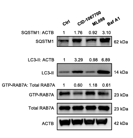
USP4 depletion-driven RAB7A ubiquitylation impairs autophagosome-lysosome fusion and aggravates periodontitis. [Abstract]2024 Dec 11:1-18. PMID: 39663592
Bafilomycin A1 purchased from MedChemExpress. Usage Cited in: Autophagy. 2024 Dec 11:1-18. [Abstract]
Western blot analysis of SQSTM1, LC3, total RAB7A, and GTP-RAB7A (immunoprecipitated by anti-GTP-RAB7A antibody) in four groups as indicated in THP-1 cells. Cells were treated with CID-1067700 (100 μM), ML098 (100 nM), or Baf A1 (50 nM) for 8 hours.
-
Autophagy
Upregulation of ISG15 induced by MAPT/tau accumulation represses autophagic flux by inhibiting HDAC6 activity: a vicious cycle in Alzheimer disease. [Abstract]2025 Apr;21(4):807-826. PMID: 39635882 -
Autophagy
CKAP4 in hepatocellular carcinoma: competitive RETREG1/FAM134B binding, reticulophagy regulation, and cancer progression. [Abstract]2025 Apr;21(4):840-859. PMID: 39689859 -
Autophagy
2024 Dec;20(12):2697-2718. PMID: 39081059 -
Autophagy
The mammalian actin elongation factor ENAH/MENA contributes to autophagosome formation via its actin regulatory function. [Abstract]2024 Aug;20(8):1798-1814. PMID: 38705725 -
Autophagy
Impaired lipophagy induced-microglial lipid droplets accumulation contributes to the buildup of TREM1 in diabetes-associated cognitive impairment. [Abstract]2023 Oct;19(10):2639-2656. PMID: 37204119 -
Autophagy
SDC1-dependent TGM2 determines radiosensitivity in glioblastoma by coordinating EPG5-mediated fusion of autophagosomes with lysosomes. [Abstract]2023 Mar;19(3):839-857. PMID: 35913916 -
Autophagy
2022 Apr 26:1-17. PMID: 35471096 -
Autophagy
TRAF6 autophagic degradation by avibirnavirus VP3 inhibits antiviral innate immunity via blocking NFKB/NF-κB activation. [Abstract]2022 Dec;18(12):2781-2798. PMID: 35266845 -
Autophagy
Protein disulfide isomerases (PDIs) negatively regulate ebolavirus structural glycoprotein expression in the endoplasmic reticulum (ER) via the autophagy-lysosomal pathway. [Abstract]2022 Oct;18(10):2350-2367. PMID: 35130104 -
Autophagy
2AB protein of Senecavirus A antagonizes selective autophagy and type I interferon production by degrading LC3 and MARCHF8. [Abstract]2022 Aug;18(8):1969-1981. PMID: 34964697 -
Autophagy
Identification of an autoinhibitory, mitophagy-inducing peptide derived from the transmembrane domain of USP30. [Abstract]2022 Sep;18(9):2178-2197. PMID: 34989313 -
Autophagy
Autophagy inhibition mediated by MCOLN1/TRPML1 suppresses cancer metastasis via regulating a ROS-driven TP53/p53 pathway. [Abstract]2022 Aug;18(8):1932-1954. PMID: 34878954 -
Autophagy
A noncanonical autophagy is involved in the transfer of Plasmodium-microvesicles to astrocytes. [Abstract]2022 Jul;18(7):1583-1598. PMID: 34747313 -
Autophagy
2022 Apr;18(4):726-744. PMID: 34282994 -
Autophagy
2021 Dec;17(12):4401-4422. PMID: 33890549 -
Autophagy
An organelle-directed chemical ligation approach enables dual-color detection of mitophagy. [Abstract]2021 Nov;17(11):3475-3490. PMID: 33435798 -
Autophagy
PRCC-TFE3 fusion-mediated PRKN/parkin-dependent mitophagy promotes cell survival and proliferation in PRCC-TFE3 translocation renal cell carcinoma. [Abstract]2021 Sep;17(9):2475-2493. PMID: 33019842 -
Autophagy
2020 Jan;16(1):106-122. PMID: 30909789 -
Autophagy
m6A mRNA methylation regulates testosterone synthesis through modulating autophagy in Leydig cells. [Abstract]2021 Feb;17(2):457-475. PMID: 31983283 -
Autophagy
YWHA/14-3-3 proteins recognize phosphorylated TFEB by a noncanonical mode for controlling TFEB cytoplasmic localization. [Abstract]2019 Jun;15(6):1017-1030. PMID: 30653408 -
Adv Sci (Weinh)
Activated Platelet-Released Heat Shock Protein 90α Triggers Autophagy-Dependent Neutrophil Extracellular Trap Formation and Amplifies Sepsis. [Abstract]2026 May;13(25):e15933. PMID: 41732887 -
Adv Sci (Weinh)
OTUD6A in Airway Epithelial Cells Exacerbates Allergic Asthma by Promoting Airway Inflammation and Airway Remodeling Through Deubiquitination of hResistin/mRELMα. [Abstract]2026 Jan 20:e16355. PMID: 41560298 -
Adv Sci (Weinh)
TBK1 Induces the Formation of Optineurin Filaments That Condensate with Polyubiquitin and LC3 for Cargo Sequestration. [Abstract]2025 Dec 17:e09927. PMID: 41405403 -
Adv Sci (Weinh)
CCDC41 Drives Oocyte Meiotic Progression by Promoting Rab11a/Rab7-Positive Vesicle Fusion with Target Membranes. [Abstract]2025 Dec 2:e04665. PMID: 41331237 -
Adv Sci (Weinh)
Punicalagin Enhances Autophagy Through Sirtuin 1/FoxO3a Axis to Inhibit Intracellular Mycobacterium Abscessus Infection. [Abstract]2025 Oct 14:e11734. PMID: 41085014 -
Adv Sci (Weinh)
BLOC1S1 Attenuates B. Melitensis 16M LPS-Triggered Autophagy by Spatial Confinement of TDP-43. [Abstract]2025 Sep 11:e05635. PMID: 40936170 -
Adv Sci (Weinh)
NAT10 Increases Lysosomal Acidification to Promote Esophageal Cancer Metastasis via ac4C Acetylation of ATP6V0E1 mRNA. [Abstract]2025 Aug;12(31):e02931. PMID: 40729677 -
Adv Sci (Weinh)
API5 Phosphorylation Promotes Antiviral Immunity by Inhibiting Degradation of Cytosolic RNA Sensor RLRs. [Abstract]2025 Jul 11:e05479. PMID: 40641422 -
Adv Sci (Weinh)
Activation of Lysosomal Retrograde Transport Triggers TPC1-IP3R1 Ca2+ Crosstalk at Lysosome-ER MCSs Leading to Lethal Depleting of ER Calcium. [Abstract]2025 Jul 25:e15313. PMID: 40709664 -
Adv Sci (Weinh)
Tryptophan Metabolite Indole-3-Aldehyde Induces AhR and c-MYC Degradation to Promote Tumor Immunogenicity. [Abstract]2025 Jun 29:e09533. PMID: 40583248 -
Adv Sci (Weinh)
Lysosome-Featured Cell Aggregate-Released Extracellular Vesicles Regulate Iron Homeostasis and Alleviate Post-Irradiation Endothelial Ferroptosis for Mandibular Regeneration. [Abstract]2025 Jun 23:e05070. PMID: 40546115 -
Adv Sci (Weinh)
Network Medicine-Based Strategy Identifies Maprotiline as a Repurposable Drug by Inhibiting PD-L1 Expression via Targeting SPOP in Cancer. [Abstract]2024 Nov 5:e2410285. PMID: 39499771 -
Adv Sci (Weinh)
Berberine Derivative B68 Promotes Tumor Immune Clearance by Dual-Targeting BMI1 for Senescence Induction and CSN5 for PD-L1 Degradation. [Abstract]2024 Dec 25:e2413122. PMID: 39721027 -
Adv Sci (Weinh)
RBBP6-Mediated ERRα Degradation Contributes to Mitochondrial Injury in Renal Tubular Cells in Diabetic Kidney Disease. [Abstract]2024 Oct 23:e2405153. PMID: 39441040 -
Adv Sci (Weinh)
Aloperine Suppresses Cancer Progression by Interacting with VPS4A to Inhibit Autophagosome-lysosome Fusion in NSCLC. [Abstract]2024 Aug;11(31):e2308307. PMID: 39166458 -
Adv Sci (Weinh)
Oncogenic Roles of Laminin Subunit Gamma-2 in Intrahepatic Cholangiocarcinoma via Promoting EGFR Translation. [Abstract]2024 Jun;11(21):e2309010. PMID: 38526177 -
Adv Sci (Weinh)
HSPA8 Activates Wnt/β-Catenin Signaling to Facilitate BRAF V600E Colorectal Cancer Progression by CMA-Mediated CAV1 Degradation. [Abstract]2024 Jan;11(3):e2306535. PMID: 37973552 -
Adv Sci (Weinh)
MCU Upregulation Overactivates Mitophagy by Promoting VDAC1 Dimerization and Ubiquitination in the Hepatotoxicity of Cadmium. [Abstract]2023 Mar;10(7):e2203869. PMID: 36642847 -
Adv Sci (Weinh)
Loss-of-Function of p21-Activated Kinase 2 Links BMP Signaling to Neural Tube Patterning Defects. [Abstract]2023 Feb;10(4):e2204018. PMID: 36504449 -
Nat Chem Biol
2025 Apr;21(4):490-500. PMID: 39215100 -
J Clin Invest
LC3-dependent intercellular transfer of phosphorylated STAT1/2 elicits CXCL9+ macrophages and enhances radiation-induced antitumor immunity. [Abstract]2025 Dec 1;135(23):e195279. PMID: 41321309 -
J Clin Invest
Parkin ubiquitinates phosphoglycerate dehydrogenase to suppress serine synthesis and tumor progression. [Abstract]2020 Jun 1;130(6):3253-3269. PMID: 32478681 -
Leukemia
Autophagosome-lysosome mediated secretion of the thrombopoietin receptor is modulated by distinct driver mutations of myeloproliferative neoplasm. [Abstract]2025 Sep;39(9):2181-2195. PMID: 40610764 -
Neuro Oncol
2023 Jan 5;25(1):82-96. PMID: 35727735 -
Theranostics
Honokiol enhances the sensitivity of cetuximab in KRASG13D mutant colorectal cancer through destroying SNX3-retromer complex. [Abstract]2024 Aug 26;14(14):5443-5460. PMID: 39310106 -
Theranostics
Repurposing Brigatinib for the Treatment of Colorectal Cancer Based on Inhibition of ER-phagy. [Abstract]2019 Jul 9;9(17):4878-4892. PMID: 31410188 -
Theranostics 2018; 8(19):5434-5451
Microglial TLR4-dependent autophagy induces ischemic white matter damage via STAT1/6 pathway. [Abstract]PMID: 30555556 -
-
J Adv Res
A novel mechanism of selenium deficiency driving aberrant Th17 cell differentiation via GPX3-targeted PIG3: mitophagy blockade exacerbates mtDNA release. [Abstract]2026 Feb 21:S2090-1232(26)00175-X. PMID: 41730416 -
J Adv Res
OGT regulates histone demethylation and alleviates osteoarthritis progression by O-GlcNAcylation of KDM6B. [Abstract]2026 Jan 2:S2090-1232(26)00003-2. PMID: 41485575 -
J Adv Res
Hyperuricemia increases susceptibility to chronic kidney injury exacerbation via autophagic flux blockade-mediated ammonia death pathway. [Abstract]2025 Nov 7:S2090-1232(25)00877-X. PMID: 41205802 -
Biomaterials
2025 Dec 18:329:123925. PMID: 41453350 -
Exp Mol Med
2025 Aug;57(8):1847-1860. PMID: 40855114 -
Biomaterials
Biodegradable polymers with tertiary amines enhance mRNA delivery of lipid nanoparticles via improved endosomal escape. [Abstract]2026 Jan:324:123541. PMID: 40644791 -
Exp Mol Med
The CREG1-FBXO27-LAMP2 axis alleviates diabetic cardiomyopathy by promoting autophagy in cardiomyocytes. [Abstract]2023 Sep;55(9):2025-2038. PMID: 37658156 -
Biomaterials
Gold nanoparticles targeting the autophagy-lysosome system to combat the inflammation-compromised osteogenic potential of periodontal ligament stem cells: From mechanism to therapy. [Abstract]2022 Sep;288:121743. PMID: 36030103
Bafilomycin A1 purchased from MedChemExpress. Usage Cited in: Biomaterials. 2022 Sep;288:121743. [Abstract]
Bafilomycin A1 (Baf; 50 nM) treatment significantly increases the percentage of both RFP+-GFP+-LC3 puncta and LC3 puncta in PDLSCs at 12 and 24 h during osteogenic induction, while there is no significant difference in the percentage of RFP+-GFP+-LC3 puncta or LC3 puncta between the Baf-treated I-PDLSCs and cells without the Baf treatment.
Bafilomycin A1 purchased from MedChemExpress. Usage Cited in: Biomaterials. 2022 Sep;288:121743. [Abstract]
In response to the Bafilomycin A1 (Baf; 50 nM) treatment, increases LC3 II expression is observed in PDLSCs, and significant differences between the Baf-treated PDLSCs and cells without the Baf treatment are observed at 6 h and 12 h during osteogenic induction.
-
Biomaterials
An optimized ionizable cationic lipid for brain tumor-targeted siRNA delivery and glioblastoma immunotherapy. [Abstract]2022 Aug:287:121645. PMID: 35779480 -
J Exp Clin Cancer Res
Membrane RRM2-positive cells represent a malignant population with cancer stem cell features in intrahepatic cholangiocarcinoma. [Abstract]2024 Sep 6;43(1):255. PMID: 39243109 -
J Exp Clin Cancer Res
The immunotoxin targeting PRLR increases tamoxifen sensitivity and enhances the efficacy of chemotherapy in breast cancer. [Abstract]2024 Jun 20;43(1):173. PMID: 38898487 -
J Nanobiotechnology
Radiotherapy-derived engineered stem cell exosomes improve anti-glioma immunotherapy by promoting the formation of tertiary lymphoid structure and improve the release of type I interferon. [Abstract]2025 Mar 22;23(1):239. PMID: 40121481 -
J Nanobiotechnology
Promoting collateral formation in type 2 diabetes mellitus using ultra-small nanodots with autophagy activation and ROS scavenging. [Abstract]2024 Mar 1;22(1):85. PMID: 38429826 -
J Nanobiotechnology
Visualization of the process of a nanocarrier-mediated gene delivery: stabilization, endocytosis and endosomal escape of genes for intracellular spreading. [Abstract]2022 Mar 9;20(1):124. PMID: 35264206 -
Sci Adv
A worm-like nucleic acid nanostructure for gene delivery and endosomal escape via ClC3 ion exchanger. [Abstract]2026 Mar 6;12(10):eadw0891. PMID: 41790895 -
Cell Discov
ASCT2 palmitoylation regulated by JNK1-ZDHHC14 axis orchestrates glutamine metabolism and NSCLC progression. [Abstract]2026 Feb 24;12(1):13. PMID: 41730846 -
Carbohydr Polym
Decoding the pharmacokinetics of intravenous lentinan: A multiscale journey from the blood to hepatic Kupffer cells' lysosomes. [Abstract]2026 Feb 15:374:124706. PMID: 41371850 -
Sci Adv
Putative PINK1/Parkin activators lower the threshold for mitophagy by sensitizing cells to mitochondrial stress. [Abstract]2025 Aug 29;11(35):eady0240. PMID: 40864725 -
Carbohydr Polym
Metabolic degradation of polysaccharides from Lentinus edodes by Kupffer cells via the Dectin-1/Syk signaling pathway. [Abstract]2023 Oct 1:317:121108. PMID: 37364942 -
Cell Discov
Dynamic O-GlcNAcylation coordinates ferritinophagy and mitophagy to activate ferroptosis. [Abstract]2022 May 3;8(1):40. PMID: 35504898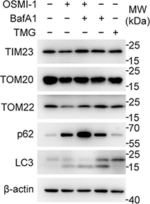
Bafilomycin A1 purchased from MedChemExpress. Usage Cited in: Cell Discov. 2022 May 3;8(1):40. [Abstract]
The levels of mitochondrial proteins including TIM23, TOM20, and TOM22 decreased along with decreased O-GlcNAcylation levels, which can be inhibited by BafA1.
-
Carbohydr Polym
Cordyceps sinensis polysaccharide inhibits colon cancer cells growth by inducing apoptosis and autophagy flux blockage via mTOR signaling. [Abstract]2020 Jun 1;237:116113. PMID: 32241434 -
Int J Oral Sci
LncRNA EUDAL shapes tumor cell response to hypoxia-induced constitutive EGFR activation and promotes chemoresistance in oral cancer. [Abstract]2025 Sep 12;17(1):64. PMID: 40940319 -
Small
Engineered Biomimetic Nanorobots Orchestrate Targeted Nose-to-Brain Delivery to Resolve Neuron-Glia Entanglement against Parkinson's Disease. [Abstract]2026 Mar;22(18):e13394. PMID: 41607240 -
Redox Biol
Redox compartmentalization drives functional heterogeneity of mature insulin secretory vesicles in pancreatic β-cells. [Abstract]2025 Aug 27:86:103847. PMID: 40902318 -
Redox Biol
Arylsulfatase K attenuates airway epithelial cell senescence in COPD by regulating parkin-mediated mitophagy. [Abstract]2025 Jul 31:86:103793. PMID: 40763656 -
Redox Biol
2025 Sep:85:103751. PMID: 40628122 -
Redox Biol
Targeting p62 by sulforaphane promotes autolysosomal degradation of SLC7A11, inducing ferroptosis for osteosarcoma treatment. [Abstract]2024 Dec 5:79:103460. PMID: 39657365 -
Redox Biol
Hydrogen sulfide attenuates disturbed flow-induced vascular remodeling by inhibiting LDHB-mediated autophagic flux. [Abstract]2024 Dec 5:79:103456. PMID: 39647238 -
Metabolism
DR region of NKAα1 is a target to ameliorate hepatic lipid metabolism disturbance in obese mice. [Abstract]2023 Aug:145:155579. PMID: 37127227 -
Redox Biol
A catalase inhibitor: Targeting the NADPH-binding site for castration-resistant prostate cancer therapy. [Abstract]2023 Jul:63:102751. PMID: 37216701 -
Redox Biol
The physiological metabolite α-ketoglutarate ameliorates osteoarthritis by regulating mitophagy and oxidative stress. [Abstract]2023 Jun:62:102663. PMID: 36924682 -
Redox Biol
Role of redox-sensitive catalytic interaction with ADAM10 in mutant-selective extracellular shedding of prion protein. [Abstract]2022 Oct:56:102456. PMID: 36041363 -
Redox Biol
2022 Feb:49:102217. PMID: 34942528 -
J Control Release
The HSP90-dependent bioorthogonal PROTAC prodrug system enables tumor-selective and enhanced protein degradation. [Abstract]2026 May 10:393:114828. PMID: 41839264 -
J Control Release
2025 Dec 30:114591. PMID: 41478376 -
Biomark Res
2025 Jan 23;13(1):17. PMID: 39849645 -
J Hazard Mater
Protection against deoxynivalenol (DON)-induced intestinal injury by sulforaphane via modulation of lysosomal function. [Abstract]2025 Dec 2:501:140687. PMID: 41352012 -
J Hazard Mater
Low-dose deoxynivalenol exposure triggers hepatic excessive ferritinophagy and mitophagy mitigated by hesperidin modulated O-GlcNAcylation. [Abstract]2024 Dec 5:480:135952. PMID: 39341193 -
J Hazard Mater
Selenium restored mitophagic flux to alleviate cadmium-induced hepatotoxicity by inhibiting excessive GPER1-mediated mitophagy activation. [Abstract]2024 Jun 13:475:134855. PMID: 38880044 -
J Hazard Mater
Benzo[a]pyrene evokes epithelial-mesenchymal transition and pulmonary fibrosis through AhR-mediated Nrf2-p62 signaling. [Abstract]2024 May 8:473:134560. PMID: 38759404 -
J Hazard Mater
Cobalt induces neurodegeneration through FTO-triggered autophagy impairment by targeting TSC1 in an m6A-YTHDF2-dependent manner. [Abstract]2023 Jul 5:453:131354. PMID: 37054644 -
-
J Hazard Mater
Systematic toxicity evaluation of polystyrene nanoplastics on mice and molecular mechanism investigation about their internalization into Caco-2 cells. [Abstract]2021 Sep 5;417:126092. PMID: 34015712 -
EBioMedicine
VIPAS39 confers ferroptosis resistance in epithelial ovarian cancer through exporting ACSL4. [Abstract]2025 Apr:114:105646. PMID: 40088627 -
EBioMedicine
F. nucleatum facilitates oral squamous cell carcinoma progression via GLUT1-driven lactate production. [Abstract]2023 Feb:88:104444. PMID: 36709580 -
EBioMedicine
2022 Apr;78:103980. PMID: 35367771 -
MedComm (2020)
5,7,4'-Trimethoxyflavone triggers cancer cell PD-L1 ubiquitin-proteasome degradation and facilitates antitumor immunity by targeting HRD1. [Abstract]2024 Jun 27;5(7):e611. PMID: 38938284 -
Cell Rep Med
Engineered oncolytic virus armed with anti-PCSK9 scFv boosts long-term CD8+ T cell immunity via rewiring MHC-I antigen presentation. [Abstract]2026 Apr 21;7(4):102724. PMID: 41923622 -
Cell Rep Med
Vesicle-mediated mitochondrial clearance presents an actionable metabolic vulnerability in triple-negative breast cancer. [Abstract]2025 Dec 16;6(12):102478. PMID: 41406946 -
J Immunother Cancer
HSP47 inhibition-induced CD155 expression through TRAF2 deubiquitination promotes tumor immune evasion. [Abstract]2025 Nov 28;13(11):e011976. PMID: 41314980 -
J Immunother Cancer
Macrophage NLRP3 activation and IL-1β release drive osimertinib-induced antitumor immunity. [Abstract]2025 Oct 5;13(10):e012182. PMID: 41052880 -
Cell Rep Med
Screening of patient-derived organoids identifies mitophagy as a cell-intrinsic vulnerability in colorectal cancer during statin treatment. [Abstract]2025 Mar 19:102039. PMID: 40154491 -
Cell Rep Med
Benzosceptrin C induces lysosomal degradation of PD-L1 and promotes antitumor immunity by targeting DHHC3. [Abstract]2024 Feb 20;5(2):101357. PMID: 38237597 -
Pharmacol Res
Histone lysine crotonylation accelerates ACSL4-mediated ferroptosis of keratinocytes via modulating autophagy in diabetic wound healing. [Abstract]2025 Jan 30:107632. PMID: 39892437 -
Pharmacol Res
Cyclopeptide RA-V from Rubia yunnanensis restores activity of Adagrasib against colorectal cancer by reducing the expression of Nrf2. [Abstract]2024 Jun 28:107252. PMID: 38945380 -
Pharmacol Res
SNX17 protects the heart from doxorubicin-induced cardiotoxicity by modulating LMOD2 degradation. [Abstract]2021 Jul:169:105642. PMID: 33933636 -
Clin Cancer Res
UBE2T-mediated HP1α ubiquitination enhances nucleolar function and promotes the progression of IDH1/TP53-mutant glioma. [Abstract]2025 Jul 8. PMID: 40627452 -
Mater Today Bio
A biomimetic multimodal nanoplatform combining neutrophil-coated two-dimensional metalloporphyrinic framework nanosheet and exendin-4 to treat obesity-related osteoporosis. [Abstract]2025 Jun 21:33:102009. PMID: 40673132 -
Cell Mol Biol Lett
The histone demethylase KDM5C enhances the sensitivity of acute myeloid leukemia cells to lenalidomide by stabilizing cereblon. [Abstract]2025 Jan 29;30(1):14. PMID: 39881283 -
J Neuroinflammation
Microglial NLRC5 drives lysosomal dysfunction to disrupt autophagic flux and promote post-stroke neuroinflammation. [Abstract]2025 Oct 31;22(1):253. PMID: 41174779 -
Mol Biomed
PARP inhibitor BMN673 triggers PARylation-mediated ATF4-GDF15 pathway to drive autophagy and ferroptosis in ataxia telangiectasia mutated gene-deficient colorectal cancer cells. [Abstract]2025 Nov 21;6(1):113. PMID: 41266732 -
Cancer Lett
2025 Apr 16:217726. PMID: 40250791 -
Cancer Lett
2025 Apr 10:217715. PMID: 40220852 -
J Neuroinflammation
Microglial progranulin differently regulates hypothalamic lysosomal function in lean and obese conditions via cleavage-dependent mechanisms. [Abstract]2025 Mar 7;22(1):68. PMID: 40055725 -
Cancer Lett
Super-enhancer mediated upregulation of MYEOV suppresses ferroptosis in lung adenocarcinoma. [Abstract]2024 May 1:589:216811. PMID: 38490328 -
J Neuroinflammation
Intermittent hypoxia therapy ameliorates beta-amyloid pathology via TFEB-mediated autophagy in murine Alzheimer's disease. [Abstract]2023 Oct 20;20(1):240. PMID: 37864249 -
Cancer Lett
2023 Nov 28:577:216435. PMID: 37806516 -
Cancer Lett
LncRNA MNX1-AS1 sustains inactivation of Hippo pathway through a positive feedback loop with USP16/IGF2BP3 axis in gallbladder cancer. [Abstract]2022 Oct 28:547:215862. PMID: 35953000 -
J Neuroinflammation
Morphine-induced microglial immunosuppression via activation of insufficient mitophagy regulated by NLRX1. [Abstract]2022 Apr 12;19(1):87. PMID: 35414088 -
J Neuroinflammation
AMPK-autophagy-mediated inhibition of microRNA-30a-5p alleviates morphine tolerance via SOCS3-dependent neuroinflammation suppression. [Abstract]2022 Jan 29;19(1):25. PMID: 35093117 -
J Neuroinflammation
Downregulating expression of OPTN elevates neuroinflammation via AIM2 inflammasome- and RIPK1-activating mechanisms in APP/PS1 transgenic mice. [Abstract]2021 Dec 3;18(1):281. PMID: 34861878 -
Cancer Lett
Prodigiosin impairs autophagosome-lysosome fusion that sensitizes colorectal cancer cells to 5-fluorouracil-induced cell death. [Abstract]2020 Jul 1;481:15-23. PMID: 32184145 -
Cancer Lett
DEPTOR inhibits cell proliferation and confers sensitivity to dopamine agonist in pituitary adenoma. [Abstract]2019 Sep 10:459:135-144. PMID: 31176743 -
Int J Biol Sci
BMSCs-derived small extracellular vesicles antagonize cerebral endothelial Caveolin-1 driven autophagic degradation of tight-junction proteins to protect blood-brain barrier post-stroke. [Abstract]2025 Jan 1;21(2):842-859. PMID: 39781452 -
Int J Biol Sci
Purinergic receptor P2Y12 boosts autoimmune hepatitis through hexokinase 2-dependent glycolysis in T cells. [Abstract]2023 Jul 9;19(11):3576-3594. PMID: 37497007 -
Int J Biol Sci
Anomanolide C suppresses tumor progression and metastasis by ubiquitinating GPX4-driven autophagy-dependent ferroptosis in triple negative breast cancer. [Abstract]2023 May 8;19(8):2531-2550. PMID: 37215985 -
Cell Death Dis
A Lamp2a-linked RNA secreted by ADSCs prevents ENO1-lactylation-glycolysis feedback and cell malignant behavior in triple-negative breast cancer. [Abstract]2026 Mar 2;17(1):288. PMID: 41771848 -
Cell Death Dis
DCAF7 recruits USP2 to facilitate hepatocellular carcinoma progression by suppressing clockophagy-induced ferroptosis. [Abstract]2025 Aug 28;16(1):654. PMID: 40877242 -
Cell Death Dis
ATP6AP1 promotes cell proliferation and tamoxifen resistance in luminal breast cancer by inducing autophagy. [Abstract]2025 Mar 25;16(1):201. PMID: 40133274 -
Cell Death Dis
2024 Nov 1;15(11):786. PMID: 39487118 -
Cell Death Dis
Phosphatase LHPP confers prostate cancer ferroptosis activation by modulating the AKT-SKP2-ACSL4 pathway. [Abstract]2024 Sep 11;15(9):665. PMID: 39261475 -
Cell Death Dis
2024 Jan 5;15(1):14. PMID: 38182563 -
Cell Death Dis
SMURF1 attenuates endoplasmic reticulum stress by promoting the degradation of KEAP1 to activate NRF2 antioxidant pathway. [Abstract]2023 Jun 14;14(6):361. PMID: 37316499 -
Cell Death Dis
Ciclopirox drives growth arrest and autophagic cell death through STAT3 in gastric cancer cells. [Abstract]2022 Nov 28;13(11):1007. PMID: 36443287 -
Cell Death Dis
The interaction between STING and NCOA4 exacerbates lethal sepsis by orchestrating ferroptosis and inflammatory responses in macrophages. [Abstract]2022 Jul 28;13(7):653. PMID: 35902564 -
Cell Death Dis
Oncogenic circTICRR suppresses autophagy via binding to HuR protein and stabilizing GLUD1 mRNA in cervical cancer. [Abstract]2022 May 20;13(5):479. PMID: 35595754 -
Cell Death Dis
2022 May 12;13(5):451. PMID: 35551175 -
Cell Death Dis
Targeting DRD2 by the antipsychotic drug, penfluridol, retards growth of renal cell carcinoma via inducing stemness inhibition and autophagy-mediated apoptosis. [Abstract]2022 Apr 23;13(4):400. PMID: 35461314 -
Cell Death Dis
Essential role of zyxin in platelet biogenesis and glycoprotein Ib-IX surface expression. [Abstract]2021 Oct 16;12(11):955. PMID: 34657146 -
Cell Death Dis
Circulating mitochondrial DNA-triggered autophagy dysfunction via STING underlies sepsis-related acute lung injury. [Abstract]2021 Jul 3;12(7):673. PMID: 34218252 -
Cell Death Dis
Exosomes derived from mesenchymal stem cells repair a Parkinson's disease model by inducing autophagy. [Abstract]2020 Apr 27;11(4):288. PMID: 32341347 -
Cell Death Dis
Critical roles of conventional dendritic cells in autoimmune hepatitis via autophagy regulation. [Abstract]2020 Jan 13;11(1):23. PMID: 31932577 -
Cell Death Dis
GIT1 contributes to autophagy in osteoclast through disruption of the binding of Beclin1 and Bcl2 under starvation condition. [Abstract]2018 Dec 13;9(12):1195. PMID: 30546041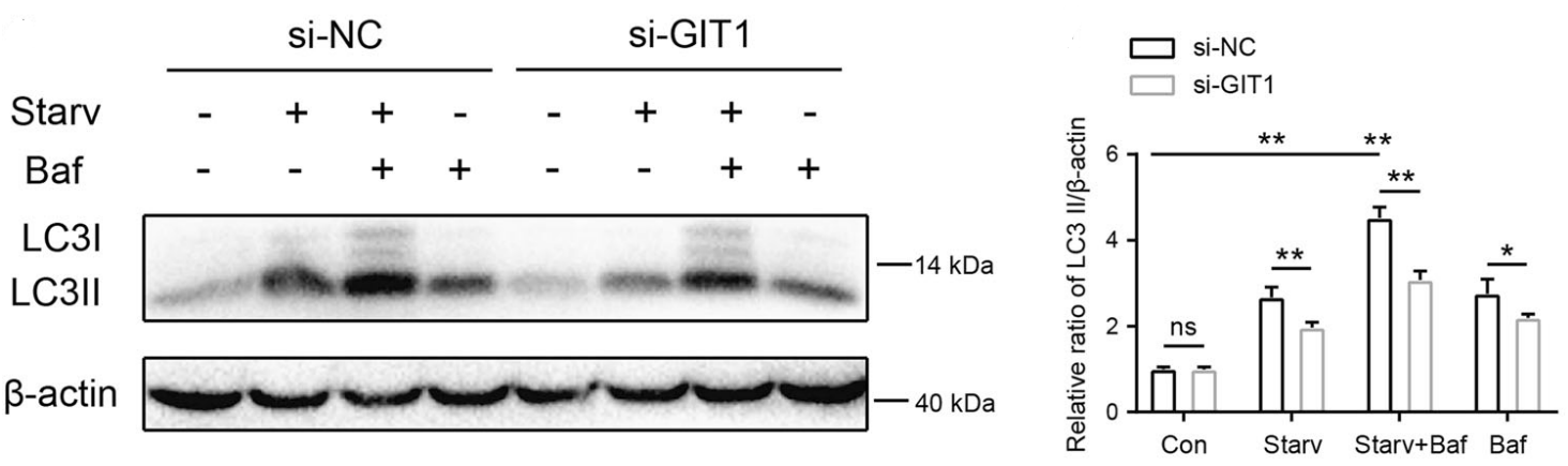
Bafilomycin A1 purchased from MedChemExpress. Usage Cited in: Cell Death Dis. 2018 Dec 13;9(12):1195. [Abstract]
Western analysis of the effect of GIT1 knockdown in lowering the LC3-II level under non-starvation or starvation conditions (1 h) with or without Bafilomycin A1 (Baf, 10 nM) in osteoclasts.
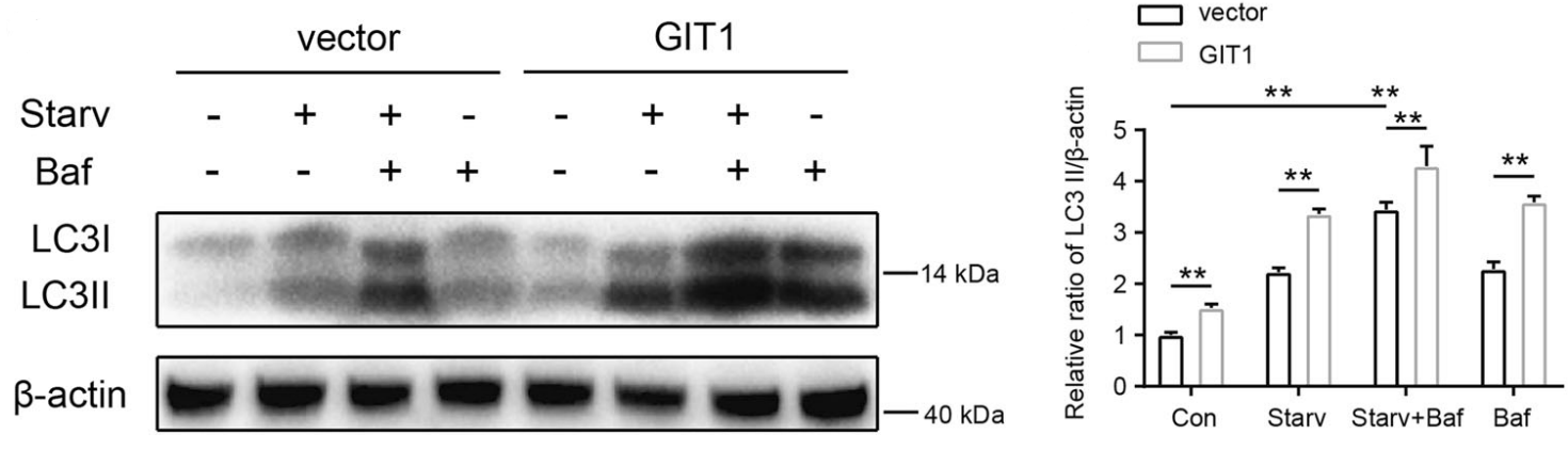
Bafilomycin A1 purchased from MedChemExpress. Usage Cited in: Cell Death Dis. 2018 Dec 13;9(12):1195. [Abstract]
Western analysis of the effect of GIT1 overexpression in increasing the LC3-II level under basal or starvation conditions with or without Bafilomycin A1 (Baf, 10 nM) in HEK293T cells.
-
Cell Death Dis
Nitazoxanide, an antiprotozoal drug, inhibits late-stage autophagy and promotes ING1-induced cell cycle arrest in glioblastoma. [Abstract]2018 Oct 9;9(10):1032. PMID: 30302016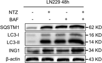
Bafilomycin A1 purchased from MedChemExpress. Usage Cited in: Cell Death Dis. 2018 Oct 9;9(10):1032. [Abstract]
Western blot showing SQSTM1 and LC3 levels in LN229 cells after treatment with 200 μM NSC 697855 (NTZ) and 200 nM Bafilomycin A1 (BAF) for 48 h.
-
Cell Death Dis
Transient receptor potential channel 6 knockdown prevents apoptosis of renal tubular epithelial cells upon oxidative stress via autophagy activation. [Abstract]2018 Oct 3;9(10):1015. PMID: 30282964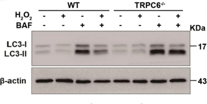
Bafilomycin A1 purchased from MedChemExpress. Usage Cited in: Cell Death Dis. 2018 Oct 3;9(10):1015. [Abstract]
Representative western blot images of LC3 (LC3I and LC3II) in primary PTC are isolated from WT and TRPC6-/- mice after treatment with H2O2 (0.5 mM 12 h) in the presence and absence of Bafilomycin A1 (BAF) (20 nM).
-
Sci China Life Sci
2026 May;69(5):1604-1619. PMID: 41793590 -
Sci China Life Sci
Receptor affinity-selective differential dynamics of membrane fusion initiation govern deltacoronavirus cross-species transmission. [Abstract]2025 Oct 24. PMID: 41144156 -
-
-
Proc Natl Acad Sci U S A
Mitochondrial ROS triggers mitophagy through activating the DNA damage response signaling pathway. [Abstract]2025 Oct 7;122(40):e2502841122. PMID: 41026812 -
Proc Natl Acad Sci U S A
Monensin suppresses EMT-driven cancer cell motility by inducing Golgi pH-dependent exocytosis of GOLIM4. [Abstract]2025 Jul 15;122(28):e2501347122. PMID: 40632561 -
Proc Natl Acad Sci U S A
2023 Feb 21;120(8):e2207425120. PMID: 36800388 -
Proc Natl Acad Sci U S A
Endocytosis triggers V-ATPase-SYK-mediated priming of cGAS activation and innate immune response. [Abstract]2022 Oct 25;119(43):e2207280119. PMID: 36252040 -
Proc Natl Acad Sci U S A
Mitochondrial outer membrane protein FUNDC2 promotes ferroptosis and contributes to doxorubicin-induced cardiomyopathy. [Abstract]2022 Sep 6;119(36):e2117396119. PMID: 36037337 -
-
Cell Commun Signal
NSP1 of Alongshan virus antagonizes type I interferon responses by promoting STUB1-mediated degradation of RIG-I. [Abstract]2026 Mar 24;24(1):262. PMID: 41877189 -
-
Cell Commun Signal
2025 Nov 12;23(1):492. PMID: 41225541 -
Cell Commun Signal
Intrinsic STING of CD8 + T cells regulates self-metabolic reprogramming and memory to exert anti-tumor effects. [Abstract]2025 Feb 19;23(1):99. PMID: 39972350 -
Cell Commun Signal
Autophagy mediated by ROS-AKT-FoxO pathway is required for intestinal regeneration in echinoderms. [Abstract]2025 Jan 7;23(1):8. PMID: 39762855 -
JACS Au
2025 May 24;5(6):2567-2579. PMID: 40575317 -
Dev Cell
A condensates-to-VPS41-associated phagic vacuoles conversion pathway controls autophagy degradation in plants. [Abstract]2024 Aug 2:S1534-5807(24)00447-7. PMID: 39111309 -
Int J Biol Macromol
HSP70 preserves brain microvascular endothelial integrity under heat stress associated with suppressed JNK-mediated apoptosis and mitophagy. [Abstract]2026 Apr:353:151239. PMID: 41791534 -
Int J Biol Macromol
Autophagic damage in senescent bone marrow mesenchymal stromal cells: Impact on Piezo1 expression during osteoporosis progression. [Abstract]2025 Sep 25;330(Pt 1):147928. PMID: 41015372 -
Int J Biol Macromol
Dynamic bidirectional regulation between Stk38 and rabies virus M protein coordinates apoptosis progression during neurotropic infection. [Abstract]2025 May 19:144398. PMID: 40398756 -
Int J Biol Macromol
Single-organelle visualization tracking natural glycosaminoglycans within mitochondria-lysosome crosstalk for inflammatory homeostasis. [Abstract]2025 Jan 27:140362. PMID: 39880268 -
Int J Biol Macromol
The role of TBC1D15 in sepsis-induced acute lung injury: Regulation of mitochondrial homeostasis and mitophagy. [Abstract]2024 Dec 29:139289. PMID: 39740704 -
Int J Biol Macromol
2025 Feb 3:140631. PMID: 39909271 -
Int J Biol Macromol
New fungal protein from Pleurotus ferulae lanzi induces AMPK-mediated autophagy and G1-phase cell cycle arrest in A549 lung cancer cells. [Abstract]2023 Jun 16;244:125453. PMID: 37330099 -
Int J Biol Macromol
Investigation of the internalization and transport mechanism of Codonopsis Radix polysaccharide both in mice and Caco-2 cells. [Abstract]2022 Aug 31:215:23-35. PMID: 35718143 -
Int J Biol Macromol
Self-stabilizing regulation of deubiquitinating enzymes in an enzymatic activity-dependent manner. [Abstract]2021 Jun 30:181:1081-1091. PMID: 33864866 -
Acta Pharmacol Sin
Hepatitis B virus core protein promotes liver cancer progression by stabilizing CANX and suppressing IRF7 transcription. [Abstract]2025 Jun 13. PMID: 40514420 -
Acta Pharmacol Sin
Inhibition of CD36 ameliorates mouse spinal cord injury by accelerating microglial lipophagy. [Abstract]2025 May;46(5):1205-1220. PMID: 39880928 -
Acta Pharmacol Sin
Capsaicin functions as a selective degrader of STAT3 to enhance host resistance to viral infection. [Abstract]2023 Nov;44(11):2253-2264. PMID: 37311796 -
Acta Pharmacol Sin
Hernandezine induces autophagic cell death in human pancreatic cancer cells via activation of the ROS/AMPK signaling pathway. [Abstract]2023 Apr;44(4):865-876. PMID: 36284209 -
Acta Pharmacol Sin
Nitidine chloride induces cardiac hypertrophy in mice by targeting autophagy-related 4B cysteine peptidase. [Abstract]2023 Mar;44(3):561-572. PMID: 35986213 -
Acta Pharmacol Sin
Autophagy inhibitors increase the susceptibility of KRAS-mutant human colorectal cancer cells to a combined treatment of 2-deoxy-D-glucose and lovastatin. [Abstract]2021 Nov;42(11):1875-1887. PMID: 33608672 -
Phytomedicine
Flavonoids of Ziziphora clinopodioides improve Alzheimer's cognitive impairment and inhibit NLRP3 inflammasome activation via autophagy-lysosome pathway. [Abstract]2026 May:154:158028. PMID: 41818943 -
Phytomedicine
Da-Bu-Yin-Wan rescues cognitive deficits in aging and Alzheimer's disease models by Wnt/β-catenin-dependent restoration of lysosomal acidification. [Abstract]2026 Apr:153:157916. PMID: 41662806 -
Phytomedicine
Ginkgetin alleviates sepsis-induced acute lung injury by promoting autophagy via inhibiting ubiquitination of Laptm5 in macrophages. [Abstract]2026 Jan 28:153:157894. PMID: 41666511 -
-
-
Phytomedicine
Salidroside mitigates Oligoasthenospermia by attenuating ferroptosis via NF-κB pathway inhibition. [Abstract]2025 Nov 29:150:157633. PMID: 41351983 -
Phytomedicine
Liquiritin in Aiduqing formula inhibits breast cancer neoangiogenesis by suppressing CTSK-mediated lysosomal degradation of CXCL1 in tumor-associated macrophages. [Abstract]2025 Oct 3:148:157358. PMID: 41072283 -
Phytomedicine
20-Deoxyingenol attenuated doxorubicin-induced cardiotoxicity by promoting autolysosome degradation through the UCHL3-TFEB pathway. [Abstract]2025 Nov:147:157220. PMID: 40916238 -
Phytomedicine
Nrf2-activated mitophagy and ferroptosis suppression synergistically mediate tangeretin's protection against hepatic ischemia-reperfusion injury. [Abstract]2025 Sep:145:157034. PMID: 40617054 -
Phytomedicine
2025 May 8:143:156835. PMID: 40378590 -
EMBO J
Interferon-induced PARP14-mediated ADP-ribosylation in p62 bodies requires the ubiquitin-proteasome system. [Abstract]2025 Apr 7. PMID: 40195501 -
EMBO J
E3 ubiquitin ligase CHIP facilitates cAMP and cGMP signalling cross-talk by polyubiquitinating PDE9A. [Abstract]2025 Feb;44(4):1249-1273. PMID: 39806097 -
Phytomedicine
LW-213, a derivative of wogonin, triggers reticulophagy-mediated cell death in NSCLC via lysosomal damage combined with NPC1 inhibition. [Abstract]2024 Aug 22:134:155958. PMID: 39241385 -
Free Radic Biol Med
SIRT1 activation restores PINK1-dependent mitophagy to reverse airway barrier dysfunction in Acinetobacter baumannii infection. [Abstract]2026 Mar 25:250:153-167. PMID: 41895413 -
Free Radic Biol Med
Optineurin enhances the chemoresistance by activating mitophagy in acute myeloid leukemia with NPM1 mutation. [Abstract]2026 May:248:162-176. PMID: 41722661 -
Free Radic Biol Med
HIF-1α translation mediated by PKCδ facilitates RSV-induced production of innate inflammatory cytokines in vitro and in vivo. [Abstract]2026 Mar 16:246:614-626. PMID: 41611036 -
Free Radic Biol Med
Dihydroartemisinin targets GPX4 to induce autophagy-dependent ferroptosis and reduce radioresistance in triple-negative breast cancer. [Abstract]2026 Mar 16:246:35-50. PMID: 41520847 -
Free Radic Biol Med
TRAF6 mediates vascular remodeling via endoplasmic reticulum stress-mitophagy in hypoxic pulmonary hypertension. [Abstract]2025 Dec 20:245:237-249. PMID: 41429276 -
Free Radic Biol Med
MiRNA-27a-5p alleviates diabetic vascular injury via modulating autophagy by targeting NOX4. [Abstract]2025 Oct 3:241:526-542. PMID: 41046944 -
Free Radic Biol Med
ENO1 blockade augment ferroptosis susceptibility in TKIs-resistant CML through GPX4 autophagic degradation. [Abstract]2025 Sep 19:S0891-5849(25)00942-6. PMID: 40976411 -
Free Radic Biol Med
Cavidine alleviates paclitaxel-induced peripheral neuropathy by promoting mitochondrial autophagy through inhibiting PKM2-mediated histone lactylation. [Abstract]2025 Sep 24:241:367-383. PMID: 41005738 -
Free Radic Biol Med
Resveratrol induces ferroptosis in triple-negative breast cancer through NEDD4L-mediated GPX4 ubiquitination and degradation. [Abstract]2025 Aug 1:235:231-247. PMID: 40316059 -
Free Radic Biol Med
Autophagy and Endoplasmic Reticulum Stress-Related Protein Homeostasis Links Palmitic Acid to Hepatic Lipotoxicity in Zebrafish (Danio rerio), Counteracted by Linoleic Acid. [Abstract]2025 Jun:233:148-161. PMID: 40089081 -
Free Radic Biol Med
Ferroptosis and PANoptosis under hypoxia pivoting on the crosstalk between DHODH and GPX4 in corneal epithelium. [Abstract]2025 Feb 16:228:173-182. PMID: 39761766 -
Free Radic Biol Med
Sulfonated albumin from hepatocytes accelerates liver fibrosis in nonalcoholic fatty liver disease through endoplasmic reticulum stress. [Abstract]2025 Feb 16:228:150-162. PMID: 39743026 -
Free Radic Biol Med
The NRF2 activator RTA-408 ameliorates chronic alcohol exposure-induced cognitive impairment and NLRP3 inflammasome activation by modulating impaired mitophagy initiation. [Abstract]2024 Aug 1:220:15-27. PMID: 38679301 -
Free Radic Biol Med
Phosphatidylethanolamine alleviates OX-LDL-induced macrophage inflammation by upregulating autophagy and inhibiting NLRP1 inflammasome activation. [Abstract]2023 Nov 1:208:402-417. PMID: 37660837 -
Free Radic Biol Med
PCSK6 attenuates cardiac dysfunction in doxorubicin-induced cardiotoxicity by regulating autophagy. [Abstract]2023 Jul:203:114-128. PMID: 37061139 -
Free Radic Biol Med
STAT3 signaling promotes cardiac injury by upregulating NCOA4-mediated ferritinophagy and ferroptosis in high-fat-diet fed mice. [Abstract]2023 May 20:201:111-125. PMID: 36940731 -
Cancer Immunol Res
Discovery of podofilox as a potent cGAMP-STING signaling enhancer with antitumor activity. [Abstract]2023 May 3;11(5):583-599. PMID: 36921097 -
Free Radic Biol Med
2021 Jun:169:271-282. PMID: 33895289 -
Free Radic Biol Med
Decreased expression of IDH1 by chronic unpredictable stress suppresses proliferation and accelerates senescence of granulosa cells through ROS activated MAPK signaling pathways. [Abstract]2021 Jun:169:122-136. PMID: 33865962 -
Free Radic Biol Med
Physalin B ameliorates nonalcoholic steatohepatitis by stimulating autophagy and NRF2 activation mediated improvement in oxidative stress. [Abstract]2021 Feb 20:164:1-12. PMID: 33388433 -
Drug Deliv
A surrogate barrier model for high-throughput blood-brain barrier permeability prediction: integrating LLC-PK1-MOCK/MDR1 Cells and lysosomal trapping correction. [Abstract]2025 Dec 31;32(1):2585612. PMID: 41307200 -
Chemosphere
Black phosphorus quantum dots induce autophagy and apoptosis of human bronchial epithelial cells via endoplasmic reticulum stress. [Abstract]2023 Jun:327:138463. PMID: 36966929 -
Sci Total Environ
Gestational exposure to carbon black nanoparticles triggered fetal growth restriction in mice: The mediation of inactivating autophagy-lysosomal degradation system in placental ferroptosis. [Abstract]2024 Dec 24:959:178167. PMID: 39721551 -
Sci Total Environ
Activation of NLRP3 signaling contributes to cadmium-induced bone defects, associated with autophagic flux obstruction. [Abstract]2023 Oct 1:893:164787. PMID: 37321488 -
J Eur Acad Dermatol Venereol
Tp47 promoted the phagocytosis of HMC3 cells though autophagy induced by endoplamic reticlum stress. [Abstract]2022 Nov;36(11):2224-2234. PMID: 35666816 -
Sci Total Environ
2021 Jul 20:779:146523. PMID: 34030247 -
Br J Pharmacol
A novel synthesised STAT3 inhibitor exerts potent anti-tumour activity by inducing lysosome-dependent cell death. [Abstract]2025 May 13. PMID: 40356419 -
Br J Pharmacol
Naringenin relieves paclitaxel-induced pain by suppressing calcitonin gene-related peptide signalling and enhances the anti-tumour action of paclitaxel. [Abstract]2024 May 7. PMID: 38715438 -
Environ Res
Carbon black nanoparticles and cadmium co-exposure aggravates bronchial epithelial cells inflammation via autophagy-lysosome pathway. [Abstract]2024 Feb 1:242:117733. PMID: 38000634 -
Br J Pharmacol
A novel sorbicillinoid compound as a potent anti-inflammation agent through inducing NLRP3 protein degradation. [Abstract]2023 Aug;180(15):1930-1948. PMID: 36788033 -
Genes Dev
USP21 deubiquitinase elevates macropinocytosis to enable oncogenic KRAS bypass in pancreatic cancer. [Abstract]2021 Oct 1;35(19-20):1327-1332. PMID: 34531315 -
Neoplasia
Synergistic effect of Chloroquine and Panobinostat in ovarian cancer through induction of DNA damage and inhibition of DNA repair. [Abstract]2021 May;23(5):515-528. PMID: 33930758 -
Br J Pharmacol
Artesunate interacts with the vitamin D receptor to reverse sepsis-induced immunosuppression in a mouse model via enhancing autophagy. [Abstract]2020 Sep;177(18):4147-4165. PMID: 32520399 -
Brain Behav Immun
Role of astrocyte senescence regulated by the non- canonical autophagy in the neuroinflammation associated to cerebral malaria. [Abstract]2024 Mar:117:20-35. PMID: 38157948 -
Biomed Pharmacother
Gingerenone A induces ferroptosis in colorectal cancer via targeting suppression of SLC7A11 signaling pathway. [Abstract]2024 Nov:180:117529. PMID: 39393329 -
Biomed Pharmacother
Substituted indole derivatives as UNC-51-like kinase 1 inhibitors: Design, synthesis and anti-hepatocellular carcinoma activity. [Abstract]2024 Aug 7:178:117260. PMID: 39116788 -
Emerg Microbes Infect
Inhibition of RAN attenuates influenza a virus replication and nucleoprotein nuclear export. [Abstract]2024 Aug 1:2387910. PMID: 39087696 -
Biomed Pharmacother
Erastin promotes random-pattern skin flaps survival by inducing mTORC1-TFEB mediated autophagy. [Abstract]2024 Jun 14:177:116918. PMID: 38878639 -
J Transl Med
Resveratrol attenuates inflammation and fibrosis in rheumatoid arthritis-associated interstitial lung disease via the AKT/TMEM175 pathway. [Abstract]2024 May 14;22(1):457. PMID: 38745204 -
Biomed Pharmacother
Brusatol induces ferroptosis in oesophageal squamous cell carcinoma by repressing GSH synthesis and increasing the labile iron pool via inhibition of the NRF2 pathway. [Abstract]2023 Nov:167:115567. PMID: 37742602 -
Biomed Pharmacother
Cinchonine exerts anti-tumor and immunotherapy sensitizing effects in lung cancer by impairing autophagic-lysosomal degradation. [Abstract]2023 Aug:164:114980. PMID: 37301135 -
Emerg Microbes Infect
Porcine deltacoronavirus resists antibody neutralization through cell-to-cell transmission. [Abstract]2023 Dec;12(1):2207688. PMID: 37125733 -
Biomed Pharmacother
Sitagliptin inhibits the survival, stemness and autophagy of glioma cells, and enhances temozolomide cytotoxicity. [Abstract]2023 Jun:162:114555. PMID: 36966667 -
Diabetes
Metrnl alleviates lipid accumulation by modulating mitochondrial homeostasis in diabetic nephropathy. [Abstract]2023 May 1;72(5):611-626. PMID: 36812572 -
J Transl Med
Alamandine/MrgD axis prevents TGF-β1-mediated fibroblast activation via regulation of aerobic glycolysis and mitophagy. [Abstract]2023 Jan 13;21(1):24. PMID: 36635651 -
Biomed Pharmacother
Autophagy is involved in Ficus carica fruit extract-induced anti-tumor effects on pancreatic cancer. [Abstract]2022 Jun:150:112966. PMID: 35427822 -
Emerg Microbes Infect
Obatoclax inhibits SARS-CoV-2 entry by altered endosomal acidification and impaired cathepsin and furin activity in vitro. [Abstract]2022 Dec;11(1):483-497. PMID: 34989664 -
Diabetes
Hepatic P38 Activation Modulates Systemic Metabolism Through Fgf21-Mediated Interorgan Communication. [Abstract]2021 Oct 21:db210240. PMID: 34675006 -
Biomed Pharmacother
Bisdemethoxycurcumin alleviates vandetanib-induced cutaneous toxicity in vivo and in vitro through autophagy activation. [Abstract]2021 Dec:144:112297. PMID: 34649218 -
Biomed Pharmacother
Berberine represses human gastric cancer cell growth in vitro and in vivo by inducing cytostatic autophagy via inhibition of MAPK/mTOR/p70S6K and Akt signaling pathways. [Abstract]2020 Aug;128:110245. PMID: 32454290 -
Biomed Pharmacother
Inhibition of autophagy enhances the anticancer effect of enzalutamide on bladder cancer. [Abstract]2019 Dec;120:109490. PMID: 31574376 -
Oncogene
TBKBP1 induces capecitabine resistance through negative regulation of type I interferon pathway in triple-negative breast cancer. [Abstract]2026 Feb;45(6):703-714. PMID: 41606294 -
Stem Cell Res Ther
MSCs promote the efferocytosis of large peritoneal macrophages to eliminate ferroptotic monocytes/macrophages in the injured endometria. [Abstract]2024 May 1;15(1):127. PMID: 38693589 -
Environ Pollut
2023 Jan 1;316(Pt 2):120627. PMID: 36370978 -
Stem Cell Res Ther
CCL20/CCR6 axis mediates macrophages to promote proliferation and migration of ESCs by blocking autophagic flux in endometriosis. [Abstract]2022 Jul 15;13(1):294. PMID: 35841069 -
Oncogene
Melatonin potentiates the cytotoxic effect of Neratinib in HER2+ breast cancer through promoting endocytosis and lysosomal degradation of HER2. [Abstract]2021 Nov;40(44):6273-6283. PMID: 34556812 -
Stem Cell Res Ther
Melatonin induces the rejuvenation of long-term ex vivo expanded periodontal ligament stem cells by modulating the autophagic process. [Abstract]2021 Apr 29;12(1):254. PMID: 33926537 -
Environ Pollut
Drp1-mediated mitochondrial fission contributes to mitophagy in paraquat-induced neuronal cell damage. [Abstract]2021 Mar 1:272:116413. PMID: 33422762 -
Cell Chem Biol
A cationic amphiphilic drug synergizes with strobilurin fungicides to control fungal-borne plant diseases. [Abstract]2025 Jun 19;32(6):872-884.e7. PMID: 40505661 -
Cell Chem Biol
2023 Mar 16;30(3):248-260.e4. PMID: 36889309 -
-
Aging Cell
2026 Feb;25(2):e70373. PMID: 41521645 -
Aging Cell
Aerobic Exercise Attenuates Autophagy-Lysosomal Flux Deficits via β2-AR-Mediated ESCRT-III Subunit CHMP4B in Mice With Human MAPT P301L. [Abstract]2025 Jul 26:e70184. PMID: 40715737 -
J Genet Genomics
2024 Oct;51(10):1020-1029. PMID: 38663479 -
Aging Cell
2020 Jul;19(7):e13171. PMID: 32662244 -
Cell Death Discov
Amlodipine exerts inhibitory effects against glioma stem cells through degrading EGFR and down-regulating its downstream pro-survival pathways. [Abstract]2025 Oct 27;11(1):492. PMID: 41145413 -
Cell Death Discov
Epirubicin induces cardiotoxicity through disrupting ATP6V0A2-dependent lysosomal acidification and triggering ferroptosis in cardiomyocytes. [Abstract]2024 Jul 24;10(1):337. PMID: 39048556 -
Cell Death Discov
MDH2 regulates the sensitivity of clear cell renal cell carcinoma to ferroptosis through its interaction with FSP1. [Abstract]2024 Aug 13;10(1):363. PMID: 39138167 -
Cell Death Discov
Iron derived from NCOA4-mediated ferritinophagy causes cellular senescence via the cGAS-STING pathway. [Abstract]2023 Nov 18;9(1):419. PMID: 37980349 -
Cell Death Discov
Engineered small extracellular vesicles loaded with miR-654-5p promote ferroptosis by targeting HSPB1 to alleviate sorafenib resistance in hepatocellular carcinoma. [Abstract]2023 Sep 30;9(1):362. PMID: 37777559 -
Cell Death Discov
An autophagy-inducing stapled peptide induces mitochondria dysfunction and triggers autotic cell death in triple-negative breast cancer. [Abstract]2023 Aug 19;9(1):303. PMID: 37598181 -
Curr Res Food Sci
Proanthocyanidin oligomers extract from hawthorn mediates cell cycle arrest, apoptosis, and lysosome vacuolation on HCT116 cells. [Abstract]2022 May 29:5:904-917. PMID: 36686366 -
Cell Death Discov
Salmonella pSLT-encoded effector SpvB promotes RIPK3-dependent necroptosis in intestinal epithelial cells. [Abstract]2022 Feb 2;8(1):44. PMID: 35110556 -
Cell Rep
Polystyrene nanoplastics readily penetrate intestine and cause sex-specific effects mediated by bile acids and microbiome. [Abstract]2026 Jan 22;45(2):116859. PMID: 41575856 -
-
Cell Rep
COPS2 coordinates biphasic autophagy hijacking for non-lytic propagation of enveloped RNA viruses. [Abstract]2025 Nov 19;44(12):116594. PMID: 41269859 -
Cell Rep
2025 Nov 25;44(11):116485. PMID: 41187061 -
Cell Rep
2025 Oct 15;44(10):116433. PMID: 41105513 -
Cell Rep
TCF25 serves as a nutrient sensor to orchestrate metabolic adaptation and cell death by enhancing lysosomal acidification under glucose starvation. [Abstract]2025 Aug 21;44(9):116186. PMID: 40844875 -
Cell Rep
The Hippo pathway kinase MST1 mediates a feedback loop to maintain NLRP3 inflammasome homeostasis. [Abstract]2025 Jul 26;44(8):116076. PMID: 40716062 -
Cell Rep
The N-degron pathway mediates the autophagic degradation of cytosolic mitochondrial DNA during sterile innate immune responses. [Abstract]2024 Dec 21;44(1):115094. PMID: 39709605 -
Cell Rep
STUB1-mediated ubiquitination and degradation of NSUN2 promotes hepatocyte ferroptosis by decreasing m5C methylation of Gpx4 mRNA. [Abstract]2024 Oct 24;43(11):114885. PMID: 39453812 -
Cell Rep
A MYC-STAMBPL1-TOE1 positive feedback loop mediates EGFR stability in hepatocellular carcinoma. [Abstract]2024 Oct 8;43(10):114812. PMID: 39388352 -
Plant Physiol
FATTY ACID DESATURASE4 enhances plant RNA virus replication and undergoes host vacuolar ATPase-mediated degradation. [Abstract]2024 Jun 27:kiae350. PMID: 38935533 -
Cell Rep
Endocytic activation and exosomal secretion of matriptase stimulate the second wave of EGF signaling to promote skin and breast cancer invasion. [Abstract]2024 Mar 27;43(4):114002. PMID: 38547126 -
Cell Rep
2023 Aug 29;42(8):112851. PMID: 37481723 -
Cell Rep
2024 Jan 23;43(1):113575. PMID: 38181788 -
Cell Rep
2023 Nov 10;42(11):113417. PMID: 37950872 -
Cell Rep
LHPP-mediated inorganic pyrophosphate hydrolysis-driven lysosomal acidification in astrocytes regulates adult neurogenesis. [Abstract]2023 Aug 11;42(8):112975. PMID: 37573508 -
Cell Rep
Sequential genome-wide CRISPR-Cas9 screens identify genes regulating cell-surface expression of tetraspanins. [Abstract]2023 Jan 31;42(2):112065. PMID: 36724073 -
Sci Data
High-content, arrayed compound screens with rhinovirus, influenza A virus and herpes simplex virus infections. [Abstract]2022 Oct 8;9(1):610. PMID: 36209289 -
Cell Rep
S-palmitoylation of PCSK9 induces sorafenib resistance in liver cancer by activating the PI3K/AKT pathway. [Abstract]2022 Aug 16;40(7):111194. PMID: 35977495 -
Neurotherapeutics
FTH1 Inhibits Ferroptosis Through Ferritinophagy in the 6-OHDA Model of Parkinson's Disease. [Abstract]2020 Oct;17(4):1796-1812. PMID: 32959272 -
J Med Chem
Hsp70-Targeting Chimeras Enable Dual Proteasomal and Lysosomal Degradation of Intracellular and Extracellular Proteins. [Abstract]2026 Apr 9;69(7):8417-8432. PMID: 41874277 -
J Med Chem
Phytohormone Strigolactams Overcomes Oxaliplatin Resistance in Colorectal Cancer by Blocking the Late-Stage Autophagy to Tip the Metabolic Balance toward Ferroptosis. [Abstract]2025 Nov 27;68(22):24546-24559. PMID: 41208142 -
J Med Chem
Discovery of TNI-97 as a Highly Selective, Orally Bioavailable HDAC6 Inhibitor for the Treatment of Triple-Negative Breast Cancer. [Abstract]2025 Jul 24;68(14):14333-14356. PMID: 40607730 -
Clin Transl Med
Vitamin K-dependent gamma-carboxyglutamic acid protein 1 promotes pancreatic ductal adenocarcinoma progression through stabilizing oncoprotein KRAS and tyrosine kinase receptor EGFR. [Abstract]2025 Jan;15(1):e70191. PMID: 39843398 -
J Med Chem
Discovery of ZLC491 as a Potent, Selective, and Orally Bioavailable CDK12/13 PROTAC Degrader. [Abstract]2024 Oct 24;67(20):18247-18264. PMID: 39388374 -
J Med Chem
2023 Sep 14;66(17):12614-12628. PMID: 37652467 -
Clin Transl Med
Loss of LncRNA DUXAP8 synergistically enhanced sorafenib induced ferroptosis in hepatocellular carcinoma via SLC7A11 de-palmitoylation. [Abstract]2023 Jun;13(6):e1300. PMID: 37337470 -
Clin Transl Med
Targeting lysosomal HSP70 induces acid sphingomyelinase-mediated disturbance of lipid metabolism and leads to cell death in T cell malignancies. [Abstract]2023 Mar;13(3):e1229. PMID: 36959764 -
J Med Chem
Development of Strigolactones as Novel Autophagy/Mitophagy Inhibitors against Colorectal Cancer Cells by Blocking the Autophagosome-Lysosome Fusion. [Abstract]2022 Jul 19. PMID: 35852796 -
Neural Regen Res
TP53 drives neuronal ferroptosis by promoting KLHL4-mediated SLC7A11 ubiquitination after spinal cord injury. [Abstract]2025 Sep 3. PMID: 40903977 -
Neural Regen Res
Enhanced autophagic clearance of amyloid-β via HDAC6-mediated V-ATPase assembly and lysosomal acidification protects against Alzheimer's disease in vitro and in vivo. [Abstract]2025 Sep 1;20(9):2633-2644. PMID: 38993141 -
Sci Signal
Degradation of HDAC10 by autophagy promotes IRF3-mediated antiviral innate immune responses. [Abstract]2022 Dec 20;15(765):eabo4356. PMID: 36538592 -
Sci Signal
SARS-CoV-2 spike protein-induced cell fusion activates the cGAS-STING pathway and the interferon response. [Abstract]2022 Apr 12;15(729):eabg8744. PMID: 35412852 -
J Anim Sci Biotechnol
AMBRA1 activation alleviates zearalenone-induced swine testicular cell ferroptosis by facilitating mitophagy. [Abstract]2026 Mar 14;17(1):49. PMID: 41826991 -
J Cell Biol
2025 May 5;224(5):e202405060. PMID: 40136051 -
Mol Med
Triolein alleviates ischemic stroke brain injury by regulating autophagy and inflammation through the AKT/mTOR signaling pathway. [Abstract]2024 Dec 6;30(1):242. PMID: 39639187 -
Mol Med
New perspectives on YTHDF2 O-GlcNAc modification in the pathogenesis of intervertebral disc degeneration. [Abstract]2024 Oct 18;30(1):180. PMID: 39425013 -
Mol Med
Improvement of ACK1-targeted therapy efficacy in lung adenocarcinoma using chloroquine or bafilomycin A1. [Abstract]2023 Jan 16;29(1):6. PMID: 36647009 -
Elife
Zbtb14 regulates monocyte and macrophage development through inhibiting pu.1 expression in zebrafish. [Abstract]2022 Oct 7;11:e80760. PMID: 36205309 -
Elife
2022 Jan 13;11:e71256. PMID: 35023829 -
Phytother Res
Punicalagin Promotes Peripheral Nerve Repair by Enhancing Mitophagy via the TLR4/MAPK Signaling Pathway and Facilitating Angiogenesis. [Abstract]2026 May;40(5):2632-2650. PMID: 41732027 -
Phytother Res
Z-Guggulsterone Inhibits Triple-Negative Breast Cancer Progression by Blocking Autophagosome-Lysosome Fusion via OGT-Mediated SNAP29 O-GlcNAcylation. [Abstract]2026 May;40(5):2364-2379. PMID: 41684288 -
J Pineal Res
Melatonin Increased Autophagy Level to Facilitate Osteogenesis of Inflamed PDLSCs Through TMEM110 Signaling Pathways. [Abstract]2025 Mar;77(2):e70039. PMID: 40065592 -
Phytother Res
Isoalantolactone exerts anti-melanoma effects via inhibiting PI3K/AKT/mTOR and STAT3 signaling in cell and mouse models. [Abstract]2024 Jun;38(6):2800-2817. PMID: 38526171 -
Phytother Res
Homoplantaginin attenuates high glucose-induced vascular endothelial cell apoptosis through promoting autophagy via the AMPK/TFEB pathway. [Abstract]2023 Jul;37(7):3025-3041. PMID: 36879478 -
Phytother Res
The alleviative effect of flavonol-type Nrf2 activator rhamnazin from Physalis alkekengi L. var. franchetii (Mast.) Makino on pulmonary disorders. [Abstract]2022 Apr;36(4):1692-1707. PMID: 35129872 -
Cell Mol Life Sci
Exercise-derived peptide confers protection against septic cardiomyopathy by enhancing CCAAT/enhancer binding protein β-mediated autophagy. [Abstract]2025 Nov 25;82(1):421. PMID: 41288736 -
EMBO Rep
SH3KBP1 promotes skeletal myofiber formation and functionality through ER/SR architecture integrity. [Abstract]2025 Apr;26(8):2166-2191. PMID: 40065183 -
EMBO Rep
RNF144B negatively regulates antiviral immunity by targeting MDA5 for autophagic degradation. [Abstract]2024 Oct;25(10):4594-4624. PMID: 39285245 -
EMBO Rep
CaMKII suppresses proteotoxicity by phosphorylating BAG3 in response to proteasomal dysfunction. [Abstract]2024 Oct;25(10):4488-4514. PMID: 39261742 -
Cell Biosci
2024 Aug 2;14(1):101. PMID: 39095802 -
Cell Mol Life Sci
Lys-63-specific deubiquitinase BRCC36 enhances the sensitivity of multiple myeloma cells to lenalidomide by inhibiting lysosomal degradation of cereblon. [Abstract]2024 Aug 13;81(1):349. PMID: 39136771 -
Cell Biosci
Enhanced liquidity of p62 droplets mediated by Smurf1 links Nrf2 activation and autophagy. [Abstract]2023 Feb 21;13(1):37. PMID: 36810259 -
EMBO Rep
Amino acid starvation-induced LDLR trafficking accelerates lipoprotein endocytosis and LDL clearance. [Abstract]2022 Feb 3;23(3):e53373. PMID: 34994492 -
J Agric Food Chem
Lysosome-Mediated Cytotoxic Autophagy Contributes to Tea Polysaccharide-Induced Colon Cancer Cell Death via mTOR-TFEB Signaling. [Abstract]2021 Jan 20;69(2):686-697. PMID: 33369397 -
Ecotoxicol Environ Saf
Phthalates promote lenvatinib resistance in hepatocellular carcinoma through the HPX-MAPK pathway. [Abstract]2025 Aug 25:303:118932. PMID: 40858030 -
JCI Insight
2025 Apr 29:e190838. PMID: 40299657 -
Ecotoxicol Environ Saf
2024 Jun 1:277:116392. PMID: 38677065 -
JCI Insight
Inhibition of retinoic acid signaling in proximal tubular epithelial cells protects against acute kidney injury. [Abstract]2023 Oct 23;8(20):e173144. PMID: 37698919 -
Prog Neurobiol
Astrocytic AT1R deficiency ameliorates Aβ-induced cognitive deficits and synaptotoxicity through β-arrestin2 signaling. [Abstract]2023 Sep:228:102489. PMID: 37355221 -
Ecotoxicol Environ Saf
Multi-locus deletion mutation induced by silver nanoparticles: Role of lysosomal-autophagy dysfunction. [Abstract]2023 Jun 1:257:114947. PMID: 37105094 -
Ecotoxicol Environ Saf
Graphene oxide induced dynamic changes of autophagy-lysosome pathway and cell apoptosis via TFEB dysregulation in F98 cells. [Abstract]2022 Nov:246:114172. PMID: 36244172 -
JCI Insight
2022 May 9; 7(9): e152481. PMID: 35531958 -
-
NPJ Aging
2025 Feb 4;11(1):6. PMID: 39905063 -
Cancer Cell Int
Deapioplatycodin D inhibits glioblastoma cell proliferation by inducing BNIP3L-mediated incomplete mitophagy. [Abstract]2025 Jan 13;25(1):11. PMID: 39800710 -
Cancer Cell Int
PCK1 activates oncogenic autophagy via down-regulation Serine phosphorylation of UBAP2L and antagonizes colorectal cancer growth. [Abstract]2023 Apr 16;23(1):68. PMID: 37062825 -
J Mol Cell Biol
Emerging role of FUS in TGFB1 and COL1A1 transcription dependent on GADD45B to induce NASH-fibrosis. [Abstract]2025 Dec 18:mjaf049. PMID: 41410503 -
Eur J Med Chem
2025 Jun 27:297:117918. PMID: 40609222 -
Cell Biol Toxicol
CUL1-neddylation contributes to K29-linked ubiquitination on p27 for autophagic degradation in sorafenib-resistant liver cancer. [Abstract]2025 Mar 20;41(1):61. PMID: 40111576 -
Cell Biol Toxicol
Targeting p38γ synergistically enhances sorafenib-induced cytotoxicity in hepatocellular carcinoma. [Abstract]2025 Jan 28;41(1):35. PMID: 39871031 -
Front Immunol
Non-Structural Protein 3 of Duck Tembusu Virus Induces Autophagy via the ERK and PI3K-AKT-mTOR Signaling Pathways. [Abstract]2022 Feb 3;13:746890. PMID: 35185869 -
J Invest Dermatol
HSPA1A Protects Cells from Thermal Stress by Impeding ESCRT-0-Mediated Autophagic Flux in Epidermal Thermoresistance. [Abstract]2021 Jan;141(1):48-58.e3. PMID: 32533962 -
Biochem Pharmacol
Targeted degradation of CD24 by a transferrin receptor-engaging bispecific degrader enhances antitumor immunity. [Abstract]2026 Apr:246:117721. PMID: 41558617 -
Biochem Pharmacol
PKCζ regulates autophagy in pancreatic ductal adenocarcinoma via BAG3: a novel therapeutic vulnerability. [Abstract]2025 Jul 16:241:117176. PMID: 40675228 -
Breast Cancer Res
Erdafitinib inhibits the tumorigenicity of MDA-MB-231 triple-negative breast cancer cells by inducing TRIM25/ubiquitin-dependent degradation of FGFR4. [Abstract]2025 Jul 9;27(1):128. PMID: 40635078 -
Biochem Pharmacol
Triflupromazine and tranylcypromine alleviate primary cisplatin resistance in lung adenocarcinoma by promoting LDHA-mediated AMBRA1 ubiquitination. [Abstract]2025 Jun 23:117073. PMID: 40562117 -
Biochem Pharmacol
TRIM21 knockout alleviates renal fibrosis by promoting autophagic degradation of mature TGF-β1. [Abstract]2025 Apr:234:116822. PMID: 39983846 -
Biochem Pharmacol
Inhibition of ferroptosis protects intrahepatic bile duct cells against ischemia-reperfusion and bile salt toxicity. [Abstract]2025 Jan 29:116788. PMID: 39890033 -
Cell Prolif
Abnormal HCK/glutamine/autophagy axis promotes endometriosis development by impairing macrophage phagocytosis. [Abstract]2024 Jul 2:e13702. PMID: 38956970 -
Biochem Pharmacol
Polydatin attenuates diabetic renal inflammatory fibrosis via the inhibition of STING pathway. [Abstract]2024 Jun 15:116373. PMID: 38885772 -
Biochem Pharmacol
Apelin-13 improves pulmonary epithelial barrier function in a mouse model of LPS-induced acute lung injury by inhibiting Chk1-mediated DNA damage. [Abstract]2024 May 25:116297. PMID: 38801925 -
Biochem Pharmacol
Canagliflozin inhibits p-gp function and early autophagy and improves the sensitivity to the antitumor effect of doxorubicin. [Abstract]2020 May;175:113856. PMID: 32061772 -
Am J Chin Med
Cepharanthine Triggers Ferroptosis in Gastric Cancer by PINK1/FUNDC1-Mediated Mitophagy-Dependent GPX4 Degradation. [Abstract]2026;54(1):329-348. PMID: 41684093 -
Pharmaceutics
3,4-Dihydroxyphenylethanol (DPE or Hydroxytyrosol) Counteracts ERK1/2 and mTOR Activation, Pro-Inflammatory Cytokine Release, Autophagy and Mitophagy Reduction Mediated by Benzo[a]pyrene in Primary Human Colonic Epithelial Cells. [Abstract]2022 Mar 17;14(3):663. PMID: 35336037 -
Pharmaceutics
2021 Nov 19;13(11):1964. PMID: 34834380 -
Pharmaceutics
Transport Mechanisms of Polymannuronic Acid and Polyguluronic Acid Across Caco-2 Cell Monolayers. [Abstract]2020 Feb 17;12(2):167. PMID: 32079270 -
Food Funct
Fucoxanthin enhances AMPK/mTOR-dependent autophagic flux and attenuates ferroptosis in Alzheimer's disease models. [Abstract]2026 Jun 23. PMID: 42333463 -
Geroscience
Autophagy revealed as a targetable vulnerability in senescent cells by cell painting phenotypic profiling: a mechanistic study of MCOPPB and related compounds. [Abstract]2026 Apr 30. PMID: 42062708 -
Geroscience
2026 Mar 5. PMID: 41781498 -
J Enzyme Inhib Med Chem
Mislocalisation of FLT3-ITD receptor contributes to MV4-11 leukaemia cell resistance to antibody-drug conjugate. [Abstract]2026 Dec;41(1):2638027. PMID: 41784256 -
World J Gastroenterol
Wogonin derivative V8 enhances bortezomib efficacy in gastric carcinoma by disrupting lysosome-mediated drug resistance. [Abstract]2026 Feb 28;32(8):113299. PMID: 41809875 -
Chem Biol Interact
2026 Mar 25:427:111940. PMID: 41616906 -
Chem Biol Interact
Thalidezine triggers Cathepsin B-mediated cell death in T-cell lymphoma by disrupting lysosomal function. [Abstract]2026 Mar 25:427:111932. PMID: 41570971 -
J Ethnopharmacol
Levistilide A from Ligusticum chuanxiong alleviates LPS-induced depression- and anxiety-like behaviors by promoting mitophagy through the SIRT3/PINK1/Parkin pathway. [Abstract]2026 Apr 24:361:121146. PMID: 41554453 -
Chem Biol Interact
Acetyl alkannin, a Shikonin monomer, inhibits the ATM/DDR pathway by targeting ATM and sensitizes cisplatin in solid tumors. [Abstract]2025 May 17:111559. PMID: 40389196 -
J Ethnopharmacol
2024 Aug 3:118658. PMID: 39103023 -
Chem Biol Interact
Targeting PKD2 aggravates ferritinophagy-mediated ferroptosis via promoting autophagosome-lysosome fusion and enhances efficacy of carboplatin in lung adenocarcinoma. [Abstract]2024 Jan 5:387:110794. PMID: 37951334 -
World J Gastroenterol
Suberoylanilide hydroxamic acid upregulates reticulophagy receptor expression and promotes cell death in hepatocellular carcinoma cells. [Abstract]2023 Sep 14;29(34):5038-5053. PMID: 37753370 -
Chem Biol Interact
HSP90/CDC37 inactivation promotes degradation of LKB1 protein to suppress AMPK signaling in bronchial epithelial cells exposed to sulfur mustard analog, 2-chloroethyl ethyl sulfide. [Abstract]2023 Sep 1:382:110643. PMID: 37481222 -
Food Funct
Lysosome-mediated mitochondrial apoptosis induced by tea polysaccharides promotes colon cancer cell death. [Abstract]2021 Nov 1;12(21):10524-10537. PMID: 34569560 -
Chem Biol Interact
Swainsonine promotes apoptosis by impairing lysosomal function and inhibiting autophagic degradation in rat primary renal tubular epithelial cells. [Abstract]2021 Feb 25;336:109319. PMID: 33186601 -
Mar Life Sci Technol
New activities of homoyessotoxin against lung cancer through the regulation of EGFR/PI3K/AKT pathway. [Abstract]2026 Feb 2;8(1):144-163. PMID: 41783560 -
Cells
2021 Sep 6;10(9):2330. PMID: 34571979 -
Cells
Trehalose Alleviates Crystalline Silica-Induced Pulmonary Fibrosis via Activation of the TFEB-Mediated Autophagy-Lysosomal System in Alveolar Macrophages. [Abstract]2020 Jan 4;9(1):122. PMID: 31947943 -
Stem Cell Reports
Integrative analysis of drug-gene signatures in human pluripotent stem cells reveals prazosin as a novel SQSTM1 regulator for ALS therapeutics. [Abstract]2026 Jun 25:102977. PMID: 42349423 -
Commun Biol
The gut microbiota metabolite trimethylamine N-oxide promotes cardiac hypertrophy by activating the autophagic degradation of SERCA2a. [Abstract]2025 Apr 10;8(1):596. PMID: 40210720 -
Drug Des Devel Ther
RET Inhibitor SPP86 Triggers Apoptosis and Activates the DNA Damage Response Through the Suppression of Autophagy and the PI3K/AKT Signaling Pathway in Melanoma Cells. [Abstract]2025 Jan 7:19:67-82. PMID: 39803607 -
Commun Biol
USP24 promotes autophagy-dependent ferroptosis in hepatocellular carcinoma by reducing the K48-linked ubiquitination of Beclin1. [Abstract]2024 Oct 8;7(1):1279. PMID: 39379617 -
Life Sci
Trehalose alleviates apoptosis by protecting the autophagy-lysosomal system in alveolar macrophages during human silicosis. [Abstract]2020 Sep 15:257:118043. PMID: 32621922 -
Life Sci
S6K1 inhibits HBV replication through inhibiting AMPK-ULK1 pathway and disrupting acetylation modification of H3K27. [Abstract]2021 Jan 15;265:118848. PMID: 33278383 -
Life Sci
p62-dependent autophagy in airway smooth muscle cells regulates metabolic reprogramming and promotes airway remodeling. [Abstract]2021 Feb 1:266:118884. PMID: 33310038 -
Life Sci
Amentoflavone suppresses cell proliferation and induces cell death through triggering autophagy-dependent ferroptosis in human glioma. [Abstract]2020 Apr 15;247:117425. PMID: 32057904 -
Life Sci
Inhibition of RELM-β prevents hypoxia-induced overproliferation of human pulmonary artery smooth muscle cells by reversing PLC-mediated KCNK3 decline. [Abstract]2020 Apr 1;246:117419. PMID: 32045592 -
CNS Neurosci Ther
SIX2-Mediated Microglial M2 Polarization and Exosomal miR-3470b Delivery Protect Dopaminergic Neurons in Parkinson's Disease. [Abstract]2026 Feb;32(2):e70756. PMID: 41700504 -
Respir Res
High matrix stiffness promotes senescence of type II alveolar epithelial cells by lysosomal degradation of lamin A/C in pulmonary fibrosis. [Abstract]2025 Apr 9;26(1):128. PMID: 40205454 -
PLoS Pathog
Bafilomycin A1 is a promising therapeutic agent against T. spiralis infection by inhibiting the heme-transporting ATP6V0C/HRG-1 complex. [Abstract]2026 Mar 16;22(3):e1014042. PMID: 41838682 -
Int J Mol Sci
2026 Jan 22;27(2):1119. PMID: 41596759 -
PLoS Pathog
Serine protease-driven entry and S2 ' cleavage flexibility of feline coronavirus during feline enterocyte infections. [Abstract]2026 Jan 8;22(1):e1013854. PMID: 41505483 -
Int J Mol Sci
Transcriptional Analysis of Spodoptera frugiperda Sf9 Cells Infected with Daphnis nerii Cypovirus-23. [Abstract]2025 Aug 2;26(15):7487. PMID: 40806613 -
Int J Mol Sci
Polyphyllin I Inhibits the Metastasis of Cervical Cancer Through the Regulation of the β-Catenin Signaling Pathway. [Abstract]2025 May 12;26(10):4630. PMID: 40429774 -
PLoS Pathog
SIRT5-mediated desuccinylation of the porcine deltacoronavirus M protein drives pexophagy to enhance viral proliferation. [Abstract]2025 May 9;21(5):e1013163. PMID: 40344161 -
Int J Mol Sci
Chloroquine Restores eNOS Signaling in Shunt Endothelial Cells via Inhibiting eNOS Uncoupling. [Abstract]2025 Feb 5;26(3):1352. PMID: 39941119 -
PLoS Pathog
Massive entry of BK Polyomavirus induces transient cytoplasmic vacuolization of human renal proximal tubule epithelial cells. [Abstract]2024 Nov 21;20(11):e1012681. PMID: 39570904 -
PLoS Pathog
Porcine reproductive and respiratory syndrome virus nonstructural protein 2 promotes the autophagic degradation of adaptor protein SH3KBP1 to antagonize host innate immune responses by enhancing K63-linked polyubiquitination of RIG-I. [Abstract]2024 Oct 28;20(10):e1012670. PMID: 39466846 -
PLoS Pathog
The HN protein of Newcastle disease virus induces cell apoptosis through the induction of lysosomal membrane permeabilization. [Abstract]2024 Feb 14;20(2):e1011981. PMID: 38354122 -
PLoS Pathog
2023 Nov 10;19(11):e1011789. PMID: 37948454 -
Int J Mol Sci
Puerarin Prevents Cadmium-Induced Neuronal Injury by Alleviating Autophagic Dysfunction in Rat Cerebral Cortical Neurons. [Abstract]2023 May 5;24(9):8328. PMID: 37176033 -
Int J Oncol
Dual targets of lethal apoptosis and protective autophagy in liver cancer with periplocymarin elicit a limited therapeutic effect. [Abstract]2023 Mar;62(3):44. PMID: 36825592 -
PLoS Pathog
2022 Oct 12;18(10):e1010907. PMID: 36223419 -
Int J Mol Sci
Antiviral Effects of ABMA and DABMA against Influenza Virus In Vitro and In Vivo via Regulating the Endolysosomal Pathway and Autophagy. [Abstract]2022 Apr 1;23(7):3940. PMID: 35409297 -
Int J Mol Sci
2022 Mar 30;23(7):3788. PMID: 35409148 -
PLoS Pathog
Bay41-4109-induced aberrant polymers of hepatitis b capsid proteins are removed via STUB1-promoted p62-mediated macroautophagy. [Abstract]2022 Jan 14;18(1):e1010204. PMID: 35030230 -
PLoS Pathog
p53 promotes ZDHHC1-mediated IFITM3 palmitoylation to inhibit Japanese encephalitis virus replication. [Abstract]2020 Oct 27;16(10):e1009035. PMID: 33108395 -
Pharm Biol
Ginkgolic acid targets HSPA8 to trigger ferroptosis in hepatocellular carcinoma via chaperone-mediated autophagy-dependent GPX4 degradation. [Abstract]2026 Dec;64(1):514-535. PMID: 41877645 -
Biomolecules
RSL3 Promotes STAT3 Ubiquitination to Induce Autophagy and Apoptosis in PARPi-Resistant Breast Cancer Cells. [Abstract]2025 Dec 18;15(12):1749. PMID: 41463402 -
Pharmaceuticals (Basel)
Therapeutic Implications of Ceritinib in Cholangiocarcinoma beyond ALK Expression and Mutation. [Abstract]2024 Feb 2;17(2):197. PMID: 38399413 -
Cell Oncol (Dordr)
Tumor-associated macrophage-derived GDNF promotes gastric cancer liver metastasis via a GFRA1-modulated autophagy flux. [Abstract]2023 Apr;46(2):315-330. PMID: 36808605 -
Pharmaceuticals (Basel)
Ciclopirox Olamine Induces Proliferation Inhibition and Protective Autophagy in Hepatocellular Carcinoma. [Abstract]2023 Jan 12;16(1):113. PMID: 36678610 -
Pharmaceuticals (Basel)
Targeting Annexin A1 as a Druggable Player to Enhance the Anti-Tumor Role of Honokiol in Colon Cancer through Autophagic Pathway. [Abstract]2023 Jan 1;16(1):70. PMID: 36678567 -
Pharmaceuticals (Basel)
Toosendanin Induces Hepatocyte Damage by Inhibiting Autophagic Flux via TFEB-Mediated Lysosomal Dysfunction. [Abstract]2022 Dec 3;15(12):1509. PMID: 36558960 -
Biomolecules
The Rabep1-Mediated Endocytosis and Activation of Trypsinogen to Promote Pancreatic Stellate Cell Activation. [Abstract]2022 Jul 31;12(8):1063. PMID: 36008957 -
Front Pharmacol
Myricetin Induces Autophagy and Cell Cycle Arrest of HCC by Inhibiting MARCH1-Regulated Stat3 and p38 MAPK Signaling Pathways. [Abstract]2021 Oct 18;12:709526. PMID: 34733155 -
Front Cell Infect Microbiol
Interaction With the Extracellular Matrix Triggers Calcium Signaling in Trypanosoma cruzi Prior to Cell Invasion. [Abstract]2021 Oct 4;11:731372. PMID: 34671568 -
Front Pharmacol
Sinensetin Reduces Osteoarthritis Pathology in the Tert-Butyl Hydroperoxide-Treated Chondrocytes and the Destabilization of the Medial Meniscus Model Mice via the AMPK/mTOR Signaling Pathway. [Abstract]2021 Jul 16:12:713491. PMID: 34335275 -
Front Bioeng Biotechnol
β-Elemene Reverses the Resistance of p53-Deficient Colorectal Cancer Cells to 5-Fluorouracil by Inducing Pro-death Autophagy and Cyclin D3-Dependent Cycle Arrest. [Abstract]2020 May 8:8:378. PMID: 32457882 -
Biomolecules
Lysosomal Fusion: An Efficient Mechanism Increasing Their Sequestration Capacity for Weak Base Drugs without Apparent Lysosomal Biogenesis. [Abstract]2020 Jan 3;10(1):77. PMID: 31947839 -
Int Immunopharmacol
Quercetin alleviates imatinib-induced premature ovarian insufficiency by regulating mitophagy via the ROS/JNK/c-JUN pathway. [Abstract]2026 Apr 15:175:116405. PMID: 41722539 -
Int Immunopharmacol
Luteolin mitigates inflammatory organ injury by targeting XIAP to block PANoptosis and mitochondrial dysfunction. [Abstract]2026 Mar 15:173:116271. PMID: 41637826 -
J Mol Cell Cardiol
DPP4-regulated endothelial cell ferroptosis modulates atherosclerosis progression by ferritinophagy. [Abstract]2026 Jan 19:213:14-30. PMID: 41565199 -
Int Immunopharmacol
Microbial metabolite tigloside alleviates osteoarthritis by repolarizing macrophages from M1 to M2 phenotype through Trafd1 destabilization and Trafd1-mediated NF-κB/STAT6 signaling pathways. [Abstract]2026 Jan 1;168(Pt 1):115747. PMID: 41192114 -
Eur J Pharmacol
Doxorubicin-mediated retardation of aggresome formation enhances Carfilzomib-induced cell death synergistically by augmenting ER stress and proapoptotic signaling. [Abstract]2025 Dec 5:1008:178303. PMID: 41183585 -
Int Immunopharmacol
Aloperine exerts anti-tumor effect and activates the tumor cell-intrinsic STING pathway in gallbladder cancer. [Abstract]2025 Sep 23:166:115590. PMID: 40991999 -
Bioorg Chem
A pomalidomide-based gefitinib PROTAC degrader effectively inhibits lung cancer progression in EGFR-TKIs-acquired resistant models by targeting EGFR degradation and ETFA-mediated ATP generation. [Abstract]2025 Aug 9:164:108864. PMID: 40815992 -
Eur J Pharmacol
VOPP1, a determinant of the sensitivity of non-small cell lung cancer cells to NAE inhibitors. [Abstract]2025 Sep 15:1003:177942. PMID: 40651787 -
Eur J Pharmacol
ROS confer resistance to 3TC in p53 mutant colorectal cancer by promoting autophagy and ER stress. [Abstract]2025 Jun 13:177837. PMID: 40517882 -
Bioorg Chem
2β-methoxy-2-deethoxyphantomolin synergistically enhances epirubicin effect against triple-negative breast cancer via targeted inhibition of AKT and HR pathways. [Abstract]2025 Apr 8:160:108453. PMID: 40253761 -
Spine J
Beyond a ferroptosis inducer: erastin can suppress nutrient deprivation induced cell death in the intervertebral disc. [Abstract]2025 Mar;25(3):597-608. PMID: 39522771 -
Bioorg Chem
The RIP3 activator C8 regulates the autophagy flux mediated by p62 and promotes the immunogenic form of cell death in human gastric cancer cells. [Abstract]2024 Nov 3:153:107937. PMID: 39520785 -
Invest Ophthalmol Vis Sci
Atg5-Mediated Lipophagy Induces Ferroptosis in Corneal Epithelial Cells in Dry Eye Disease. [Abstract]2024 Dec 2;65(14):12. PMID: 39636725 -
mBio
SIRT7 remodels the cytoskeleton via RAC1 to enhance host resistance to Mycobacterium tuberculosis. [Abstract]2024 Oct 16;15(10):e0075624. PMID: 39287444 -
Eur J Pharmacol
Loureirin B ameliorates cholestatic liver fibrosis via AKT/mTOR/ATG7-mediated autophagy of hepatic stellate cells. [Abstract]2024 May 15:971:176552. PMID: 38580181 -
Am J Physiol Cell Physiol
Metformin Mitigates Adipogenesis of Fibro-adipogenic Progenitors After Rotator Cuff Tears Via Activating mTOR/ULK1 Mediated Autophagy. [Abstract]2024 Jun 1;326(6):C1590-C1603. PMID: 38586878 -
Int Immunopharmacol
Arabinose confers protection against intestinal injury by improving integrity of intestinal mucosal barrier. [Abstract]2024 Jan 5:126:111188. PMID: 37995573 -
Int Immunopharmacol
Gitogenin suppresses lung cancer progression by inducing apoptosis and autophagy initiation through the activation of AMPK signaling. [Abstract]2022 Oct:111:108806. PMID: 35914447 -
Int Immunopharmacol
Costunolide ameliorates colitis via specific inhibition of HIF1α/glycolysis-mediated Th17 differentiation. [Abstract]2021 Aug:97:107688. PMID: 33932695 -
J Med Virol
Influenza A Virus Induces Autophagosome by Inhibiting LTCC/Calpain 2/LC3A Signaling to Promote Viral Replication. [Abstract]2025 May;97(5):e70393. PMID: 40358023 -
Hepatol Commun
HBV core protein enhances WDR46 stabilization to upregulate NUSAP1 and promote HCC progression. [Abstract]2025 May 6;9(5):e0680. PMID: 40366140 -
J Med Virol
E3 ubiquitin ligase FBXO22 inhibits SARS-CoV-2 replication via promoting proteasome-dependent degradation of NSP5. [Abstract]2024 Sep;96(9):e29891. PMID: 39223933 -
Neuropharmacology
Roflupram alleviates autophagy defects and reduces mutant hSOD1-induced motor neuron damage in cell and mouse models of amyotrophic lateral sclerosis. [Abstract]2024 Apr 1:247:109812. PMID: 38218579 -
Neuropharmacology
Defective autophagy contributes to bupivacaine-induced aggravation of painful diabetic neuropathy in db/db mice. [Abstract]2024 Mar 1:245:109814. PMID: 38104768 -
Molecules
Resveratrol Ameliorates Fibrosis in Rheumatoid Arthritis-Associated Interstitial Lung Disease via the Autophagy-Lysosome Pathway. [Abstract]2022 Dec 2;27(23):8475. PMID: 36500562 -
J Med Virol
2023 Jan;95(1):e28175. PMID: 36163413 -
Nanomedicine
Repurposing pentamidine using hyaluronic acid-based nanocarriers for skeletal muscle treatment in myotonic dystrophy. [Abstract]2023 Jan:47:102623. PMID: 36309185 -
Molecules
Nigrosporins B, a Potential Anti-Cervical Cancer Agent, Induces Apoptosis and Protective Autophagy in Human Cervical Cancer Ca Ski Cells Mediated by PI3K/AKT/mTOR Signaling Pathway. [Abstract]2022 Apr 9;27(8):2431. PMID: 35458629 -
Mol Oncol
Repurposing antitussive benproperine phosphate against pancreatic cancer depends on autophagy arrest. [Abstract]2020 Nov 23;15(2):725-738. PMID: 33226737 -
Cancers (Basel)
Proteome Responses to Acute Inhibition of De Novo Sphingolipid Synthesis Suggest Cancer Combination Therapies. [Abstract]2026 Jun 2;18(11):1827. PMID: 42279409 -
Cancers (Basel)
Triptolide Triggers Protective Autophagy via ROS Induction in NSCLC: Therapeutic Synergy with Autophagy Inhibition. [Abstract]2026 Mar 11;18(6):902. PMID: 41899507 -
Cancers (Basel)
Cancer Cells Evade Stress-Induced Apoptosis by Promoting HSP70-Dependent Clearance of Stress Granules. [Abstract]2022 Sep 25;14(19):4671. PMID: 36230594 -
Cancers (Basel)
Deregulation of Exo70 Facilitates Innate and Acquired Cisplatin Resistance in Epithelial Ovarian Cancer by Promoting Cisplatin Efflux. [Abstract]2021 Jul 11;13(14):3467. PMID: 34298686 -
Mol Neurobiol
OSBP Participates in Neural Damage Repair by Regulating Lysosome Transport Under Oxidative Stress. [Abstract]2025 Jun;62(6):7557-7575. PMID: 39915357 -
Mol Neurobiol
AQP4 Endocytosis-Lysosome Degradation Mediated by MMP-9/β-DG Involved in Diabetes Cognitive Impairment. [Abstract]2024 Oct;61(10):8438-8453. PMID: 38512439 -
Mol Neurobiol
Uric Acid Mitigates Cognitive Deficits via TFEB-Mediated Microglial Autophagy in Mice Models of Alzheimer's Disease. [Abstract]2024 Jun;61(6):3678-3696. PMID: 38008888 -
ACS Omega
Rapamycin Inhibits Osteoclastogenesis and Prevents LPS-Induced Alveolar Bone Loss by Oxidative Stress Suppression. [Abstract]2023 May 29;8(23):20739-20754. PMID: 37323396 -
Mol Neurobiol
Transmission of NLRP3-IL-1β Signals in Cerebral Ischemia and Reperfusion Injury: from Microglia to Adjacent Neuron and Endothelial Cells via IL-1β/IL-1R1/TRAF6. [Abstract]2023 May;60(5):2749-2766. PMID: 36717480 -
Front Cell Dev Biol
Blockade of GCH1/BH4 Axis Activates Ferritinophagy to Mitigate the Resistance of Colorectal Cancer to Erastin-Induced Ferroptosis. [Abstract]2022 Feb 10:10:810327. PMID: 35223839 -
Front Cell Dev Biol
Flubendazole Elicits Antitumor Effects by Inhibiting STAT3 and Activating Autophagy in Non-small Cell Lung Cancer. [Abstract]2021 Aug 26:9:680600. PMID: 34513827 -
Front Cell Dev Biol
Metformin-Enhanced Cardiac AMP-Activated Protein Kinase/Atrogin-1 Pathways Inhibit Charged Multivesicular Body Protein 2B Accumulation in Ischemia-Reperfusion Injury. [Abstract]2021 Feb 5:8:621509. PMID: 33614629 -
Front Cell Dev Biol
Structural and Functional Characterization of Fibronectin in Extracellular Vesicles From Hepatocytes. [Abstract]2021 Mar 18:9:640667. PMID: 33816490 -
Eur J Nutr
6-Shogaol from ginger shows anti-tumor effect in cervical carcinoma via PI3K/Akt/mTOR pathway. [Abstract]2021 Aug;60(5):2781-2793. PMID: 33416981 -
Poult Sci
ERAD inhibitor Eeyarestatin I (EerI) suppresses duck plague virus by restoring STING-mediated antiviral immunity. [Abstract]2026 Mar 17;105(6):106808. PMID: 41865653 -
Biochim Biophys Acta Mol Basis Dis
2026 Jan 27;1872(4):168170. PMID: 41604740 -
Lipids Health Dis
MLN4924 inhibits macrophage foam cell formation by enhancing autophagy and regulating metabolic phenotype. [Abstract]2025 Oct 2;24(1):307. PMID: 41039585 -
Poult Sci
Exploring ANT1 dysfunction as a pathological driver of wooden breast myopathy in broiler chickens. [Abstract]2025 Aug 26;104(11):105730. PMID: 40972420 -
FASEB J
Deubiquitinase OTUD4 Stabilizes SLC5A2 to Promote Pancreatic Cancer Proliferation and Migration Through Enchaining Glycolysis-Mediated Autophagy. [Abstract]2025 Jul 31;39(14):e70880. PMID: 40719816 -
Biochim Biophys Acta Mol Basis Dis
Deficiency in the conserved ECHS1 gene causes Leigh syndrome by impairing mitochondrial respiration efficiency and suppressing ADRB2-PKA signaling. [Abstract]2025 May 28:167930. PMID: 40446940 -
Lipids Health Dis
2025 Apr 12;24(1):138. PMID: 40221727 -
FEBS J
A high cholesterol diet aggravates experimental colitis through SREBP2-modulated endocytosis and degradation of occludin and Zo-1 proteins. [Abstract]2025 Mar;292(5):1052-1069. PMID: 39279038 -
Poult Sci
Up-regulated Lnc BTU promotes the production of duck plague virus DNA polymerase and inhibits the activation of JAK-STAT pathway to facilitate duck plague virus replication. [Abstract]2024 Dec;103(12):104238. PMID: 39383668 -
Biochim Biophys Acta Mol Basis Dis
Oleanonic acid ameliorates mutant Aβ precursor protein-induced oxidative stress, autophagy deficits, ferroptosis, mitochondrial damage, and ER stress in vitro. [Abstract]2024 Aug 10:167459. PMID: 39134286 -
FASEB J
Hypoxia-inducible factor prolyl hydroxylase inhibitor alleviates heatstroke-induced acute kidney injury by activating BNIP3-mediated mitophagy. [Abstract]2024 Jun 30;38(12):e23723. PMID: 38865198 -
FASEB J
Inhibition of NLRP3 inflammasome by MCC950 under hypoxia alleviates photoreceptor apoptosis via inducing autophagy in Müller glia. [Abstract]2024 May 31;38(10):e23671. PMID: 38752538 -
FASEB J
Amphiregulin improves ventricular remodeling after myocardial infarction by modulating autophagy and apoptosis. [Abstract]2024 Feb 29;38(4):e23488. PMID: 38358359 -
Biochim Biophys Acta Mol Basis Dis
USP46 enhances tamoxifen resistance in breast cancer cells by stabilizing PTBP1 to facilitate glycolysis. [Abstract]2024 Mar;1870(3):167011. PMID: 38176460 -
FEBS J
CD74-ROS1 L2026M mutant enhances autophagy through the MEK/ERK pathway to promote invasion, metastasis and crizotinib resistance in non-small cell lung cancer cells. [Abstract]2024 Mar;291(6):1199-1219. PMID: 38148635 -
Exp Neurol
Resolvin D2 activates anti-inflammatory microglia via restoring autophagy flux and alleviate neuropathic pain following spinal cord injury in rats. [Abstract]2023 Dec:370:114573. PMID: 37858697 -
J Cell Mol Med
Microglial FoxO3a deficiency ameliorates ferroptosis-induced brain injury of intracerebral haemorrhage via regulating autophagy and heme oxygenase-1. [Abstract]2024 Jan;28(1):e18007. PMID: 37890842 -
J Cell Mol Med
2024 Mar;28(5):e17856. PMID: 37461162 -
FASEB J
2023 May;37(5):e22933. PMID: 37093709 -
FASEB J
Extracellular HSP90 promotes differentiation of lens epithelial cells to fiber cells by activating LRP1-YAP-PROX1 axis. [Abstract]2023 Feb;37(2):e22783. PMID: 36705056 -
FASEB J
2023 Feb;37(2):e22749. PMID: 36688808 -
Mediators Inflamm
Lipopolysaccharide Inhibits Autophagy and Promotes Inflammatory Responses via p38 MAPK-Induced Proteasomal Degradation of Atg13 in Hepatic Stellate Cells. [Abstract]2022 Sep 13;2022:9603989. PMID: 36148140 -
J Cell Mol Med
MgIG exerts therapeutic effects on crizotinib-induced hepatotoxicity by limiting ROS-mediated autophagy and pyroptosis. [Abstract]2022 Aug;26(16):4492-4505. PMID: 35855570 -
Bioengineered
MiR-7-5p suppresses invasion via downregulation of the autophagy-related gene ATG7 and increases chemoresistance to cisplatin in BCa. [Abstract]2022 Mar;13(3):7328-7339. PMID: 35300572 -
Mol Nutr Food Res
Morin Acts as a USP7 Inhibitor to Hold Back the Migration of Rheumatoid Arthritis Fibroblast-Like Synoviocytes in a "Prickle1-mTORC2" Dependent Manner. [Abstract]2021 Oct;65(19):e2100367. PMID: 34331380 -
J Cell Mol Med
Gastrodin induces lysosomal biogenesis and autophagy to prevent the formation of foam cells via AMPK-FoxO1-TFEB signalling axis. [Abstract]2021 Jun;25(12):5769-5781. PMID: 33973365 -
J Cell Mol Med
Manganese activates autophagy to alleviate endoplasmic reticulum stress-induced apoptosis via PERK pathway. [Abstract]2020 Jan;24(1):328-341. PMID: 31639278 -
-
-
iScience
CCT3-mediated regulation of XPO1/RB1 axis stability promotes cellular senescence and tumor progression in clear cell renal carcinoma. [Abstract]2026 Jan 29;29(3):114840. PMID: 41732260 -
iScience
ATP6V0D2 drives triple-negative breast cancer progression and inhibits cisplatin sensitivity by promoting PHLPP2 lysosomal degradation. [Abstract]2025 Oct 27;28(11):113853. PMID: 41312388 -
Biogerontology
2025 Jul 21;26(4):147. PMID: 40689999 -
iScience
Inhibition of cathepsin L ameliorates inflammation through the A20/NF-κB pathway in endotoxin-induced acute lung injury. [Abstract]2024 Oct 4;27(11):111024. PMID: 39559762 -
iScience
Targeted down-regulation of SRSF1 exerts anti-cancer activity in OSCC through impairing lysosomal function and autophagy. [Abstract]2023 Oct 27;26(12):108330. PMID: 38025785 -
iScience
Irradiation combined with PD-L1-/- and autophagy inhibition enhances the antitumor effect of lung cancer via cGAS-STING-mediated T cell activation. [Abstract]2022 Jun 30;25(8):104690. PMID: 35847556 -
Virol Sin
Adaptor protein complex 1 gamma 1 subunit is an important host factor involved in both Zika virus and dengue virus infections. [Abstract]2025 Aug 5:S1995-820X(25)00107-5. PMID: 40759332 -
Antiviral Res
2025 Sep:241:106242. PMID: 40701276 -
J Neurochem
2025 Mar;169(3):e70026. PMID: 40026213 -
J Integr Med
Jiedu recipe, a compound Chinese herbal medicine, suppresses hepatocellular carcinoma metastasis by inhibiting the release of tumor-derived exosomes in a hypoxic microenvironment. [Abstract]2024 Nov;22(6):696-708. PMID: 39521704 -
Virol Sin
PRRSV degrades MDA5 via dual autophagy receptors P62 and CCT2 to evade antiviral innate immunity. [Abstract]2024 Apr;39(2):264-276. PMID: 38272236 -
Virol Sin
Hsp90 β is Critical for the Infection of Severe Fever with Thrombocytopenia Syndrome Virus. [Abstract]2024 Feb;39(1):113-122. PMID: 38008382 -
-
Virol Sin
Autophagy induced by human adenovirus B7 structural protein VI inhibits viral replication. [Abstract]2023 Oct;38(5):709-722. PMID: 37549881 -
Virol Sin
2023 Aug;38(4):520-530. PMID: 37156297 -
Antiviral Res
CDK4/6 inhibitor palbociclib promotes SARS-CoV-2 cell entry by down-regulating SKP2 dependent ACE2 degradation. [Abstract]2023 Apr:212:105558. PMID: 36806814 -
AAPS PharmSciTech
Transcellular Transport Behavior of the Intact Polymeric Mixed Micelles with Different Polymeric Ratios. [Abstract]2023 Feb 15;24(2):69. PMID: 36792796 -
Virol Sin
2022 May 3;S1995-820X(22)00077-3. PMID: 35504537 -
Front Cell Neurosci
Inhibition of Brd4 by JQ1 Promotes Functional Recovery From Spinal Cord Injury by Activating Autophagy. [Abstract]2020 Sep 2;14:555591. PMID: 32982695 -
J Biol Chem
Andrographolide targets syndecan4 to impair its interaction with syntenin and inhibits the biogenesis of small extracellular vesicles. [Abstract]2026 Jan 20:111182. PMID: 41570990 -
Sci Rep
Lysosomal homeostasis regulates myocardial ischemia-reperfusion injury through autophagy pathway. [Abstract]2025 Oct 22;15(1):36952. PMID: 41125732 -
Sci Rep
Pyrotinib promotes the antitumor effect of T-DM1 by increasing drug endocytosis in HER2-positive breast cancer. [Abstract]2025 May 28;15(1):18625. PMID: 40437017 -
Sci Rep
2024 Dec 4;14(1):30163. PMID: 39627455 -
Sci Rep
Downregulation of Lnc-ABCA12-3 modulates UBQLN1 expression and protein homeostasis pathways in amyotrophic lateral sclerosis. [Abstract]2024 Sep 13;14(1):21383. PMID: 39271939 -
Sci Rep
Genipin promotes the apoptosis and autophagy of neuroblastoma cells by suppressing the PI3K/AKT/mTOR pathway. [Abstract]2024 Aug 30;14(1):20231. PMID: 39215133 -
Sci Rep
2024 Aug 16;14(1):18974. PMID: 39152142 -
J Biol Chem
MGST3 regulates BACE1 protein translation and amyloidogenesis by controlling the RGS4-mediated AKT signaling pathway. [Abstract]2024 Jul 4:107530. PMID: 38971310 -
Sci Rep
The roles of autophagy, ferroptosis and pyroptosis in the anti-ovarian cancer mechanism of harmine and their crosstalk. [Abstract]2024 Mar 18;14(1):6504. PMID: 38499622 -
Sci Rep
The effect of genetic variants of SLC22A18 on proliferation, migration, and invasion of colon cancer cells. [Abstract]2024 Feb 16;14(1):3925. PMID: 38366023 -
J Biol Chem
Diarylheptanoid 35d overcomes EGFR TKI resistance by inducing hsp70-mediated lysosomal degradation of EGFR in EGFR-mutant lung adenocarcinoma. [Abstract]2023 Jun;299(6):104814. PMID: 37178919 -
Biomedicines
PPARα Inhibits Astrocyte Inflammation Activation by Restoring Autophagic Flux after Transient Brain Ischemia. [Abstract]2023 Mar 21;11(3):973. PMID: 36979952 -
Sci Rep
2022 Dec 24;12(1):22282. PMID: 36566289 -
J Biol Chem
Channel HCN4 mutation R666Q associated with sporadic arrhythmia decreases channel electrophysiological function and increases protein degradation. [Abstract]2022 Nov;298(11):102599. PMID: 36244448 -
J Biol Chem
Inhibitor of glutamine metabolism V9302 promotes ROS-induced autophagic degradation of B7H3 to enhance antitumor immunity. [Abstract]2022 Apr;298(4):101753. PMID: 35189139 -
Aging
Isoquercitrin induces apoptosis and autophagy in hepatocellular carcinoma cells via AMPK/mTOR/p70S6K signaling pathway. [Abstract]2020 Nov 29;12(23):24318-24332. PMID: 33260158 -
Aging
Autophagy is induced in human keratinocytes during human papillomavirus 11 pseudovirion entry. [Abstract]2020 Nov 16;12(22):23017-23028. PMID: 33197887 -
J Biol Chem
Autolysosomal degradation of cytosolic chromatin fragments antagonizes oxidative stress-induced senescence. [Abstract]2020 Apr 3;295(14):4451-4463. PMID: 32047109 -
J Virol
MARCH2 inhibits avian leukosis virus replication by targeting gp85 for ubiquitination and degradation. [Abstract]2025 Dec 23;99(12):e0161625. PMID: 41334913 -
J Virol
SUMOylation-dependent degradation of nucleocapsid is responsible for Pestivirus uncoating. [Abstract]2025 Nov 25:e0164825. PMID: 41288382 -
Microbiol Spectr
2025 Oct 7;13(10):e0136225. PMID: 40910953 -
J Virol
2025 Aug 11:e0077125. PMID: 40788012 -
J Virol
PEDV Nsp14 induces mitophagy-mediated degradation of MAVS to antagonize host innate immunity and facilitate viral proliferation. [Abstract]2025 Aug 19;99(8):e0049825. PMID: 40631894 -
Virol J
Bisbenzylisoquinoline alkaloids inhibit influenza virus replication by disrupting endosomal acidification. [Abstract]2025 Jun 4;22(1):181. PMID: 40468427 -
J Virol
2025 Jun 18:e0074125. PMID: 40530850 -
Virol J
Usutu virus NS4A induces autophagy and is targeted by the selective autophagy receptor p62/SQSTM1 for degradation. [Abstract]2025 Apr 17;22(1):103. PMID: 40247289 -
J Virol
2025 Mar 18;99(3):e0198024. PMID: 39976465 -
J Virol
FBXO45 restricts HIV-1 replication by inducing SQSTM1/p62-mediated autophagic degradation of Tat. [Abstract]2025 Feb 12:e0191224. PMID: 39936917 -
J Virol
2024 Aug 30:e0133524. PMID: 39212385 -
ACS Infect Dis
Pec 1 of Pseudomonas aeruginosa Inhibits Bacterial Clearance of Host by Blocking Autophagy in Macrophages. [Abstract]2024 Aug 9;10(8):2741-2754. PMID: 39047963 -
Microbiol Spectr
CSFV restricts necroptosis to sustain infection by inducing autophagy/mitophagy-targeted degradation of RIPK3. [Abstract]2024 Jan 11;12(1):e0275823. PMID: 38100396 -
J Virol
2023 Jul 27;97(7):e0051223. PMID: 37347173 -
J Virol
The SFTSV Nonstructural Proteins Induce Autophagy to Promote Viral Replication via Interaction with Vimentin. [Abstract]2023 Apr 27;97(4):e0030223. PMID: 37039677 -
Virol J
2023 Jan 31;20(1):18. PMID: 36721152 -
J Virol
The A137R Protein of African Swine Fever Virus Inhibits Type I Interferon Production via the Autophagy-Mediated Lysosomal Degradation of TBK1. [Abstract]2022 May 11;96(9):e0195721. PMID: 35412346 -
Virol J
Inhibitors of endosomal acidification suppress SARS-CoV-2 replication and relieve viral pneumonia in hACE2 transgenic mice. [Abstract]2021 Feb 27;18(1):46. PMID: 33639976 -
J Virol
White Spot Syndrome Virus Benefits from Endosomal Trafficking, Substantially Facilitated by a Valosin-Containing Protein, To Escape Autophagic Elimination and Propagate in the Crustacean Cherax quadricarinatus. [Abstract]2020 Nov 23;94(24):e01570-20. PMID: 32967962 -
J Virol
Interferon-Induced Transmembrane Protein 3 Is a Virus-Associated Protein Which Suppresses Porcine Reproductive and Respiratory Syndrome Virus Replication by Blocking Viral Membrane Fusion. [Abstract]2020 Nov 23;94(24):e01350-20. PMID: 32999030 -
Cell Signal
Mechanical stretch accelerates endothelial cell injury by inducing endoplasmic reticulum stress-mediated, autophagy-dependent ferroptosis in endothelial cells. [Abstract]2026 Feb:138:112271. PMID: 41285224 -
Cell Signal
Estradiol alleviates disease phenotypes caused by m.3635G > a mutations by activating mitochondrial biogenesis and PINK1-Parkin mediated mitophagy in iPSC-derived retinal pigment epithelium cells. [Abstract]2025 Sep 8:136:112121. PMID: 40930474 -
Cell Signal
Gambogic acid induces ferroptosis and suppresses colorectal cancer progression by modulating the m6A modification of p62. [Abstract]2025 Nov:135:112024. PMID: 40721064 -
PLoS Genet
2025 Jul 10;21(7):e1011794. PMID: 40638681 -
Cell Signal
LAPTM5 confers cisplatin resistance in NSCLC by suppressing LAMP1 ubiquitination to stabilize lysosomal membranes and sustain autophagic flux. [Abstract]2025 Apr 23:132:111834. PMID: 40280227 -
Cell Signal
HDAC inhibitor enhances ferroptosis susceptibility of AML cells by stimulating iron metabolism. [Abstract]2025 Jan 3:111583. PMID: 39756501 -
Mol Cell Biochem
Cytoglobin augments ferroptosis through autophagic degradation of ferritin in colorectal cancer cells. [Abstract]2025 May;480(5):2881-2892. PMID: 39503803 -
Biochim Biophys Acta Mol Cell Res
2024 Dec;1871(8):119853. PMID: 39353470 -
ACS Pharmacol Transl Sci
Bone Morphogenetic Protein Signaling Agonist SB4 (BMPSB4) Inhibits Corticotroph Pituitary Neuroendocrine Tumors by Activation of Autophagy via a BMP4/SMADs-Dependent Pathway. [Abstract]2024 Jun 24;7(7):1951-1970. PMID: 39022361 -
Biochim Biophys Acta Mol Cell Res
Ambra1 in exosomes secreted by HK-2 cells damaged by supersaturated oxalate induce mitophagy and autophagy-ferroptosis in normal HK-2 cells to participate in the occurrence of kidney stones. [Abstract]2023 Oct 6;1871(1):119604. PMID: 37806389 -
Mol Cell Biochem
Inhibition of STIM1 alleviates high glucose-induced proliferation and fibrosis by inducing autophagy in mesangial cells. [Abstract]2024 Sep;479(9):2365-2379. PMID: 37736800 -
Cell Signal
2021 Jun:82:109956. PMID: 33607258 -
Cell Signal
Autophagy participants in the dedifferentiation of mouse 3T3-L1 adipocytes triggered by hypofunction of insulin signaling. [Abstract]2021 Apr:80:109911. PMID: 33422645 -
Mol Cell Biochem
OP16 induces deadly autophagy and apoptosis of cells by inhibiting Akt in esophageal squamous cell carcinoma. [Abstract]2020 Sep;472(1-2):219-230. PMID: 32671513 -
Cell Signal
2019 Jun:58:119-130. PMID: 30849520 -
J Proteome Res
Lipidomic Network Analysis Reveals Amyloid-β-Induced Lysosomal Lipid Accumulation in the Cortex and Hippocampus of 5xFAD Mice. [Abstract]2025 Jul 4;24(7):3389-3398. PMID: 40552726 -
Heliyon
Queen bee acid pretreatment attenuates myocardial ischemia/reperfusion injury by enhancing autophagic flux. [Abstract]2024 Jun 21;10(12):e33371. PMID: 39021954 -
Heliyon
eIF2α-mediated integrated stress response links multiple intracellular signaling pathways to reprogram vascular smooth muscle cell fate in carotid artery plaque. [Abstract]2024 Feb 23;10(5):e26904. PMID: 38434290 -
Bone
2023 Feb:167:116643. PMID: 36513279 -
Stem Cells
2023 Jan 30;41(1):77-92. PMID: 36208284 -
Viruses
ASFV MGF110-7L Inhibits eIF4G1 Expression via Endoplasmic Reticulum Stress to Block Host Translation. [Abstract]2026 Feb 12;18(2):229. PMID: 41754572 -
Exp Cell Res
Membrane attack complex impairs hepatocyte autophagy in alcohol-related liver disease by regulating the SIRT1-FOXO3 signaling. [Abstract]2025 Aug 15;451(2):114713. PMID: 40834969 -
Viruses
DNAJ Homolog Subfamily C Member 11 Stabilizes SARS-CoV-2 NSP3 to Promote Double-Membrane Vesicle Formation. [Abstract]2025 Jul 22;17(8):1025. PMID: 40872740 -
Vet Res
Foot-and-mouth disease virus activates glycolysis and hijacks HK2 to inhibit innate immunity and promote viral replication. [Abstract]2025 Apr 1;56(1):71. PMID: 40170113 -
Vet Res
The cholesterol metabolite 25-hydroxycholesterol suppresses porcine deltacoronavirus via lipophagy inhibition and mTORC1 modulation. [Abstract]2025 Jan 31;56(1):23. PMID: 39891192 -
Microb Pathog
2024 Dec:197:107046. PMID: 39433139
Bafilomycin A1 purchased from MedChemExpress. Usage Cited in: Microb Pathog. 2024 Dec:197:107046. [Abstract]
BMDMs pretreated with degarelix (1 μM), aminoguanidine hydrochloride (AGHS, 100 μM), BafA1 (100 nM), NAC (5 mM), Z-VAD (20 μM), or 3-MA (5 mM) were infected with H37Rv (MOI = 2). At different time points after infection, bacterial survival was calculated by dividing the CFU at 24 h by the CFU at 3 h.
-
Food Chem Toxicol
Cell death induced by nicotine in human neuroblastoma SH-SY5Y cells is mainly attributed to cytoplasmic vacuolation originating from the trans-Golgi network. [Abstract]2024 Mar:185:114431. PMID: 38176581 -
Exp Cell Res
Upregulated Tβ4 expression in inflammatory bowel disease impairs the intestinal mucus barrier by inhibiting autophagy in mice. [Abstract]2024 Jan 1;434(1):113871. PMID: 38049080 -
Front Med
NFIL3 Facilitates Neutrophil Autophagy, Neutrophil Extracellular Trap Formation and Inflammation During Gout via REDD1-Dependent mTOR Inactivation. [Abstract]2021 Sep 30;8:692781. PMID: 34660620 -
Microb Pathog
Classical swine fever virus infection suppresses claudin-1 expression to facilitate its replication in PK-15 cells. [Abstract]2021 Aug:157:105012. PMID: 34062228 -
Exp Cell Res
Sodium butyrate causes α-synuclein degradation by an Atg5-dependent and PI3K/Akt/mTOR-related autophagy pathway. [Abstract]2020 Feb 1;387(1):111772. PMID: 31836471 -
Cardiovasc Ther
Trimethylamine-N-Oxide Impedes Late Endothelial Progenitor Cell-Mediated Revascularization by Triggering Mitochondrial Apoptosis via Suppression of MnSOD. [Abstract]2025 Jun 18:2025:9910333. PMID: 40568449 -
J Immunol
Zebrafish fkbp5 attenuates antiviral innate immunity by autophagic degradation of transcription factor irf7. [Abstract]2025 May 20:vkaf089. PMID: 40391431 -
J Immunol
IRF7 Exacerbates Candida albicans Infection by Compromising CD209-Mediated Phagocytosis and Autophagy-Mediated Killing in Macrophages. [Abstract]2024 Jun 15;212(12):1932-1944. PMID: 38709167 -
J Neurosci Res
Autophagy inhibitors 3-MA and BAF may attenuate hippocampal neuronal necroptosis after global cerebral ischemia-reperfusion injury in male rats by inhibiting the interaction of the RIP3/AIF/CypA complex. [Abstract]2024 Feb;102(2):e25301. PMID: 38361405 -
Toxicol Appl Pharmacol
The skin photoprotective effect of trilinolein: Induction of cellular autophagy via the AMPK-mTOR signaling pathway. [Abstract]2024 Feb:483:116836. PMID: 38272316 -
J Immunol
Duck MARCH8 Negatively Regulates the RLR Signaling Pathway through K29-Linked Polyubiquitination of MAVS. [Abstract]2023 Mar 15;210(6):786-794. PMID: 36715497 -
Am J Physiol Renal Physiol
Metformin ameliorates bladder dysfunction in a rat model of partial bladder outlet obstruction. [Abstract]2021 May 1;320(5):F838-F858. PMID: 33645317 -
Toxicol Appl Pharmacol
Cu(II) disrupts autophagy-mediated lysosomal degradation of oligomeric Aβ in microglia via mTOR-TFEB pathway. [Abstract]2020 Aug 15;401:115090. PMID: 32512069 -
Biochim Biophys Acta Mol Cell Biol Lipids
Sphinganine-induced lysosomal membrane permeabilization: Interplay with subcellular oxidative levels. [Abstract]2026 Jan;1871(1):159705. PMID: 41213325 -
Appl Biochem Biotechnol
NSUN6 Promotes Gastric Cancer Progression by Stabilizing CEBPZ mRNA in a m5C-Dependent Manner. [Abstract]2025 Sep 10. PMID: 40928700 -
Chem Asian J
Selective Protein Degradation through Tetrazine Ligation of Genetically Incorporated Unnatural Amino Acids. [Abstract]2024 Sep 2:e202400824. PMID: 39221720 -
Psychopharmacology (Berl)
Ketamine induces rapid antidepressant effects via the autophagy-NLRP3 inflammasome pathway. [Abstract]2022 Oct;239(10):3201-3212. PMID: 35925279 -
Front Oncol
Solasonine Causes Redox Imbalance and Mitochondrial Oxidative Stress of Ferroptosis in Lung Adenocarcinoma. [Abstract]2022 May 18:12:874900. PMID: 35664792 -
Psychopharmacology (Berl)
Dopamine D1 receptors mediate methamphetamine-induced dopaminergic damage: involvement of autophagy regulation via the AMPK/FOXO3A pathway. [Abstract]2022 Mar;239(3):951-964. PMID: 35190859 -
J Pharm Pharmacol
2024 May 3;76(5):567-578. PMID: 38271051 -
IUBMB Life
N-acetylneuraminic acid modulates SQSTM1/p62 sialyation-mediated ubiquitination degradation contributing to vascular endothelium dysfunction in experimental atherosclerosis mice. [Abstract]2024 Mar;76(3):161-178. PMID: 37818680 -
J Cancer
Upregulation of V-ATPase by STAT3 Activation Promotes Anoikis Resistance and Tumor Metastasis. [Abstract]2021 Jun 11;12(16):4819-4829. PMID: 34234852 -
Adipocyte
WDR4-mediate tRNA m7G modification to promote mitophagy and browning of white adipose tissue for ameliorating obesity in male mice. [Abstract]2025 Dec;14(1):2588888. PMID: 41292047 -
Hum Cell
ODC1 loss upon KLF6 upregulation promotes macrophage pyroptosis and acute kidney injury in sepsis. [Abstract]2025 Sep 17;38(6):163. PMID: 40962975 -
Cancer Med
Hepatitis B Virus X Protein Upregulates SREBP2 to Modulate Autophagy in Hepatocellular Carcinoma. [Abstract]2025 Jun;14(11):e70916. PMID: 40476478 -
mSphere
Effect of urolithin A on intracellular survival of Mycobacterium tuberculosis by regulating AKT-FOXO1-mediated autophagy. [Abstract]2025 Apr 10:e0006125. PMID: 40207917 -
Cell Biol Int
2024 Oct;48(10):1559-1572. PMID: 38953242 -
Funct Integr Genomics
2023 May 5;23(2):147. PMID: 37145301 -
J Tradit Complement Med
Salvianic acid A promotes osteogenic differentiation of bone marrow mesenchymal stem cells in senile osteoporosis through bromodomain-containing protein 4/Ariadne RBR E3 ubiquitin-protein ligase 1/Rubicon axis. [Abstract]2025 Feb 7;15(4):434-445. PMID: 40677544 -
Curr Issues Mol Biol
Modulation of Autophagy-Lysosome Axis by African Swine Fever Virus and Its Encoded Protein pEP153R. [Abstract]2024 Oct 7;46(10):11236-11254. PMID: 39451547 -
Ren Fail
TXNIP deficiency attenuates renal fibrosis by modulating mTORC1/TFEB-mediated autophagy in diabetic kidney disease. [Abstract]2024 Dec;46(1):2338933. PMID: 38616177 -
Genomics
Mitophagy and mitochondrion-related expression profiles in response to physiological and pathological hypoxia in the corneal epithelium. [Abstract]2023 Oct 31;115(6):110739. PMID: 37918455 -
Mol Immunol
DUSP5 (dual-specificity protein phosphatase 5) suppresses BCG-induced autophagy via ERK 1/2 signaling pathway. [Abstract]2020 Oct;126:101-109. PMID: 32795663 -
Free Radic Res
Dioscin induces ferroptosis to suppress the metastasis of gastric cancer through the SLC7A11/GPX4 axis. [Abstract]2025 Jun 3:1-18. PMID: 40460255 -
Free Radic Res
2022 Sep-Oct;56(9-10):651-665. PMID: 36592372 -
Toxicol Lett
Swainsonine-induced vacuolar degeneration is regulated by mTOR-mediated autophagy in HT22 cells. [Abstract]2023 Jan 15:373:41-52. PMID: 36402259 -
Mol Brain
High glucose and palmitic acid induces neuronal senescence by NRSF/REST elevation and the subsequent mTOR-related autophagy suppression. [Abstract]2022 Jul 18;15(1):61. PMID: 35850767 -
Am J Cancer Res
δ-Catenin promotes cell migration and invasion via Bcl-2-regulated suppression of autophagy in prostate cancer cells. [Abstract]2022 Jan 15;12(1):108-122. PMID: 35141007 -
Front Cardiovasc Med
Downregulation of Cullin 3 Ligase Signaling Pathways Contributes to Hypertension in Preeclampsia. [Abstract]2021 Apr 13:8:654254. PMID: 33928137 -
Oral Dis
Vitamin D decreases Porphyromonas gingivalis internalized into macrophages by promoting autophagy. [Abstract]2021 Oct;27(7):1775-1788. PMID: 33098722 -
Am J Cancer Res
Epimedokoreanin C, a prenylated flavonoid isolated from Epimedium koreanum, induces non-apoptotic cell death with the characteristics of methuosis in lung cancer cells. [Abstract]2021 Jul 15;11(7):3496-3514. PMID: 34354857 -
J Cell Biochem
Deficiency of HSF4 Increases the Secretion of Small Extracellular Vesicles via Upregulation of Chaperone-Mediated Autophagy. [Abstract]2025 Apr;126(4):e70031. PMID: 40211689 -
J Appl Toxicol
Bafi A1 inhibits nano-copper oxide-induced mitochondrial damage by reducing the release of copper from lysosomes. [Abstract]2024 Aug;44(8):1257-1268. PMID: 38700028 -
Mol Biol Rep
Mechanisms of autophagy and endoplasmic reticulum stress in the reversal of platinum resistance of epithelial ovarian cancer cells by naringin. [Abstract]2023 Aug;50(8):6457-6468. PMID: 37326754 -
J Biochem Mol Toxicol
Inhibition of autophagy reduces the rate of fluoride-induced LS8 apoptosis via regulating ATG5 and ATG7. [Abstract]2023 Mar;37(3):e23280. PMID: 36536498 -
Int J Biochem Cell Biol
Trehalose induces B cell autophagy to alleviate myocardial injury via the AMPK/ULK1 signalling pathway in acute viral myocarditis induced by Coxsackie virus B3. [Abstract]2022 May;146:106208. PMID: 35381374 -
Innate Immun
Down-regulation of hsa_circ_0045474 induces macrophage autophagy in tuberculosis via miR-582-5p/TNKS2 axis. [Abstract]2022 Jan;28(1):11-18. PMID: 34861798 -
Nanotechnology
Autophagic blockage by bismuth sulfide nanoparticles inhibits migration and invasion of HepG2 cells. [Abstract]2020 Nov 13;31(46):465102. PMID: 32857735 -
Int J Biochem Cell Biol
Resveratrol induces depletion of TRAF6 and suppresses prostate cancer cell proliferation and migration. [Abstract]2020 Jan;118:105644. PMID: 31712163 -
Vet Microbiol
SEC10 suppresses KLF15-mediated transcriptional activation of JAK1 and promotes BoHV-1 replication. [Abstract]2026 May:316:111004. PMID: 41903488 -
Vet Microbiol
Porcine reproductive and respiratory syndrome virus nsp2 protects viral RNA-dependent RNA polymerase from autophagic degradation. [Abstract]2026 May:316:110993. PMID: 41861694 -
Exp Eye Res
Tacrolimus exerts anti-inflammatory effects in moderate to severe dry eye through activation of autophagy-lysosomal pathway. [Abstract]2026 Feb:263:110777. PMID: 41338298 -
Vet Microbiol
ENTR1 stabilizes MAVS by inhibiting NIX-mediated mitophagy to restrict BPIV3 and VSV replication. [Abstract]2025 Dec 31:313:110865. PMID: 41494271 -
Vet Microbiol
Sanguinarine inhibits bovine parainfluenza virus type 3 replication through CD97 suppression. [Abstract]2025 Oct 20:311:110776. PMID: 41135374 -
Vet Microbiol
2025 Sep 6:310:110721. PMID: 40925072 -
Vet Microbiol
Sec6 suppresses BEFV-triggered type I IFN responses by promoting P62-mediated MAVS degradation. [Abstract]2025 Sep:308:110658. PMID: 40780029 -
Vet Microbiol
VAPA suppresses BEFV and VSV-induced type I IFNs signaling response by targeting JAK1 for NEDD4-mediated ubiquitin-proteasome degradation. [Abstract]2025 May:304:110456. PMID: 40080976 -
Vet Microbiol
STAT6 promotes innate immunity against BEFV and VSV by inhibiting STUB1 and NIX-mediated MAVS degradation. [Abstract]2024 Nov:298:110290. PMID: 39471658 -
Toxicol In Vitro
Isobavachin induces autophagy-mediated cytotoxicity in AML12 cells via AMPK and PI3K/Akt/mTOR pathways. [Abstract]2024 Aug 16:105919. PMID: 39154867 -
J Assist Reprod Genet
BNIP3-mediated autophagy via the mTOR/ULK1 pathway induces primordial follicle loss after ovarian tissue transplantation. [Abstract]2023 Mar;40(3):491-508. PMID: 36869237 -
Vet Microbiol
Autophagy is a defense mechanism controlling Streptococcus suis serotype 2 infection in murine microglia cells. [Abstract]2021 Jul:258:109103. PMID: 33991788 -
PLoS One
Interferon Regulatory Factor-1 (IRF1) activates autophagy to promote liver ischemia/reperfusion injury by inhibiting β-catenin in mice. [Abstract]2020 Nov 2;15(11):e0239119. PMID: 33137133 -
Clin Exp Pharmacol Physiol
Dapagliflozin alleviates high glucose-induced injury of endothelial cells via inducing autophagy. [Abstract]2024 Apr;51(4):e13846. PMID: 38382536 -
J Cardiovasc Transl Res
The SGLT2 Inhibitor Canagliflozin Reduces Atherosclerosis by Enhancing Macrophage Autophagy. [Abstract]2023 Oct;16(5):999-1009. PMID: 37126209 -
Traffic
AP2S1 regulates APP degradation through late endosome-lysosome fusion in cells and APP/PS1 mice. [Abstract]2023 Jan;24(1):20-33. PMID: 36412210 -
Tissue Cell
Autophagy, lysosome dysfunction and mTOR inhibition in MNU-induced photoreceptor cell damage. [Abstract]2019 Dec;61:98-108. PMID: 31759414 -
Gene
Ethanol extract of Vanilla planifolia stem induces autophagy and alters transcriptome networks in pancreatic ductal adenocarcinoma cells. [Abstract]2026 Sep 20:1005:150258. PMID: 42242329 -
Amino Acids
HECTD2 as a target for veratric acid in the regulation of ferroptosis in renal cell carcinoma. [Abstract]2024 Sep 30;56(1):57. PMID: 39343853 -
BMC Nephrol
KLHL3-dependent WNK4 degradation affected by potassium through the neddylation and autophagy pathway. [Abstract]2023 Jul 22;24(1):217. PMID: 37481568 -
Exp Ther Med
Meteorin‑like/meteorin‑β protects against cardiac dysfunction after myocardial infarction in mice by inhibiting autophagy. [Abstract]2024 May 22;28(1):293. PMID: 38827476 -
Thorac Cancer
SLC34A2 promotes cell proliferation by activating STX17-mediated autophagy in esophageal squamous cell carcinoma. [Abstract]2024 May 8. PMID: 38720472 -
-
Biochem Biophys Res Commun
Evaluation of autophagy effector proteins for targeted protein degradation using chemically induced proximity. [Abstract]2026 Jul 2:820:153847. PMID: 42054895 -
Biochem Biophys Res Commun
Engineered MAP30ER: A plant toxin-derived platform for EGFR-Targeted delivery of protein and chemotherapeutic payloads. [Abstract]2025 Jun 10:776:152184. PMID: 40517671 -
Biochem Biophys Res Commun
Structure-based artificial intelligence-aided design of MYC-targeting degradation drugs for cancer therapy. [Abstract]2025 Apr 21:766:151870. PMID: 40288261 -
Biochem Biophys Res Commun
Downregulation of O-GlcNAc transferase activity impairs basal autophagy and late endosome positioning under nutrient-rich conditions in human colon cells. [Abstract]2024 Jun 3:724:150198. PMID: 38852504 -
Biochim Biophys Acta Gen Subj
An integrative method for evaluating the biological effects of nanoparticle-protein corona. [Abstract]2023 Mar;1867(3):130300. PMID: 36577488 -
Biochem Biophys Res Commun
2022 May 7:603:130-137. PMID: 35287054 -
Biochem Biophys Res Commun
Daurisoline inhibits hepatocellular carcinoma progression by restraining autophagy and promoting cispaltin-induced cell death. [Abstract]2021 Jan 1;534:1083-1090. PMID: 33213840 -
Biochem Biophys Res Commun
Exploring the mechanism of cisplatin resistance by transcriptome sequencing and reversing the chemoresistance by autophagy inhibition in small cell lung cancer. [Abstract]2020 Dec 10;533(3):474-480. PMID: 32977950 -
Biochem Bioph Res Co
2020 Sep 3;529(4):1045-1052. PMID: 32819563 -
MethodsX
2024 Jul 11:13:102860. PMID: 39105088 -
Biol Pharm Bull
RSK Inhibition Induces Apoptosis by Downregulating Protein Synthesis in a Variety of Acute Myeloid Leukemia Cell Lines. [Abstract]2021 Dec 1;44(12):1843-1850. PMID: 34602526 -
Am J Transl Res
2020 Sep 15;12(9):5252-5268. PMID: 33042417 -
-
Biosci Biotechnol Biochem
Kangfuxin liquid reduces the ultraviolet B-induced photodamage of HaCaT cells by regulating autophagy. [Abstract]2023 Nov 21;87(12):1485-1494. PMID: 37682519 -
-
bioRxiv
Myofibroblast- specific autophagy drives cyst growth in autosomal dominant polycystic kidney disease. [Abstract]2026 Jun 9:2026.06.05.730503. PMID: 42327149 -
-
bioRxiv
2026 May 29:2026.05.26.727520. PMID: 42244578 -
-
bioRxiv
Sacituzumab Govitecan as an Effective Strategy for Sensitizing Chemoresistant HNSCC Cells to Senolytic Intervention. [Abstract]2026 Apr 15:2026.04.13.718209. PMID: 42039381 -
-
bioRxiv
Ischemia-Induced Post-Translational Modifications of GLT-1 Mediate Aberrant Trafficking and Impaired Glutamate Uptake. [Abstract]2026 Mar 17:2026.03.13.711603. PMID: 41890068 -
Res Sq
2026 Mar 4:rs.3.rs-8437418. PMID: 41858825 -
-
-
-
-
-
-
-
-
bioRxiv
Ribosome remodeling drives translation adaptation during viral infection and cellular stress. [Abstract]2025 Oct 24:2025.10.24.684008. PMID: 41279401 -
bioRxiv
Colorectal cancer-associated PCBP1 mutations disrupt protein stability in a dominant negative manner. [Abstract]2025 Sep 25:2025.09.23.678070. PMID: 41040312 -
-
-
-
-
-
-
-
-
-
bioRxiv
2025 Feb 1:2025.01.30.635746. PMID: 39975205 -
-
-
bioRxiv
Dispersion indices for universal quantification of fluorescently-labelled subcellular structure spatial distributions. [Abstract]2024 Aug 19:2024.08.18.608451. PMID: 39229224 -
-
-
-
-
-
-
-
-
-
-
-
-
-
-
bioRxiv
2023 Jun 1:2023.06.01.541136. PMID: 37398081 -
bioRxiv
Inhibition of Retinoic Acid Signaling in Proximal Tubular Epithelial cells Protects against Acute Kidney Injury by Enhancing Kim-1-dependent Efferocytosis. [Abstract]2023 Jun 15:2023.06.15.545113. PMID: 37398101 -
-
-
-
-
-
-
-
-
-
-
-
-
-
-
J Oncol
2022 Jun 24:2022:7104592. PMID: 36245991 -
Oxid Med Cell Longev
2022 Apr 5:2022:1898844. PMID: 36046681 -
Oxid Med Cell Longev
Human Papillomavirus Type 16 Early Protein E7 Activates Autophagy through Inhibition of Dual-Specificity Phosphatase 5. [Abstract]2022 Mar 10:2022:1863098. PMID: 35368866 -
-
Evid Based Complement Alternat Med
Tanshinone IIa Induces Autophagy and Apoptosis via PI3K/Akt/mTOR Axis in Acute Promyelocytic Leukemia NB4 Cells. [Abstract]2021 Oct 15:2021:3372403. PMID: 34691211 -
-
-
Oxid Med Cell Longev
Promoting Nrf2/Sirt3-Dependent Mitophagy Suppresses Apoptosis in Nucleus Pulposus Cells and Protects against Intervertebral Disc Degeneration. [Abstract]2021 Jun 9:2021:6694964. PMID: 34211633 -
-
Oxid Med Cell Longev
Fucoidan Inhibits NLRP3 Inflammasome Activation by Enhancing p62/SQSTM1-Dependent Selective Autophagy to Alleviate Atherosclerosis. [Abstract]2020 Aug 6;2020:3186306. PMID: 33505579 -
-
Oxid Med Cell Longev
Mangiferin Prevents TBHP-Induced Apoptosis and ECM Degradation in Mouse Osteoarthritic Chondrocytes via Restoring Autophagy and Ameliorates Murine Osteoarthritis. [Abstract]2019 Oct 15;2019:8783197. PMID: 31885823


Solvent & Solubility
DMSO : ≥ 100 mg/mL (160.56 mM; Hygroscopic DMSO has a significant impact on the solubility of product, please use newly opened DMSO)
* "≥" means soluble, but saturation unknown.
Please refer to the solubility information to select the appropriate solvent. Once prepared, please aliquot and store the solution to prevent product inactivation from repeated freeze-thaw cycles.
Storage method and period of stock solution: -80°C, 6 months; -20°C, 1 month (protect from light). When stored at -80°C, please use it within 6 months. When stored at -20°C, please use it within 1 month.
Concentration (start) × Volume (start) = Concentration (final) × Volume (final)
Select the appropriate dissolution method based on your experimental animal and administration route.
- For the following dissolution methods, please ensure to first prepare a clear stock solution using an In Vitro approach and then sequentially add co-solvents:
- To ensure reliable experimental results, the clarified stock solution can be appropriately stored based on storage conditions. As for the working solution for In Vivo experiments, it is recommended to prepare freshly and use it on the same day.
- The percentages shown for the solvents indicate their volumetric ratio in the final prepared solution. If precipitation or phase separation occurs during preparation, heat and/or sonication can be used to aid dissolution.
Add each solvent one by one: 10% DMSO 40% PEG300 5% Tween-80 45% Saline
Solubility: 2.5 mg/mL (4.01 mM); Suspended solution; Need ultrasonic
This protocol yields a suspended solution of 2.5 mg/mL. Suspended solution can be used for oral and intraperitoneal injection.
Taking 1 mL working solution as an example, add 100 μL DMSO stock solution (25.0 mg/mL) to 400 μL PEG300, and mix evenly; then add 50 μL Tween-80 and mix evenly; then add 450 μL Saline to adjust the volume to 1 mL.
Preparation of Saline: Dissolve 0.9 g sodium chloride in ddH₂O and dilute to 100 mL to obtain a clear Saline solution.
Please enter the basic information of animal experiments:
-
-
-
-
Recommended: Prepare an additional quantity of animals to account for potential losses during experiments.
Please enter your animal formula composition:
-
%DMSO +
Recommended: Keep the proportion of DMSO in working solution below 2% if your animal is weak.
-
%+
-
+%Tween-80 + +
-
%Saline +
The co-solvents required include: DMSO, . All of co-solvents are available by MedChemExpress (MCE). , Tween 80. All of co-solvents are available by MedChemExpress (MCE).
Working solution concentration: 0.22 mg/mL
Method for preparing stock solution: mg drug dissolved in μL DMSO. Stock solution concentration: mg/mL. * In solvent : -80°C, 6 months; -20°C, 1 month (protect from light)
1. Take μL DMSO stock solution;
2. Add μL .
μL , mix evenly;
3. Then add μL Tween 80, mix evenly;
4. Then add μL
Please ensure that the stock solution in the first step is dissolved to a clear state, and add co-solvents in sequence. You can use ultrasonic heating (ultrasonic cleaner, recommended frequency 20-40 kHz), vortexing, etc. to assist dissolution.

Protocol
Cells are harvested using 0.05% trypsin and suspended in culture medium containing 10% FCS, and 200 µL suspension is added to each well of a 96-well plate. Cells are cultured for 20 h for adhesion. Bafilomycin A1 is added to the wells at the final concentrations of 200, 400 and 800 nM, in triplicate. At 24, 48 and 72 h, 20 µl WST-1 is added to the cells. Following incubation at 37°C for 4 h, the plates are read to determine the optical density (OD) at 435 nm with 675 nm reference using a spectrophotometer[2].
MedChemExpress (MCE) has not independently confirmed the accuracy of these methods. They are for reference only.
Mice: Tumor-bearing mice are divided randomly into three experimental groups: a low-dose Bafilomycin A1 (0.1 mg/kg per day)-treated group (n=5), a high-dose Bafilomycin A1 (1 mg/kg per day)-treated group (n=5),and a control group (n=5). Tumor size is measured and tumor volume doubling time is calculated[4].
MedChemExpress (MCE) has not independently confirmed the accuracy of these methods. They are for reference only.


Purity & Documentation
-
Data Sheet (281 KB)
-
SDS (394 KB)
- English - EN (394 KB)
- Français - FR (394 KB)
- Deutsch - DE (394 KB)
- Norwegian - NO (394 KB)
- Español - ES (394 KB)
- Swedish - SV (394 KB)
- Italian - IT (394 KB)
- Korean - KR (394 KB)
- Portuguese - PT (394 KB)
-
Handling Instructions (2659 KB)
References
[1]. Bowman EJ, et al. Bafilomycins: a class of inhibitors of membrane ATPases from microorganisms, animal cells, and plant cells. Proc Natl Acad Sci U S A. 1988;85(21):7972-7976. [Content Brief]
[2]. Mauvezin C, et al. Bafilomycin A1 disrupts autophagic flux by inhibiting both V-ATPase-dependent acidification and Ca-P60A/SERCA-dependent autophagosome-lysosome fusion. Autophagy. 2015;11(8):1437-1438. [Content Brief]
[3]. Yuan N, et al. Bafilomycin A1 targets both autophagy and apoptosis pathways in pediatric B-cell acute lymphoblastic leukemia. Haematologica. 2015;100(3):345-356. [Content Brief]
[4]. Yoshimori T, et al. Bafilomycin A1, a specific inhibitor of vacuolar-type H(+)-ATPase, inhibits acidification and protein degradation in lysosomes of cultured cells. J Biol Chem. 1991;266(26):17707-17712 [Content Brief]
[5]. Yuan N, et al. Bafilomycin A1 targets both autophagy and apoptosis pathways in pediatric B-cell acute lymphoblastic leukemia. Haematologica. 2015 Mar;100(3):345-56. [Content Brief]
[6]. Lu X, et al. Bafilomycin A1 inhibits the growth and metastatic potential of the BEL-7402 liver cancer and HO-8910 ovarian cancer cell lines and induces alterations in their microRNA expression. Exp Ther Med. 2015 Nov;10(5):1829-1834. [Content Brief]
[7]. Ohkuma S, et al. Inhibition of cell growth by bafilomycin A1, a selective inhibitor of vacuolar H(+)-ATPase. In Vitro Cell Dev Biol Anim. 1993 Nov;29A(11):862-6. [Content Brief]
[8]. Ohta T, et al. Bafilomycin A1 induces apoptosis in the human pancreatic cancer cell line Capan-1. J Pathol. 1998 Jul;185(3):324-30. [Content Brief]
[9]. Cattani L, et al. Bafilomycin A1 and intracellular multiplication of Legionella pneumophila. Antimicrob Agents Chemother. 1997;41(1):212-214. [Content Brief]
[10]. Yuan N, et al. Bafilomycin A1 targets both autophagy and apoptosis pathways in pediatric B-cell acute lymphoblastic leukemia. Haematologica. 2015 Mar;100(3):345-56. [Content Brief]


Complete Stock Solution Preparation Table
Please refer to the solubility information to select the appropriate solvent. Once prepared, please aliquot and store the solution to prevent product inactivation from repeated freeze-thaw cycles.
Storage method and period of stock solution: -80°C, 6 months; -20°C, 1 month (protect from light). When stored at -80°C, please use it within 6 months. When stored at -20°C, please use it within 1 month.
| Optional Solvent | Concentration Solvent Mass | 1 mg | 5 mg | 10 mg | 25 mg |
|---|---|---|---|---|---|
| DMSO | 1 mM | 1.6056 mL | 8.0279 mL | 16.0557 mL | 40.1394 mL |
| 5 mM | 0.3211 mL | 1.6056 mL | 3.2111 mL | 8.0279 mL | |
| 10 mM | 0.1606 mL | 0.8028 mL | 1.6056 mL | 4.0139 mL | |
| 15 mM | 0.1070 mL | 0.5352 mL | 1.0704 mL | 2.6760 mL | |
| 20 mM | 0.0803 mL | 0.4014 mL | 0.8028 mL | 2.0070 mL | |
| 25 mM | 0.0642 mL | 0.3211 mL | 0.6422 mL | 1.6056 mL | |
| 30 mM | 0.0535 mL | 0.2676 mL | 0.5352 mL | 1.3380 mL | |
| 40 mM | 0.0401 mL | 0.2007 mL | 0.4014 mL | 1.0035 mL | |
| 50 mM | 0.0321 mL | 0.1606 mL | 0.3211 mL | 0.8028 mL | |
| 60 mM | 0.0268 mL | 0.1338 mL | 0.2676 mL | 0.6690 mL | |
| 80 mM | 0.0201 mL | 0.1003 mL | 0.2007 mL | 0.5017 mL | |
| 100 mM | 0.0161 mL | 0.0803 mL | 0.1606 mL | 0.4014 mL |


 Powered by Bioz