




Immune checkpoint inhibitors targeting PD-1/PD-L1 and CTLA-4 have demonstrated that tumor-mediated immune suppression can be therapeutically reversed. However, durable responses remain limited to a subset of patients, and emerging checkpoints such as LAG-3, TIM-3, TIGIT and VISTA have not yet achieved the broad clinical impact of established targets[1]. This limitation reflects the complexity of tumor immune escape. Rather than relying on a single inhibitory pathway, tumors establish adaptive and interconnected immune resistance networks that evolve under therapeutic pressure.
This article examines tumor immune evasion through impaired immune recognition, restricted immune-cell infiltration and suppressed effector function, and explores how genetic, epigenetic, oncogenic, microenvironmental, RNA and protein-level regulation shape checkpoint activity. It further discusses the shift from single-target blockade towards mechanism-driven combinations and dynamic patient stratification.
- How Tumors Build Multi-Layered Immune Evasion
- Immune Checkpoints: A Dynamic Network, Not an On‑Off Switch
- The Evolving Landscape of Immune Checkpoint Therapy
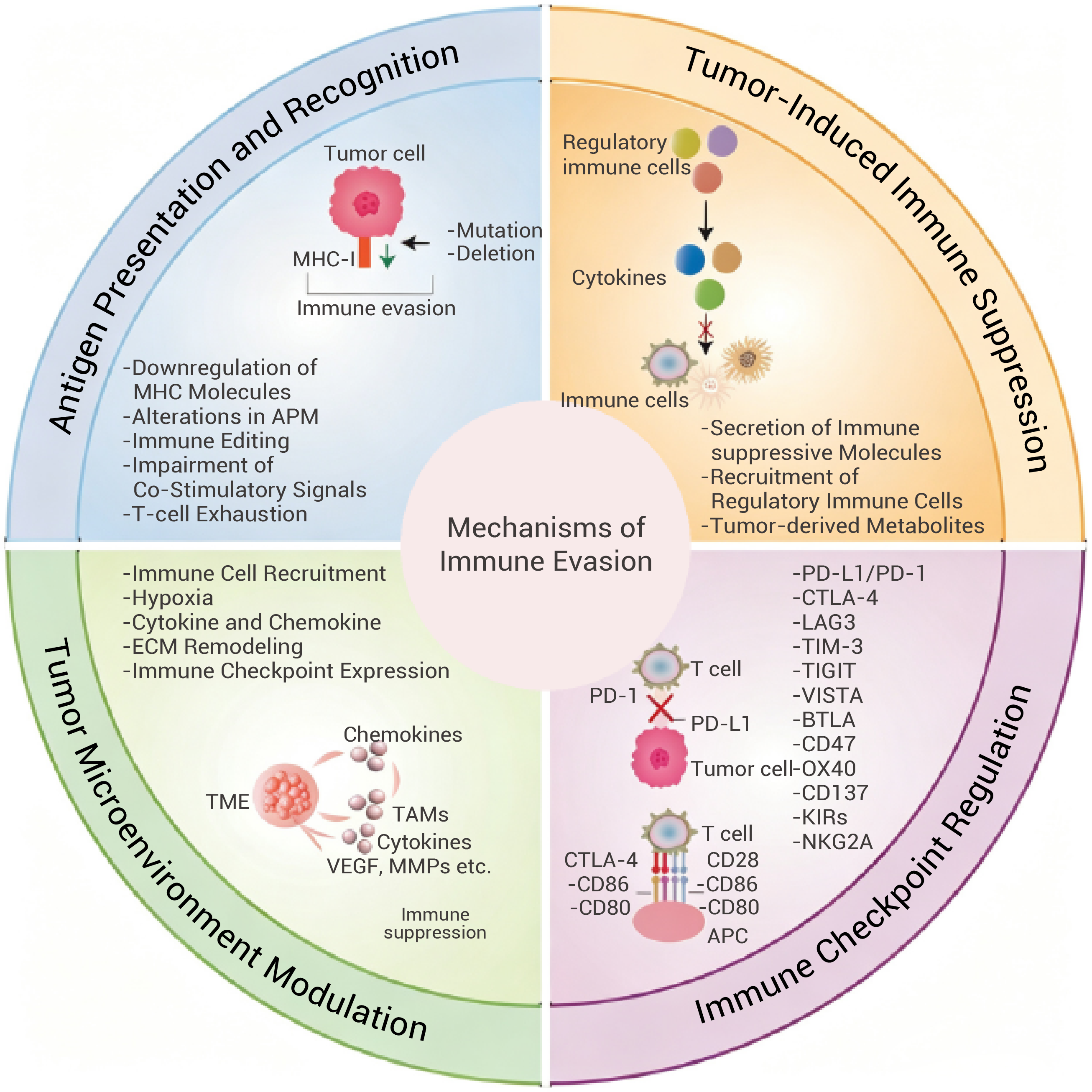
Figure 1. Mechanisms of immune evasion in cancer[3].
Effective anti-tumor immunity requires a coordinated sequence of events, including tumor antigen release and presentation, T-cell priming, immune-cell trafficking, tumor infiltration, target recognition and cytotoxic elimination[2]. Disruption of any individual step can compromise immune control, whereas simultaneous impairment of multiple processes can render single-agent checkpoint blockade insufficient.
Therefore, tumor immune escape should not be viewed as the activation of a single dominant inhibitory pathway, but rather as the establishment of a multilayered ecosystem involving tumor cells, immune populations, stromal components and metabolic constraints.

Figure 2. The CD58-CD2 axis is co-regulated with PD-L1 via CMTM6 and shapes anti-tumor immunity[5].
The first requirement for immune-mediated tumor elimination is the recognition of tumor-associated antigens. Tumors can reduce their immune visibility by losing immunogenic antigens, decreasing major histocompatibility complex (MHC) expression or disrupting antigen processing and presentation pathways. Under these conditions, PD-1 pathway inhibition may be insufficient because T cells remain unable to efficiently recognise malignant cells.
Beyond antigen presentation, effective T-cell activation also requires co-stimulatory signals. Interactions such as CD28-CD80/CD86 and CD2-CD58 reinforce immune synapse formation, T-cell expansion and cytotoxic activity[4]. Consequently, tumors can escape immune attack not only by increasing inhibitory signals, but also by disrupting programmes required for immune activation.
Loss of CD58 provides a representative example. CD58, the ligand for CD2, contributes to stable interactions between tumor cells and T cells[5]. In several malignancies, including lymphomas, CD58 inactivation through mutation or deletion reduces immune-cell engagement and compromises anti-tumor responses. CD58 loss has been associated with reduced persistence of CAR-T cell responses and may also influence sensitivity to immune checkpoint blockade and TCR-based therapies[6].
These findings highlight that immune escape is not always achieved through increased expression of inhibitory checkpoints. In some cases, tumors eliminate the signals required for immune activation itself. When antigen recognition and co-stimulation are impaired, removal of a single inhibitory checkpoint may not be sufficient to restore T-cell function.
Immune recognition alone does not guarantee tumor elimination. Effector T cells must migrate through abnormal vasculature and tumor stroma before reaching malignant cells. However, many solid tumors develop physical and molecular barriers that prevent effective immune infiltration, resulting in an immune-excluded phenotype[7].
Transforming growth factor-β (TGF-β) is a central regulator of this process. It promotes fibroblast activation, extracellular matrix deposition and stromal remodelling, while simultaneously limiting cytotoxic lymphocyte recruitment and inducing inhibitory immune programmes[3]. Through these effects, TGF-β influences not only checkpoint expression but also the spatial distribution and functional state of immune cells.
Checkpoint regulation also extends beyond direct interactions between tumor cells and lymphocytes. Tumor-associated endothelial cells can express PD-L1 and restrict cytotoxic T-cell entry across the vascular barrier, creating an "immune checkpoint at the gate"[8]. Similarly, PD-L1 expression by cancer-associated fibroblasts and other stromal populations may contribute to immune exclusion.
This concept helps explain why some patients with limited PD-L1 expression on tumor cells can still respond to PD-1/PD-L1 blockade: therapeutic activity may involve checkpoint regulation across the broader tumor ecosystem rather than solely tumor cell–T-cell interactions.
Therefore, checkpoint evaluation should move beyond asking whether tumor cells express PD-L1 and instead consider which cells express checkpoint molecules, where these cells are located and when these interactions occur during immune responses.
Even after entering tumors, T cells may fail to eliminate malignant cells. Persistent antigen exposure, inflammatory signals and metabolic stress can drive tumor-infiltrating lymphocytes towards dysfunctional or exhausted states, characterized by the expression of multiple inhibitory receptors including PD-1, TIM-3, LAG-3 and TIGIT[1].
The tumor microenvironment further reinforces this state through regulatory T cells, tumor-associated macrophages and myeloid-derived suppressor cells. These populations suppress immune activity through inhibitory cytokines, checkpoint ligands, metabolic enzymes and immunoregulatory pathways. For example, IL-10 can promote B7-H4 expression on macrophages and enhance inhibitory receptor expression on CD8+ T cells, whereas TGF-β can drive coordinated suppressive programmes[9].
Tumor metabolism represents another major layer of immune regulation. Rapidly proliferating tumor cells consume glucose, amino acids and oxygen, creating nutrient limitation and metabolic stress for immune cells. Lactate accumulation and extracellular acidification impair T-cell and NK-cell activity and can promote PD-L1 induction through inflammatory pathways[10]. Additional metabolites, including kynurenine, extracellular adenosine, prostaglandin E2 (PGE2) and reactive oxygen species, further shape suppressive immune states.
Thus, tumor-infiltrating T cells may simultaneously experience metabolic restriction, suppressive cellular interactions and broad checkpoint engagement. The presence of immune cells within tumors therefore does not necessarily indicate effective anti-tumor immunity.

Figure 3. Overview of the regulatory mechanisms of PD-L1 expression[11].
Historically, immune checkpoint research has focused on whether a specific ligand or receptor is expressed and whether blocking this interaction can restore T-cell activity. Increasing evidence, however, indicates that checkpoint expression represents the endpoint of a complex regulatory system rather than an isolated molecular event.
A single tumor may achieve PD-L1 upregulation through diverse mechanisms, including genomic alterations, inflammatory signalling, oncogenic pathway activation, enhanced mRNA stability or impaired protein degradation[11]. These distinct regulatory states may differ substantially in biological function, persistence and therapeutic sensitivity.
Checkpoint abundance alone therefore does not fully define immune resistance. Understanding how checkpoints are induced, maintained and spatially organized is essential for predicting therapeutic response.
Tumors can establish immune resistance through genetic alterations that directly enhance checkpoint activity or disrupt immune activation pathways.
In classical Hodgkin's lymphoma and primary mediastinal large B-cell lymphoma, amplification of the 9p24.1 region containing CD274 and PDCD1LG2 drives increased PD-L1 and PD-L2 expression. Because this region also includes JAK2, amplification can further enhance checkpoint transcription through JAK-STAT signalling, creating a tumor-intrinsic immune escape programme[12].
Structural alterations affecting regulatory regions provide another mechanism. Disruption of the CD274 3' untranslated region (3'-UTR) can remove regulatory elements and microRNA-binding sites that normally restrict PD-L1 mRNA stability or translation[13]. Such alterations can enable persistent PD-L1 expression independently of external immune stimulation.
Compared with genetic alterations, epigenetic regulation provides a more flexible mechanism for tumor adaptation. DNA methylation, histone modifications and chromatin remodelling collectively determine whether immune-related genes remain transcriptionally accessible.
Hypomethylation of inhibitory checkpoint loci, including CD274, CD276 and TIGIT, can promote sustained expression, whereas epigenetic silencing of co-stimulatory genes such as CD28, CD80 and CD86 may reduce immune activation capacity[14].
Chromatin regulators, including SWI/SNF complexes, ARID1A, BRD4, DNMTs, HDACs, EZH2 and LSD1, influence checkpoint programmes by altering chromatin accessibility and transcription factor recruitment[15].
These mechanisms suggest that immune escape can acquire a form of molecular memory: even after the initial inflammatory stimulus subsides, epigenetic programmes may maintain an immunosuppressive state. At the same time, the reversibility of epigenetic regulation provides an opportunity to restore tumor immunogenicity through targeted chromatin modulation.
Immune checkpoint regulation is closely integrated with pathways that control tumor growth, survival and metabolic adaptation. Many oncogenic programmes simultaneously promote malignant progression and immune resistance.
Major signalling pathways, including RAS-RAF-MEK-ERK, PI3K-AKT-mTOR, JAK-STAT and NF-κB, regulate checkpoint expression at multiple levels, from transcription to translation and protein stability. For example, MYC can directly promote expression of PD-L1 and CD47, enabling tumor cells to suppress both T-cell-mediated killing and macrophage-mediated phagocytosis. KRAS-associated signalling can regulate a broader immune escape network involving PD-L1, CD47, B7-H3, FGL1 and CD155.
These observations expand the role of oncogenic drivers beyond tumor proliferation. Genetic alterations that determine how aggressively a cancer grows may also influence how effectively it evades immune surveillance. Accordingly, targeting oncogenic pathways may influence tumor immunity by weakening mechanisms that support immune escape.
Signals from the tumor microenvironment further integrate into this regulatory network. Among them, interferon-γ (IFN-γ) represents one of the best-characterized examples of adaptive immune resistance. Produced by activated T cells and NK cells, IFN-γ promotes antigen presentation and immune activation. However, it also activates the JAK-STAT1-IRF1 pathway, inducing PD-L1 expression in tumor and myeloid cells. Thus, elevated PD-L1 may indicate not a complete absence of immunity, but rather an active response to immune pressure. This creates a paradoxical feedback loop in which stronger immune attack can simultaneously trigger stronger inhibitory signalling. Such adaptive resistance represents a fundamental principle underlying dynamic checkpoint regulation.
Additional microenvironmental factors, including TGF-β, IL-10, hypoxia, lactate, kynurenine and PGE2, further remodel checkpoint activity. Hypoxia-driven HIF signalling can enhance the expression of PD-L1, CD47 and VISTA; kynurenine activates the aryl hydrocarbon receptor (AhR) pathway; and PGE2 can influence tumor cells, myeloid populations and lymphocytes simultaneously.
Checkpoint molecules therefore function as integration nodes connecting tumor genotype, inflammatory signalling, metabolism and cellular composition. Blocking one checkpoint may disrupt a single inhibitory pathway, but alternative mechanisms can compensate and restore immune suppression.
Checkpoint regulation extends beyond transcription. Tumors can rapidly alter surface checkpoint abundance through RNA processing, translation control and protein homeostasis.
Alternative splicing can generate membrane-bound, truncated or soluble checkpoint isoforms with distinct biological functions. These variants may retain inhibitory activity, act as decoys or alter interactions with therapeutic antibodies and endogenous ligands. PD-1, PD-L1, CTLA-4 and LAG-3 are all subject to regulation through RNA processing or proteolytic shedding.
Non-coding RNAs, including microRNAs, long non-coding RNAs and circular RNAs, regulate checkpoint expression by controlling mRNA degradation, translation efficiency or competitive RNA interactions. RNA modifications such as N6-methyladenosine (m6A) further influence transcript stability and translation.
Under metabolic stress or therapeutic pressure, tumors can selectively maintain immune escape proteins through translational programmes such as mTOR-eIF4F signalling.
Following protein synthesis, checkpoint molecules undergo extensive post-translational regulation, including glycosylation, phosphorylation, ubiquitination, deubiquitination, palmitoylation, trafficking, internalization and recycling. These processes determine protein stability, localization and functional activity.

Figure 4. Transcriptional regulation of PD-L1 expression[16].
A key example is CMTM6, which binds PD-L1 at the plasma membrane and within recycling endosomes, limiting lysosomal degradation and promoting PD-L1 recycling to the cell surface. Interestingly, CMTM6 can also regulate the stability of the co-stimulatory ligand CD58, suggesting that a single protein-homeostasis regulator may simultaneously influence inhibitory and activating immune interactions.
PD-L1 glycosylation enhances protein stability and affects PD-1 binding, whereas ubiquitination and deubiquitination regulate its degradation[16]. Membrane trafficking and lipid modification further regulate surface availability.
Therefore, measuring CD274 mRNA alone is insufficient to define PD-L1 functional status. A tumor with unchanged PD-L1 transcription may still maintain immune suppression through prolonged protein stability.
Future checkpoint profiling will likely need to assess not only whether a checkpoint is expressed, but also how its expression is maintained, where the protein is located and whether it remains functionally active.

Figure 5. Clinical trajectories of adaptive resistance and the seven challenges in the immunotherapy response cycle[17].
The success of PD-1/PD-L1 and CTLA-4 blockade established immune checkpoints as clinically actionable regulators of anti-tumor immunity. However, limited response durability and the emergence of resistance have shifted the field from targeting individual inhibitory receptors towards understanding and modulating broader immune-regulatory networks.
Future immunotherapy strategies will likely depend on identifying the dominant resistance mechanisms within each tumor context and selecting combinations that address specific immune escape programmes rather than applying universal checkpoint combinations.
PD-1/PD-L1 and CTLA-4 remain the foundation of immune checkpoint therapy, but they regulate distinct stages of immune activation. CTLA-4 primarily limits early T-cell priming by competing with CD28 for co-stimulatory ligands, whereas PD-1 predominantly regulates effector T-cell function within peripheral tissues and the tumor microenvironment. Their complementary biological roles provide a mechanistic rationale for combined blockade.
Beyond these established targets, several emerging checkpoints have entered clinical development, including LAG-3, TIM-3, TIGIT, VISTA, B7-H3, B7-H4, BTLA-HVEM, PVRIG-CD112, CD47-SIRPα and CD73-adenosine pathways. These molecules regulate diverse interactions among T cells, NK cells, macrophages, myeloid suppressor populations and tumor cells.
Among these emerging targets, LAG-3 has achieved the most advanced clinical translation following PD-1 and CTLA-4. LAG-3 interacts with multiple ligands, including MHC class II and fibrinogen-like protein 1 (FGL1), and is regulated through proteolytic cleavage and soluble isoforms. Its frequent co-expression with PD-1 on dysfunctional T cells provides a mechanistic basis for combined blockade.
However, emerging checkpoints are unlikely to simply replace PD-1 as universally applicable therapeutic platforms. Unlike PD-L1, which is often induced as part of adaptive resistance within the tumour microenvironment, many other checkpoint pathways involve ligands that are broadly expressed in normal tissues, potentially narrowing their therapeutic windows.
Moreover, checkpoint expression does not necessarily indicate functional dependency. Some molecules may represent markers of terminal T-cell dysfunction rather than dominant drivers of immune suppression. Blocking such pathways may therefore have limited capacity to reverse established dysfunctional states.
Consequently, future checkpoint combinations are expected to become increasingly context-dependent, guided by tumor type, immune-cell state, spatial organisation and underlying resistance mechanisms.
An alternative therapeutic strategy is to target the regulatory mechanisms that establish checkpoint expression rather than directly blocking checkpoint molecules themselves. This approach offers several potential advantages. First, upstream regulators often control both tumor progression and immune escape. Targeting these nodes may simultaneously impair malignant fitness and weaken immunosuppressive protection. Second, interfering with checkpoint-inducing pathways may limit adaptive resistance before inhibitory programmes become fully established. Third, restoring endogenous immune activation programmes, such as co-stimulatory signalling, may provide a more physiologically balanced strategy than systemic immune activation through agonistic antibodies.
Oncogenic signalling pathways represent one example of this approach. KRAS and its downstream networks regulate multiple immunemodulatory molecules and microenvironmental features[18]. Combining KRAS inhibition with immune checkpoint blockade may therefore exert dual effects by directly suppressing tumor growth while reshaping tumor immune states. However, therapeutic benefit is likely to be influenced by mutation subtype, tumor lineage, treatment history and baseline immune context.
Epigenetic regulators represent another promising but complex class of immune modulators. DNMT and HDAC inhibition can restore the expression of antigen presentation machinery, CD80/CD86 and other immune activation programmes, thereby increasing tumor immunogenicity. However, epigenetic therapies may also induce PD-L1 expression, highlighting the need for rational combinations rather than indiscriminate immune activation.
TGF-β inhibition represents a strategy aimed at overcoming immune exclusion. By reducing stromal barriers, enhancing T-cell infiltration and limiting the induction of inhibitory receptors, TGF-β blockade may improve responses to PD-1/PD-L1 inhibition. Nevertheless, inconsistent results across models and clinical studies suggest that benefit may be restricted to tumors with strong TGF-β activity and immune-excluded phenotypes.
Additional pathways, including COX-PGE2 signalling, purinergic metabolism, IDO-kynurenine-AhR signalling, hypoxia pathways and tumor metabolic regulation, remain attractive combination targets. However, their broad physiological functions create challenges in achieving sufficient tumor selectivity and limiting toxicity.
Thus, the most promising upstream regulators are likely to share three characteristics:
· They regulate multiple complementary immune escape mechanisms.
· They exhibit tumor or microenvironmental selectivity.
· Pharmacological tools are available to support mechanistic and translational investigation.
Targets such as mutant KRAS, IDH1/2, DNMTs, HDACs, TGF-β and PGE2 signalling remain actively investigated because they fulfil these requirements to varying degrees.
A central challenge in immunotherapy is determining which patients require which immune intervention and at what stage of treatment. Current biomarkers, including PD-L1 immunohistochemistry, tumor mutational burden (TMB) and microsatellite instability (MSI), have improved patient selection but remain incomplete.
PD-L1 expression shows substantial temporal and spatial heterogeneity, meaning that a single biopsy captures only a limited snapshot of tumor immune status. High TMB does not necessarily generate immunogenic neoantigens, and although MSI-associated biomarkers can predict response in selected cancers, they apply to a relatively limited patient population.
Future biomarkers may need to assess not simply baseline checkpoint abundance, but rather the capacity of tumors to induce and maintain immune resistance. For example, a tumor with low baseline PD-L1 but intact IFN-γ-JAK-STAT signalling may rapidly induce PD-L1 following immune activation. Conversely, a tumor with high PD-L1 expression driven by genomic amplification, regulatory-region disruption or enhanced protein stability may represent a biologically distinct state requiring different therapeutic strategies.
Certain genetic alterations provide mechanistic insights into immune resistance. The 9p24.1 amplification observed in classical Hodgkin lymphoma promotes PD-L1 and PD-L2 expression and contributes to sensitivity to PD-1 blockade. CD274 3'-UTR disruption may sustain PD-L1 expression, whereas CD58 loss may indicate impaired co-stimulatory capacity and reduced sensitivity to T-cell-based therapies. However, most of these alterations remain limited to specific tumor contexts and require prospective validation before routine clinical application.
Beyond genomic markers, integrated immune profiling can provide a broader view of tumor states. IFN-γ-related gene signatures reflect ongoing immune activation, whereas exhaustion-associated transcriptional programmes and co-expression patterns of PD-1, TIM-3, LAG-3 and TIGIT may distinguish different forms of T-cell dysfunction.
Spatial organisation is equally important. PD-L1 expression on tumor cells, myeloid populations, endothelial cells or fibroblasts may represent fundamentally different immune escape mechanisms. Identical overall expression levels may have distinct biological consequences depending on whether checkpoints are concentrated within tumor cores, invasive margins or vascular niches.
Emerging technologies, including single-cell sequencing, spatial transcriptomics, spatial proteomics and multiplex imaging, are enabling increasingly detailed maps of checkpoint networks across cellular compartments. Functional genomic screens can further identify regulators controlling checkpoint induction, protein stability and therapeutic resistance.
Combined with circulating tumor DNA, peripheral immune monitoring, serial biopsies and longitudinal imaging, these approaches may transform immunotherapy from a one-time biomarker-based decision into an adaptive treatment strategy.
Ultimately, next-generation biomarkers should not simply answer "how much checkpoint is present?" but rather:
· Which immune escape programme is the tumor currently using?
· How will this programme evolve under treatment pressure?
· Is immune dysfunction reversible?
· Which therapeutic intervention can best disrupt the dominant resistance mechanism at this stage?
Immune checkpoint blockade has shown that tumor-mediated immune suppression can be reversed, but tumors rarely rely on a single inhibitory pathway. Instead, they establish dynamic immune escape networks involving antigen presentation, co-stimulatory signalling, immune-cell infiltration, stromal and metabolic regulation, and checkpoint control at genetic, epigenetic, RNA and protein levels.
Future immunotherapy will therefore need to move beyond isolated checkpoint blockade towards mechanism-guided combinations and dynamic patient stratification. The key challenge is to identify which immune escape pathways dominate in each tumor context, how they evolve under treatment pressure and how they can be effectively disrupted.
| Cat. No. | Product Name | Description |
|---|---|---|
| HY-16438 | RRx-001 | A dual small molecule checkpoint inhibitor by downregulating CD47 and SIRP-α. |
| HY-102011 | BMS-1166 | A potent PD-1/PD-L1 immune checkpoint inhibitor. |
| HY-101093 | CA-170 | An orally delivered dual inhibitor of VISTA and PD-L1. |
| HY-120635 | BMS-1001 (hydrochloride) | An orally active human PD-L1/PD-1 immune checkpoint inhibitor. |
| HY-150879 | BMS-37 | A PD-1/PD-L1 immune checkpoint inhibitor with an IC50 towards the PD-L1/PD-1 complex in the range of 18 to 200 nM. |
| HY-138407 | Evixapodlin | A human PD-1/PD-L1 protein/protein interaction inhibitor with an IC50 of 0.213 nM. |
| HY-134884 | INCB086550 | A potent, oral, small-molecule PD-L1 inhibitor with IC50s value of 3.1, 4.9 and 1.9 nM for human, cynomolgus, and rat, respectively. |
| HY-145388 | AU-15330 | A proteolysis-targeting chimera (PROTAC) degrader of the SWI/SNF ATPase subunits, SMARCA2 and SMARCA4. |
| HY-114268 | BRD-K98645985 | A BAF (mammalian SWI/SNF) transcriptional repression inhibitor with an EC50 of ~2.37 μM. |
| HY-133897 | (Rac)-Indoximod | An indoleamine 2,3-dioxygenase (IDO) inhibitor. |
| HY-100438 | Hydronidone | An inhibitor of phosphodiesterase-4 (PDE4) and cyclo-oxygenase (COX) as well as TGF-β. |
Note: MCE can provide products for research use only. We do not sell to patients.
 LinkedIn
 Researchgate
 Instagram
 X
X Facebook
 WhatsApp
 Reddit
 Mail
Mail- [1]. Eris C, et al. Nat Rev Cancer. 2026 Aug;26(8):560-585.
- [2]. Chen DS, Mellman I. Immunity. 2013 Jul 25;39(1):1-10. [Content Brief]
- [3]. Tufail M, et al. Signal Transduct Target Ther. 2025 Jul 31;10(1):227.
- [4]. Robbie G, et al. Blood. 2020 Nov(136): 53-54.
- [5]. Ho P, et al. Cancer Cell. 2023 Jul 10;41(7):1207-1221.e12.
- [6]. Yan X, et al. Blood Adv. 2022 Nov 22;6(22):5844-5856.
- [7]. Fang J, et al. Cell Death Dis. 2023 Sep 4;14(9):586.
- [8]. Piao W, et al. Nat Commun. 2022 Apr 21;13(1):2176.
- [9]. O'Malley G, et al. Cancer Immunol Res. 2018 Nov;6(11):1426-1441.
- [10]. Apostolova P, et al. Trends Immunol. 2022 Dec;43(12):969-977.
- [11]. Sun C, et al. Immunity. 2018 Mar 20;48(3):434-452.
- [12]. Green MR, et al. Blood. 2010 Oct 28;116(17):3268-77.
- [13]. Kataoka K, et al. Nature. 2016 Jun 16;534(7607):402-6. [Content Brief]
- [14]. Saadi W, et al. Cancers (Basel). 2022 Dec 28;15(1):184.
- [15]. Perrier A, et al. Biomolecules. 2020 Jul 16;10(7):1061.
- [16]. Yamaguchi H, et al. Nat Rev Clin Oncol. 2022 May;19(5):287-305. [Content Brief]
- [17]. Yang K, et al. Cell Mol Immunol. 2026 Jul;23(7):743-772.
- [18]. Parikh K, et al. J Hematol Oncol. 2022 Oct 25;15(1):152.

