



Apoptosis and Necrosis Assay Kit
Based on 5 publication(s) in Google Scholar
MCE Apoptosis and Necrosis Assay Kit provides a rapid and convenient method to detect cell apoptosis and necrosis.
-
Storage :
4°C, 6 months, -20°C, 1 year. Protect from light.


To place orders, for customer services and technical support, please contact: MedChemExpress USA
Tel: 609-228-6898 E-mail: [email protected] [email protected]







Description & Advantages
MCE Apoptosis and Necrosis Assay Kit provides a rapid and convenient method to detect cell apoptosis and necrosis. This kit contains two ready-to-use dyes bound to DNA. Hoechst 33342, a type of blue fluorescence dye (excitation/emission maxima ≈ 352/461 nm when bound to DNA), stains the condensed chromatin in apoptotic cells more brightly than the chromatin in normal cells. Propidium Iodide (PI) is a cell-membrane impermeable dye with characteristic excitation maximum at 535 nm and emission maximum at 617 nm which intercalates with nucleic acids. The simultaneous use of these two dyes makes it possible to distinguish normal, apoptotic, and dead cell populations by flow cytometer or fluorescence microscope. Normal cells show weak blue fluorescence + weak red fluorescence, apoptotic cells show strong blue fluorescence + weak red fluorescence, necrotic cells show strong blue fluorescence + strong red fluorescence.
This kit takes only 20-30 minutes to complete one-step staining and there is no need for dilution or preparation of other solutions when detected by flow cytometer. Each kit contains enough reagents for 100 samples.


Protocol
Suspension cells or flow cytometry
1. Collect approximately 1 × 10⁵-1 × 10⁶ cells and wash once with PBS.
2. Resuspend cells in 1 mL Cell Stain Buffer.
3. Add 5 μL Hoechst 33342 Stain and 5 μL PI Stain and mix gently.
4. Incubate protected from light for 20-30 min.
5. Analyze directly by flow cytometry without additional staining-solution preparation. Detect Hoechst 33342 using a UV/near-405 nm excitation channel and PI using a standard red-fluorescence channel.
Adherent cells / fluorescence microscopy
1. After treatment, wash cells gently and add sufficient Cell Stain Buffer to cover the sample.
2. Add 5 μL Hoechst 33342 Stain and 5 μL PI Stain per 1 mL Cell Stain Buffer and incubate protected from light for 20-30 min.
3. Observe by fluorescence microscopy. Normal cells show weak blue/weak red fluorescence, apoptotic cells strong blue/weak red fluorescence, and necrotic cells strong blue/strong red fluorescence.


Publications
-
Journal Impact Factor
-
Most Recent
-
Pharmacol ResTransfer of miR-877-3p via extracellular vesicles derived from dental pulp stem cells attenuates neuronal apoptosis and facilitates early neurological functional recovery after cerebral ischemia-reperfusion injury through the Bclaf1/p53 signaling pathway. [Abstract]2024 Aug:206:107266. PMID: 38878918
-
Eur J Med ChemDiscovery of Sibiriline derivatives as novel receptor-interacting protein kinase 1 inhibitors. [Abstract]2023 Mar 15:250:115190. PMID: 36801518
-
FASEB JLipoxin A4 Regulates M2 Macrophage-Derived Exosomal miR-25-5p to Protect Cell Pyroptosis in Bronchopulmonary Dysplasia. [Abstract]2025 Nov 30;39(22):e71218. PMID: 41223004
Storage
4°C, 6 months, -20°C, 1 year.
Protect from light.


Components
| Components | HY-K1070-100T |
|---|---|
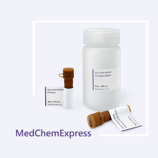
| Cell Stain Buffer | 100 mL |
| Hoechst 33342 Stain | 0.5 mL |
| PI Stain | 0.5 mL |


 Please enter the correct Lot Number
