



Protein A Plus Magnetic Agarose Beads
Based on 1 publication(s) in Google Scholar
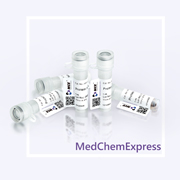
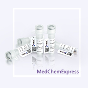
MCE Protein A Plus Magnetic Agarose Beads can be used for the detection and purification of IgG from serum, ascites fluid, cell culture supernatant and other antibody samples.
-
Storage :
4℃, 2 years. Do not dry or freeze.


To place orders, for customer services and technical support, please contact: MedChemExpress USA
Tel: 609-228-6898 E-mail: [email protected] [email protected]






Description & Advantages
Protein A is a bacterial cell wall protein isolated from Staphylococcus aureus and binds to most mammalian IgGs mainly through Fc regions. Native Protein A contains 5 IgG binding domains and many other domains with unknown functions. The latter have been eliminated from recombinant Protein A to reduce nonspecific binding.
MCE Protein A Plus Magnetic Agarose Beads is produced through the covalent coupling of recombinant protein A with magnetic agarose beads. It has high loading capacity, exceptional specificity, and stability, can be used for the detection and purification of IgG from serum, ascites fluid, cell culture supernatant and other antibody samples.
The specifications of the product correspond to the actual beads volume, with the beads content of 50%.


Protocol
Recommended Buffers (Not Provided)
| Buffer | Composition |
|---|---|
| Binding/Wash Buffer | 20 mM Na2HPO4, 0.15 M NaCl, pH 7.0 |
| Elution Buffer | 0.1 M glycine, pH 3.0 |
| Neutralization Buffer | 1 M Tris-HCl, pH 8.5 |
| Storage Buffer | 1× PBS containing 20% ethanol |
Note: Prepare buffers with high-purity water and filter them through a 0.22 μm or 0.45 μm membrane.
Sample Preparation
Centrifuge or filter antibody-containing samples such as serum, ascites, or culture supernatant. Dilute or dialyze against binding/wash buffer when necessary to adjust ionic strength and pH.
Antibody Purification with Protein A Plus Magnetic Agarose Beads
1. Bead Pretreatment: Thoroughly resuspend the Protein A Plus Magnetic Agarose Beads, transfer the required volume into a tube, perform magnetic separation, discard the storage solution, and wash with 5 bead volumes of binding/wash buffer 2-3 times.
2. Antibody Binding: Add the prepared antibody sample and incubate on a rotator at room temperature for 1-2 h or at 4°C overnight. Perform magnetic separation and collect the flow-through.
3. Washing: Wash with 5-10 bead volumes of binding/wash buffer 3-5 times, until the OD280 of the wash supernatant approaches baseline.
4. Elution: Add 3-5 bead volumes of elution buffer, incubate at room temperature for 5-10 min, perform magnetic separation, and collect the supernatant. Immediately add neutralization buffer at 1/10 of the eluate volume. Repeat 2-3 times if necessary.
5. SDS-PAGE Analysis: Analyze the original sample, flow-through, wash, and elution fractions.
6. Regeneration and Storage: Wash thoroughly with binding/wash buffer and ddH2O, resuspend in storage buffer, and store at 2-8°C. Do not freeze or dry.


Publications
-
Journal Impact Factor
-
Most Recent
Storage
4℃, 2 years.
Do not dry or freeze.


Components
| Components | HY-K0242-1 mL | HY-K0242-5 mL | HY-K0242-10 mL |
|---|---|---|---|
| Protein A Plus Magnetic Agarose Beads | 1 mL | 5 mL | 10 mL |


 Please enter the correct Lot Number
