



IR-780
Based on 7 publication(s) in Google Scholar
IR-780 is a near-infrared fluorescent probe for in vivo imaging of tumor cells. IR-780 is transported into tumor cells via OATPs and ABCB10, with uptake dependent on glycolytic activity and plasma membrane potential. IR-780 preferentially accumulates in tumor cell mitochondria, including those of drug-resistant cancer cells, without chemical conjugation. IR-780 generates reactive oxygen species (ROS), induces hyperthermia and apoptosis, inhibits tumor growth and recurrence, and modulates HSP70 expression upon ultrasound or 808 nm laser exposure. IR-780 acts as a sonosensitizer, photodynamic and photothermal agent, and drug delivery carrier, with low acute imaging-dose toxicity and rapid vital organ clearance. IR-780 can be used for the research of cancer, such as breast cancer, lung cancer, and non-small cell lung cancer (NSCLC).
For research use only. We do not sell to patients.
- Purity : 99.94%
- CAS No.: 207399-07-3
- Formula: C36H44ClIN2
- Molecular Weight:667.11
-
Storage:
-20°C, sealed storage, away from moisture and light
* In solvent : -80°C, 6 months; -20°C, 1 month (sealed storage, away from moisture and light)

To place orders, for customer services and technical support, please contact: MedChemExpress USA
Tel: 609-228-6898 E-mail: [email protected] [email protected]


-
 Biological Activity
-
Chemical Information
-
Solvent & Solubility
- Protocols
- Purity & Documentation
- References
-
 Help & FAQs


-
HY-139109
IR-783Anticancer Dye -
HY-D1711
MHI-148Tumor Targeted Dye -
HY-136886
IR-820Dye -
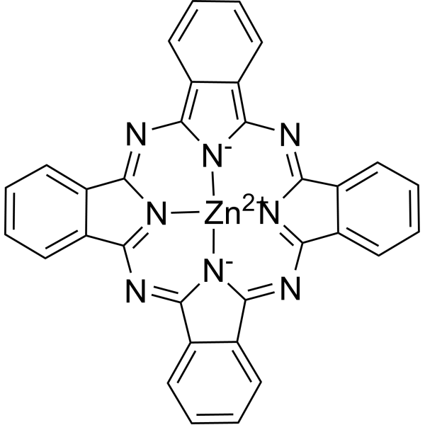
HY-19204
Zinc phthalocyaninePhotooxidant -
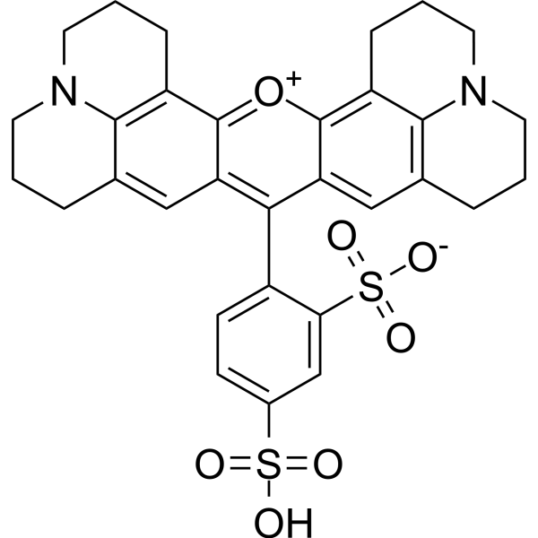
HY-101878
Texas RedRed Fluorescent Dye -
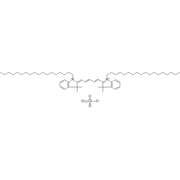
HY-D1028
DiD perchlorateDye -
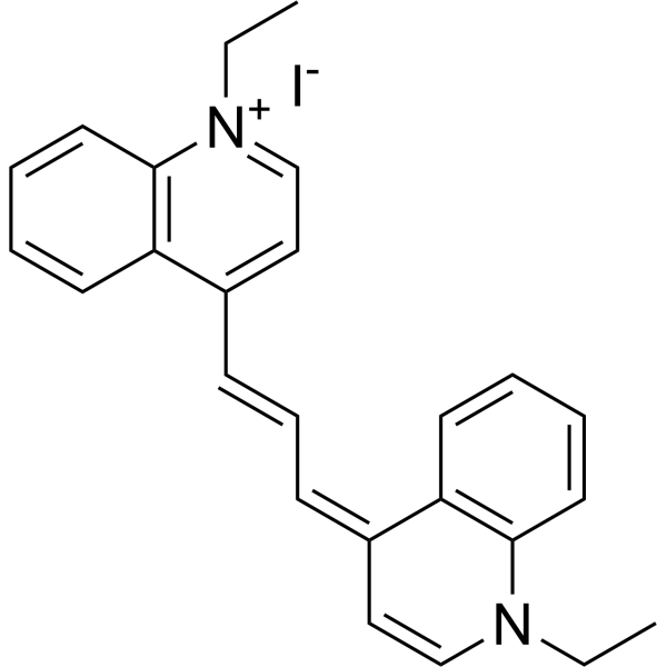
HY-W248587
CryptocyanineFluorescent Dye -
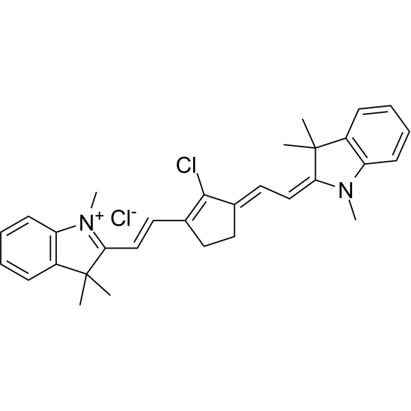
HY-D1504
IR-797 chlorideNIR Dye -
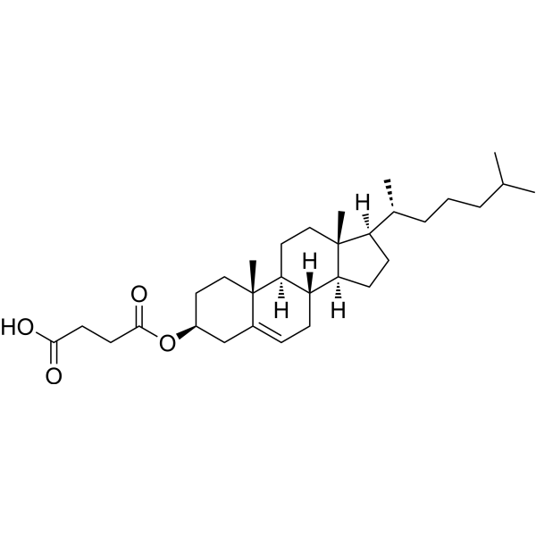
HY-W010800
Cholesteryl hemisuccinateTopoisomerase Inhibitor -
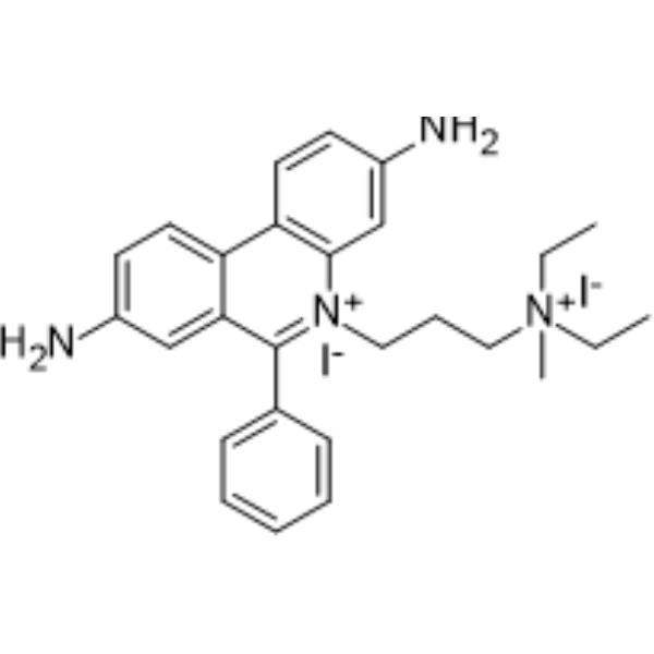
HY-D0815
Propidium IodideFluorochrome -
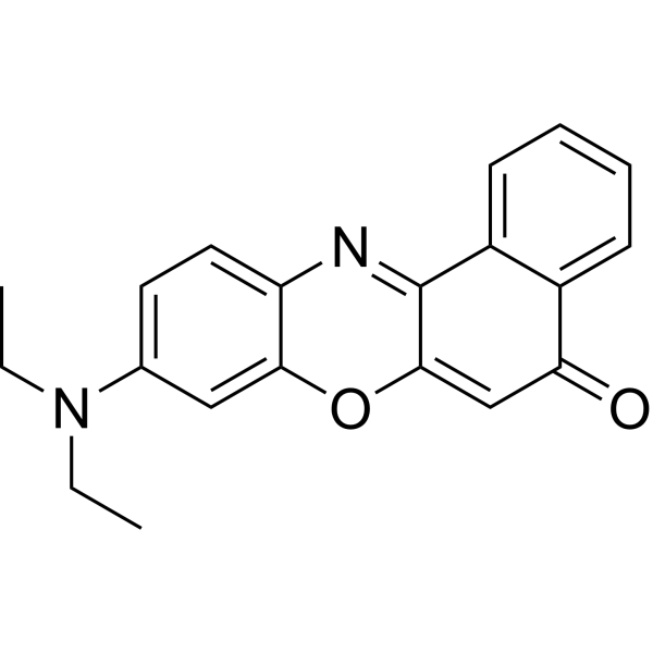
HY-D0718
Nile RedHydrophobic Fluorescent Stain -
HY-W040258
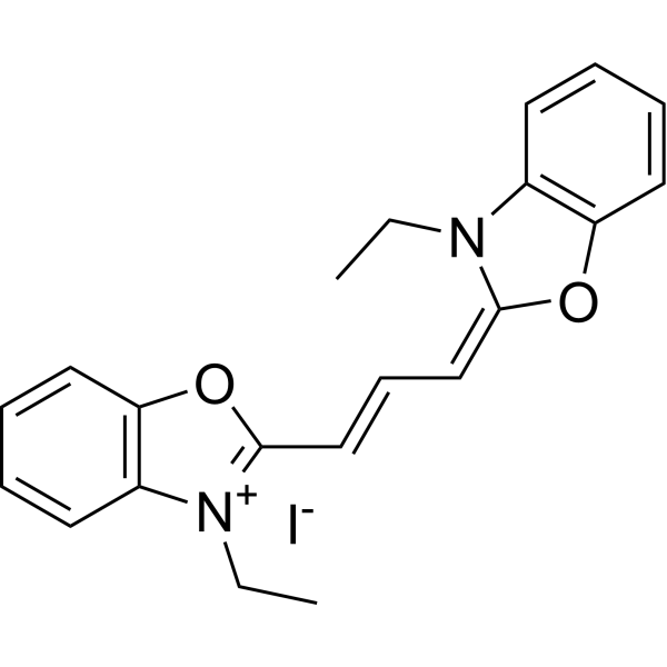
3,3'-Diethyloxacarbocyanine iodideMicroviscosity Fluorescent Probe
Publications Citing Use of MedChemExpress (MCE) IR-780
More- Adv Funct Mater. 2025 May 15.
- Nat Commun. 2025 Jul 15;16(1):6532. [Abstract]
- Cell Rep Med. 2026 Mar 17;7(3):102640. [Abstract]
- Int J Biol Macromol. 2026 Jul:370:152860. [Abstract]
- ACS Appl Mater Interfaces. 2025 Dec 31. [Abstract]
- Cancer Nanotechnol. 2025 Oct 21;16(1):1-20.
- Am J Cancer Res. 2023 Nov 15;13(11):5368-5381. [Abstract]
 Customer Validation & Images

-
In Vivo Imaging
-
In Vivo Imaging
-
Cell Proliferation/Viability Assay
-
Cell Imaging/Staining
-
Cell Migration/Invasion Assay


Biological Activity
Description
In Vitro
IR-780 (10 μM; 15 min at 37°C) preferentially accumulates in the mitochondria of human MCF-7, HeLa, and MG-63 tumor cells via organic anion transporter peptide-mediated uptake[1].
IR-780 acts as an SDT agent in 4T1 breast cancer cells, generating increased ROS upon US irradiation[2].
IR-780 selectively accumulates in the mitochondria of A549/DR drug-resistant lung cancer cells, inhibiting cell growth and promoting apoptosis by disrupting mitochondrial function[2].
IR-780 (4-16 μM; 1-3 h (flow cytometry); 1 h (confocal microscopy)) exhibited dose- and time-dependent uptake by 4T1 breast cancer cells, with higher concentrations and longer incubation times leading to greater uptake[3].
IR-780 (10 μM; 3 h (pre-incubation); 20 s US irradiation)-induced sonodynamic cell damage in 4T1 breast cancer cells is inhibited by histidine and SOD but not mannitol[3].
IR-780 (2.5-40 μM; 48-72 h) inhibits the viability of human cancer cell lines (A549, H460, HepG2, U251, MCF-7) with A549 cells being the most susceptible[4].
IR-780 (10 μM; 4 h) significantly inhibits the clone formation ability of A549/DR cells[4].
IR-780 (10 μM; 12 h) significantly inhibits the migration ability of A549/DR cells[4].
IR-780 (20 μM; 20 min) increases ROS production in A549/DR cells[4].
IR-780 (10-20 μM; 24 h) decreases the mitochondrial membrane potential of A549/DR cells, with significant reduction at 20 μM[4].
IR-780 (20 μM; 24 h) induces apoptosis in over 60% of A549/DR cells after 24 h[4].
IR-780 (4-16 μM; 20-40 s ultrasound irradiation) acted as a sonosensitizer to significantly reduce 4T1 murine breast cancer cell viability and induce apoptosis/necrosis in a dose- and ultrasound time-dependent manner, with higher concentrations and longer ultrasound exposure causing greater cytotoxicity[9].
IR-780 (5 μg/mL equivalent free IR-780; 10-120 min) loaded into heparin-folic acid nanoparticles exhibits time-dependent fluorescence activation in MCF-7 cells, with fluorescence intensity increasing significantly over 120 min of incubation, indicating cellular uptake and release of active IR-780[11].
MedChemExpress (MCE) has not independently confirmed the accuracy of these methods. They are for reference only.
-
Cell Line:A549, H460, HepG2, U251, MCF-7
-
Concentration:0, 2.5, 5, 10, 20, 40 μM
-
Incubation Time:48 h, 72 h
-
Result:Displayed anticancer activity across all tested cell lines; identified A549 cells as the most susceptible. Calculated IC50 values for each cell line at 48 h and 72 h.
-
Cell Line:A549, A549/DR
-
Concentration:0, 2.5, 5, 10, 15, 20 μM
-
Incubation Time:48 h
-
Result:Showed concentration-dependent cytotoxicity on A549 cells, eliminating nearly all cells at 20 μM. Exhibited similar cytotoxicity to both A549 and A549/DR cells across concentrations, and effectively suppressed A549/DR cells, nearly killing all at 20 μM.
-
Cell Line:A549/DR
-
Concentration:0, 10, 20 μM
-
Incubation Time:24 h
-
Result:Induced apoptosis in more than 60% of A549/DR cells after incubation with 20 μM IR-780 for 24 h, compared to 7% in the control group.
In Vivo
IR-780 iodide (0.2 mg/kg; i.v.; single dose) preferentially accumulates in chemically induced lung tumors in immune-intact C57BL/6 mice, enabling clear tumor visualization via near-infrared fluorescence imaging[1].
IR-780 (80 μg; intratumoral injection; followed by US irradiation at 2 W/cm2 for 4 minutes) as a sonosensitizer combined with US significantly inhibits breast tumor growth and increases tumor cell apoptosis in BALB/c mice[3].
IR-780 (5.0 mg/kg; i.p.; every two days; 4-5 times) exhibits potent tumoricidal activity and inhibits tumor recurrence in a mouse LLC xenograft model, with a tumor formation rate of 40% in secondary mice[4].
IR-780 (5.74 μmol/kg; i.v.; every three days; 15 days; 808 nm laser irradiation) exerts tumor growth inhibition in Hep1-6 tumor-bearing BALB/c mice[6].
IR-780 (80 μg; intratumoral injection; followed by ultrasound irradiation at 2 W/cm2 for 4 minutes) combined with US irradiation induces significant breast tumor growth inhibition in BALB/c mice, reducing mean tumor volume to 40.7 mm3 at day 30 and achieving a 50.9 % tumor cell apoptotic index, with no significant body weight changes[9].
IR-780 (0.7 mg/kg; i.v.; single dose) accumulates in MCF-7 breast cancer xenografts, enabling NIR fluorescence imaging, though with lower tumor retention than IR-780 delivered via HF-IR-780 NPs[11].
MedChemExpress (MCE) has not independently confirmed the accuracy of these methods. They are for reference only.
-
Animal Model:Athymic nude mice (5-6 weeks old, 20-25 g)[1]
-
Dosage:0.2 mg/kg
-
Administration:i.v.; single dose
-
Result:Visualized intense, specific near-infrared fluorescence signals from MCF-7, HeLa, and MG-63 tumor xenografts with low background interference at 48 hours post-injection. Detected fluorescence signal persisting in tumors for at least 20 days, with contrast index (CI) values ranging from 4 to 14 and an average CI of 7 on day 20.
-
Animal Model:BALB/c (female, 6 to 10 week old) injected with 4T1 breast carcinoma cells.[3]
-
Dosage:80 μg
-
Administration:intratumoral injection; followed by US irradiation at 2 W/cm² for 4 minutes
-
Result:Achieved mean tumor size of 40.7 mm3 at day 30; showed significant tumor growth inhibition; induced 50.9% apoptotic tumor cells (TUNEL-positive); caused significantly increased degree of cell necrosis.
-
Animal Model:C57 BL/6 (male, 8 weeks old, 20-25 g)[4]
-
Dosage:5.0 mg/kg
-
Administration:i.p.; every two days; 5 times; 4 times
-
Result:Significantly suppressed tumor growth compared to control group and ADM group.
Reduced tumor weight compared to control, CTX, and ADM groups.
Produced much smaller tumors than ADM group at day 16.
Induced a 40% tumor formation rate in secondary mice from treated xenograft cells.
-
Animal Model:BALB/c (male, 6-8 weeks, 18-22 g, Hep1-6 tumor model)[6]
-
Dosage:5.74 μmol/kg
-
Administration:i.v.; every three days; 15 days; 808 nm NIR laser (1 W/cm²) irradiation for 5 min at 6 h post-injection
-
Result:Exerted tumor growth inhibition (a small number of trypan blue-stained cells observed in in vitro correlate studies), though less potent than the IR780-LA/CPT-ss-CPT NPs + laser group
-
Animal Model:BALB/c (female, 6-8 weeks old; 4T1 breast carcinoma xenograft)[9]
-
Dosage:80 μg
-
Administration:intratumoral injection; followed by ultrasound irradiation at 2 W/cm² for 4 minutes
-
Result:Achieved mean tumor volume of 40.7 mm3 at day 30. Induced 50.9% apoptotic tumor cells via TUNEL staining. Observed significant tumor cell necrosis via histological analysis. Detected no significant changes in mouse body weight during the study.
- RNA extraction experimental
- Annexin V plus membrane-impermeant dye apoptosis staining
- ROS/oxidative-stress fluorescent staining
- Patient-Derived Orthotopic Xenograft (PDOX)
- Breast Cancer Modeling
- Detection of Apoptosis
- TUNEL staining for apoptotic DNA fragmentation
- Fluorescent plasma-membrane potential dye assay
- Patient-Derived Xenograft (PDX)
- Protocol for Pharmacokinetic Study
- Apoptosis Solutions
- Bioluminescent/Fluorescent Imaging Xenograft
- Subcutaneous Cell-Line-Derived Xenograft
- Orthotopic Cell-Line Xenograft


Chemical Information
-
CAS No. 207399-07-3
-
Appearance Solid
-
Molecular Weight 667.11
-
Formula C36H44ClIN2
-
Color Light yellow to green yellow
-
SMILES
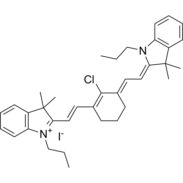
CCC[N+]1=C(/C=C/C2=C(Cl)/C(CCC2)=C/C=C3N(CCC)C4=C(C=CC=C4)C\3(C)C)C(C)(C)C5=C1C=CC=C5.[I-]
-
Shipping
Room temperature in continental US; may vary elsewhere.
-
Storage
-20°C, sealed storage, away from moisture and light
* In solvent : -80°C, 6 months; -20°C, 1 month (sealed storage, away from moisture and light)


Publications (7)
-
Journal Impact Factor
-
Most Recent
-
-
Nat Commun
Living therapeutics of nonpathogenic bacteria as biosynthesis factory and active carriers for enhancing tumor-targeted therapy. [Abstract]2025 Jul 15;16(1):6532. PMID: 40664636
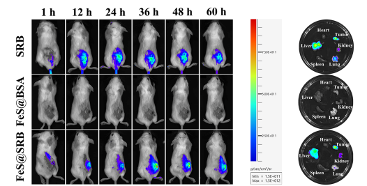
IR-780 purchased from MedChemExpress. Usage Cited in: Nat Commun. 2025 Jul 15;16(1):6532. [Abstract]
Representative fluorescence image of subcutaneous 4T1 tumor-bearing mice at different time after intravenous injection with IR-780 (IR780; 0.5 mg/mL; 37 °C for 15 min; IV; 100 μL)-labeled SRB, FeS@BSA and FeS@SRB, respectively.
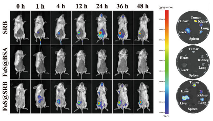
IR-780 purchased from MedChemExpress. Usage Cited in: Nat Commun. 2025 Jul 15;16(1):6532. [Abstract]
Representative fluorescence images of orthotopic 4T1 tumor-bearing mice at different time after intravenous injection with IR-780 (IR780; 0.5 mg/mL; 37 °C for 15 min; IV; 100 μL)-labeled SRB, FeS@BSA and FeS@SRB, respectively, and the corresponding fluorescence images of tumors and organs at 48 h.
-
Cell Rep Med
Hierarchically collapsible nanoactuator modulates mitochondrial ferroptosis-bioenergetic homeostasis cascade to decouple ischemic stroke. [Abstract]2026 Mar 17;7(3):102640. PMID: 41742403 -
Int J Biol Macromol
Programmable core-shell microneedles with marine chondroitin sulfate core and chito-oligosaccharide shell disrupt the pathological microenvironment cycle for enhanced healing of infected burn wounds. [Abstract]2026 Jul:370:152860. PMID: 42235776 -
ACS Appl Mater Interfaces
Mitochondrial Rapid Accumulation and Self-Enhanced Penetrating Nanomedicine for Tumor Multi-role Treatment. [Abstract]2025 Dec 31. PMID: 41476371
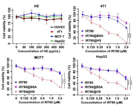
IR-780 purchased from MedChemExpress. Usage Cited in: ACS Appl Mater Interfaces. 2025 Dec 31. [Abstract]
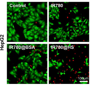
MTT cytotoxicity profiles of IR-780 (0-2.5 μM), IR780@BSA, and IR780@HS at varying concentrations (48 h treatment) in HepG2, MCF-7, and 4T1, HS cell lines.
IR-780 purchased from MedChemExpress. Usage Cited in: ACS Appl Mater Interfaces. 2025 Dec 31. [Abstract]
Viability assessment via Calcein-AM/PI dual staining in HepG2 cells following a 24 h treatment with IR780 ( 0.25 μM)@HS.
IR-780 purchased from MedChemExpress. Usage Cited in: ACS Appl Mater Interfaces. 2025 Dec 31. [Abstract]
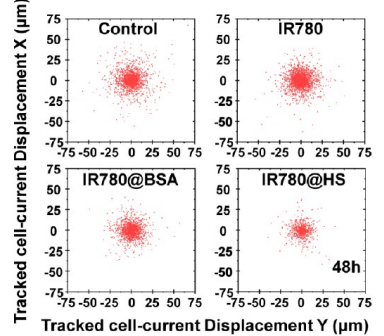
Wound-healing and migration assays indicated that IR780 ( 0.25 μM)@HS markedly suppressed HepG2 cell migration relative to the control groups.
IR-780 purchased from MedChemExpress. Usage Cited in: ACS Appl Mater Interfaces. 2025 Dec 31. [Abstract]
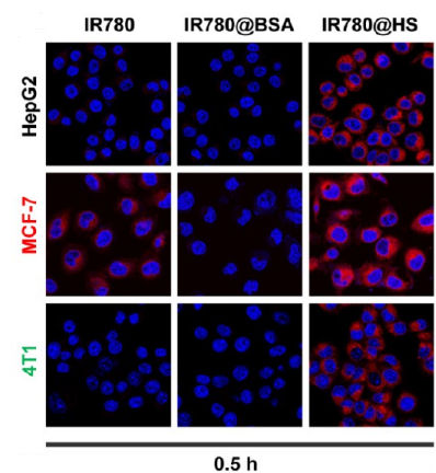
Cellular internalization mechanisms of IR780@HS. (0.5 h) Confocal microscopy images.
-
-
Am J Cancer Res
Exosomes with IR780 and Lenvatinib loaded on GPC3 single-chain scFv antibodies for targeted hyperthermia and chemotherapy in hepatocellular carcinoma therapy. [Abstract]2023 Nov 15;13(11):5368-5381. PMID: 38058824


Solvent & Solubility
In Vitro:
DMSO : 12.5 mg/mL (18.74 mM; ultrasonic and warming and heat to 60°C; Hygroscopic DMSO has a significant impact on the solubility of product, please use newly opened DMSO)
Please refer to the solubility information to select the appropriate solvent. Once prepared, please aliquot and store the solution to prevent product inactivation from repeated freeze-thaw cycles.
Storage method and period of stock solution: -80°C, 6 months; -20°C, 1 month (sealed storage, away from moisture and light). When stored at -80°C, please use it within 6 months. When stored at -20°C, please use it within 1 month.
Please refer to the solubility information to select the appropriate solvent. Once prepared, please aliquot and store the solution to prevent product inactivation from repeated freeze-thaw cycles.
Storage method and period of stock solution: -80°C, 6 months; -20°C, 1 month (sealed storage, away from moisture and light). When stored at -80°C, please use it within 6 months. When stored at -20°C, please use it within 1 month.
Concentration (start) × Volume (start) = Concentration (final) × Volume (final)


Purity & Documentation
-
Data Sheet (292 KB)
-
SDS (394 KB)
- English - EN (394 KB)
- Français - FR (394 KB)
- Deutsch - DE (394 KB)
- Norwegian - NO (394 KB)
- Español - ES (394 KB)
- Swedish - SV (394 KB)
- Italian - IT (394 KB)
- Korean - KR (394 KB)
- Portuguese - PT (394 KB)
-
Handling Instructions (2659 KB)
References
[1]. Zhang C, et al. A near-infrared fluorescent heptamethine indocyanine dye with preferential tumor accumulation for in vivo imaging. Biomaterials. 2010;31(25):6612-6617. [Content Brief]
[3]. Li Y, et al. IR-780 Dye as a Sonosensitizer for Sonodynamic Therapy of Breast Tumor. Sci Rep. 2016;6:25968. Published 2016 May 13. [Content Brief]
[4]. Wang Y, et al. Preferential accumulation of the near infrared heptamethine dye IR-780 in the mitochondria of drug-resistant lung cancer cells. Biomaterials. 2014;35(13):4116-4124. [Content Brief]
[5]. Zhang E, et al. Mechanistic study of IR-780 dye as a potential tumor targeting and drug delivery agent. Biomaterials. 2014;35(2):771-778. [Content Brief]
[6]. He W, et al. A versatile strategy to create an active tumor-targeted chemo-photothermal therapy nanoplatform: A case of an IR-780 derivative co-assembled with camptothecin prodrug. Acta Biomater. 2019;84:356-366. [Content Brief]
[7]. Lu YJ, et al. Liposomal IR-780 as a Highly Stable Nanotheranostic Agent for Improved Photothermal/Photodynamic Therapy of Brain Tumors by Convection-Enhanced Delivery. Cancers (Basel). 2021;13(15):3690. Published 2021 Jul 22. [Content Brief]
[8]. Yue C, et al. IR-780 dye loaded tumor targeting theranostic nanoparticles for NIR imaging and photothermal therapy. Biomaterials. 2013;34(28):6853-6861. [Content Brief]
[9]. Baeten J, et al. Development of fluorescent materials for Diffuse Fluorescence Tomography standards and phantoms. Opt Express. 2007;15(14):8681-8694. [Content Brief]


Complete Stock Solution Preparation Table
Please refer to the solubility information to select the appropriate solvent. Once prepared, please aliquot and store the solution to prevent product inactivation from repeated freeze-thaw cycles.
Storage method and period of stock solution: -80°C, 6 months; -20°C, 1 month (sealed storage, away from moisture and light). When stored at -80°C, please use it within 6 months. When stored at -20°C, please use it within 1 month.
| Optional Solvent | Concentration Solvent Mass | 1 mg | 5 mg | 10 mg | 25 mg |
|---|---|---|---|---|---|
| DMSO | 1 mM | 1.4990 mL | 7.4950 mL | 14.9900 mL | 37.4751 mL |
| 5 mM | 0.2998 mL | 1.4990 mL | 2.9980 mL | 7.4950 mL | |
| 10 mM | 0.1499 mL | 0.7495 mL | 1.4990 mL | 3.7475 mL | |
| 15 mM | 0.0999 mL | 0.4997 mL | 0.9993 mL | 2.4983 mL |


IR-780 Related Classifications
HY-D1063 Related Classifications
 Powered by Bioz