


D-Luciferin
Based on 126 publication(s) in Google Scholar
D-luciferin is the natural substrate of the enzyme luciferase (Luc) that catalyzes the production of the typical yellowgreen light of fireflies. The 560 nm chemiluminescence from this reaction peaks within seconds, with light output that is proportional to luciferase concentration when the substrate luciferin is present in excess. The luciferase (luc) gene is a popular reporter gene for research and agent screening. Chemiluminescent techniques are virtually background-free, making the luc reporter gene ideal for detecting low-level gene expression. As little as 0.02 pg of luciferase can be reliably measured in a standard scintillation counter. In addition to its role as a reporter of gene expression, luciferase is commonly used in an extremely sensitive assay for ATP. We of er the firefly luciferase (HY-P1004A), luciferin free acid (HY-12591A), as well as its water-soluble sodium salts (HY-12591) and potassium salts (HY-12591B) .
Nur für Forschungszwecke. Wir verkaufen nicht an Patienten.
- Reinheit: 99.21%
- CAS. Nr.: 2591-17-5
- Formel: C11H8N2O3S2
- Molecular Weight:280.33
-
Speicherung:
4°C, protect from light
* In solvent : -80°C, 6 months; -20°C, 1 month (protect from light)

To place orders, for customer services and technical support, please contact: MedChemExpress USA
Tel: 609-228-6898 E-mail: [email protected] [email protected]


-
 Biologische Aktivität
-
Chemical Information
-
Lösungsmittel & Löslichkeit
- Protokoll
- Reinheit & Dokumentation
- Verweise
-
 Help & FAQs


Publications Citing Use of MedChemExpress (MCE) D-Luciferin
More- Cancer Cell. 2026 Mar 9;44(3):658-675.e12. [Abstract]
- Science. 2026 Jun 25;392(6805):eadz3624. [Abstract]
- Mol Cancer. 2025 Jan 24;24(1):29. [Abstract]
- Nat Nanotechnol. 2026 Jun 15. [Abstract]
- Cell Metab. 2025 Sep 2;37(9):1907-1925.e14. [Abstract]
- Cell Metab. 2022 Nov 1;34(11):1843-1859.e11. [Abstract]
- Adv Mater. 2025 Mar;37(9):e2412430. [Abstract]
- Mil Med Res. 2023 Jul 25;10(1):34. [Abstract]
- Cancer Commun (Lond). 2025 Nov 10. [Abstract]
- Cell Stem Cell. 2026 Apr 2;33(4):660-675.e5. [Abstract]
- Sci Bull. 2025 Apr 15;70(7):1126-1138. [Abstract]
- Adv Funct Mater. 2023 Sep 15.
- Autophagy. 2024 Mar;20(3):541-556. [Abstract]
- Autophagy. 2023 Jun;19(6):1745-1763. [Abstract]
- Nat Commun. 2026 May 11;17(1):4190. [Abstract]
- Nat Commun. 2025 Nov 18;16(1):10096. [Abstract]
- Nat Commun. 2025 Oct 1;16(1):8750. [Abstract]
- Nat Commun. 2025 Oct 9;16(1):8990. [Abstract]
- Nat Commun. 2025 Mar 1;16(1):2104. [Abstract]
- ACS Nano. 2025 Oct 21;19(41):36353-36372. [Abstract]
- ACS Nano. 2024 Dec 10;18(49):33366-33380. [Abstract]
- J Nanobiotechnology. 2025 Nov 18;23(1):717. [Abstract]
- J Nanobiotechnology. 2025 Jun 6;23(1):421. [Abstract]
- J Nanobiotechnology. 2025 Mar 28;23(1):252. [Abstract]
- J Nanobiotechnology. 2023 Oct 19;21(1):383. [Abstract]
- Acta Pharm Sin B. 2023 Dec;13(12):5074-5090. [Abstract]
- Acta Pharm Sin B. 2023 Mar;13(3):967-981. [Abstract]
- J Clin Invest. 2026 May 15;136(10):e199285. [Abstract]
- J Exp Clin Cancer Res. 2025 Feb 19;44(1):60. [Abstract]
- Adv Sci (Weinh). 2026 Apr;13(20):e15431. [Abstract]
- Adv Sci (Weinh). 2025 Oct 23:e11573. [Abstract]
- Adv Sci (Weinh). 2025 Apr;12(13):e2415229. [Abstract]
- Compos B Eng. 2024 Feb 15, 271, 111162.
- Sci Adv. 2026 May 15;12(20):eaed0154. [Abstract]
- Sci Adv. 2025 Mar 28;11(13):eads2295. [Abstract]
- Sci Adv. 2024 Aug 16;10(33):eado1533. [Abstract]
- Plant Commun. 2024 Dec 9;5(12):101076. [Abstract]
- Plant Commun. 2024 Aug 12;5(8):100937. [Abstract]
- Biomaterials. 2025 Nov:322:123391. [Abstract]
- Plant Cell. 2026 May 11:koag137. [Abstract]
- Plant Cell. 2025 Jun 4;37(6):koaf098. [Abstract]
- Carbohydr Polym. 2026 Mar 1:375:124760. [Abstract]
- Asian J Pharm Sci. 2022 Jul;17(4):557-570. [Abstract]
- J Control Release. 2025 Sep 10:385:113970. [Abstract]
- Pharmacol Res. 2026 Feb:224:108088. [Abstract]
- Pharmacol Res. 2021 Apr:166:105527. [Abstract]
- Cell Death Dis. 2020 Sep 17;11(9):765. [Abstract]
- Rare Metals. 2025 Oct 12.
- Cancer Lett. 2023 Oct 28:575:216403. [Abstract]
- J Immunother Cancer. 2026 May 12;14(5):e012347. [Abstract]
- J Immunother Cancer. 2025 Apr 15;13(4):e011108. [Abstract]
- Mol Ther. 2026 Jun 2:S1525-0016(26)00470-3. [Abstract]
- Phytomedicine. 2025 Nov 25:148:157389. [Abstract]
- Phytomedicine. 2025 Dec:149:157559. [Abstract]
- Adv Healthc Mater. 2025 Aug;14(22):e2405061. [Abstract]
- J Hazard Mater. 2024 Dec 5:480:136351. [Abstract]
- Acta Biomater. 2026 Jul:218:244-260. [Abstract]
- Acta Biomater. 2025 May 1:197:431-443. [Abstract]
- Acta Pharmacol Sin. 2022 Apr;43(4):1046-1058. [Abstract]
- J Transl Med. 2024 Aug 28;22(1):792. [Abstract]
- Sci China Life Sci. 2022 Feb;65(2):341-361. [Abstract]
- Hortic Res. 2025 Sep 17.
- Hortic Res. 2022 Jan 20:9:uhab088. [Abstract]
- Cell Chem Biol. 2022 Mar 17;29(3):517-529.e5. [Abstract]
- Int J Nanomedicine. 2026 Mar 20:21:577606. [Abstract]
- New Phytol. 2025 Dec 7. [Abstract]
- Int J Biol Macromol. 2025 Dec 15:149699. [Abstract]
- Int J Biol Macromol. 2025 Dec;333(Pt 2):148750. [Abstract]
- New Phytol. 2025 Nov 3. [Abstract]
- New Phytol. 2023 Sep;239(5):1887-1902. [Abstract]
- New Phytol. 2022 Dec;236(5):1796-1808. [Abstract]
- Plant Physiol. 2026 Jun 18:kiag396. [Abstract]
- Plant Physiol. 2024 Dec 2;196(4):2784-2794. [Abstract]
- Plant Physiol. 2023 Oct 26;193(3):2105-2121. [Abstract]
- Plant Physiol. 2021 Jun 11;186(2):1302-1317. [Abstract]
- Cell Mol Gastroenterol Hepatol. 2021;12(3):839-856. [Abstract]
- Free Radic Biol Med. 2026 Mar 16:246:93-106. [Abstract]
- ACS Appl Mater Interfaces. 2026 Jun 24;18(24):33542-33558. [Abstract]
- ACS Appl Mater Interfaces. 2024 Dec 11;16(49):67192-67202. [Abstract]
- Cell Rep. 2026 Jun 25;45(7):117606. [Abstract]
- J Med Chem. 2024 Nov 28;67(22):20100-20117. [Abstract]
- Int J Oncol. 2020 Mar;56(3):761-771. [Abstract]
- Plant Cell Environ. 2023 Jun;46(6):1921-1934. [Abstract]
- Plant Cell Environ. 2023 May;46(5):1726-1742. [Abstract]
- J Agric Food Chem. 2025 Jul 9;73(27):16787-16803. [Abstract]
- Mol Ther Nucleic Acids. 2025 Nov 27;37(1):102785. [Abstract]
- J Mater Chem B. 2026 May 15. [Abstract]
- Breast Cancer Res. 2025 Dec 18;27(1):217. [Abstract]
- Biosensors (Basel). 2024 May 16;14(5):252. [Abstract]
- Colloids Surf B Biointerfaces. 2026 Jan:257:115180. [Abstract]
- J Integr Agric. 2025 Mar 27.
- Cell Oncol (Dordr). 2026 Apr 20;49(3):77. [Abstract]
- Int Immunopharmacol. 2026 Mar 1:172:116170. [Abstract]
- Int Immunopharmacol. 2024 Nov 7;143(Pt 3):113573. [Abstract]
- mBio. 2025 Apr 9;16(4):e0366524. [Abstract]
- Sci Rep. 2025 Dec 17;15(1):43979. [Abstract]
- Sci Rep. 2025 Apr 1;15(1):11040. [Abstract]
- Neoplasia. 2025 Mar:61:101123. [Abstract]
- Cell Signal. 2025 Oct 18:112174. [Abstract]
- iScience. 2025 Sep 24;28(11):113647. [Abstract]
- Nanomedicine. 2025 Oct:69:102849. [Abstract]
- Antiviral Res. 2024 Dec:232:106036. [Abstract]
- Front Biosci (Landmark Ed). 2025 Sep 25;30(9):43761. [Abstract]
- BMC Cancer. 2023 Oct 31;23(1):1047. [Abstract]
- J Cell Commun Signal. 2025 Aug 24;19(3):e70040. [Abstract]
- Vet Res. 2021 Jun 26;52(1):95. [Abstract]
- Pathol Res Pract. 2025 Aug:272:156051. [Abstract]
- Parasit Vectors. 2023 Apr 24;16(1):139. [Abstract]
- Arch Biochem Biophys. 2026 May 15:110866. [Abstract]
- Bioorg Med Chem. 2025 Nov 19:133:118489. [Abstract]
- Immun Inflamm Dis. 2024 Sep;12(9):e70005. [Abstract]
- J Leukoc Biol. 2019 Nov;106(5):1089-1100. [Abstract]
- Gastro Hep Adv. 2022 Nov 8;2(3):412-423. [Abstract]
- Plasma Process Polym. 2024 Oct 22.
- J Gastrointest Oncol. 2025 Apr 30;16(2):599-614. [Abstract]
- J Vis Exp. 2025 Oct 17:(224). [Abstract]
- bioRxiv. 2026 Mar 29.
- bioRxiv. 2025 February 27.
- bioRxiv. 2024 August 16.
- Icahn School of Medicine at Mount Sinai. 2024.
- SSRN. 2023 Sep 15.
- bioRxiv. 2023 Apr 10.
- SSRN. 2023 Mar 20.
- Research Square Print. 2023 Feb 3.
- Research Square Print. December 19th, 2022.
- Biomed Pharmacother. 2019 Sep:117:109126. [Abstract]
 Customer Validation & Images

-
In Vivo Imaging
-
In Vivo Efficacy Study
-
In Vivo Imaging
-
In Vivo Imaging
-
In Vivo Imaging


Biologische Aktivität
|
Cell Line
|
Type | Value | Description | References |
|---|---|---|---|---|
| HT-29 | EC50 |
12.9 μM
Compound: 10
|
Agonist activity at human GPR35 expressed in human HT29 cells by DMR assay
Agonist activity at human GPR35 expressed in human HT29 cells by DMR assay
|
[PMID: 23888932] |
| HT-29 | EC50 |
277 μM
Compound: 10
|
Agonist activity at human GPR35 expressed in HT29 cells by beta-arrestin translocation assay
Agonist activity at human GPR35 expressed in HT29 cells by beta-arrestin translocation assay
|
[PMID: 23888932] |
Guide (Following is our recommended protocol. This protocol only provides a guideline, and should be modified according to your specific needs).
1. Example protocol for in vitro bioluminescent image assays
1.1 Prepare D-luciferin stock solution in DMSO. Mix well. Use immediately, or make single use aliquots, and store at -20 °C, avoid freeze-thaw cycles, avoid exposure to the light.
1.2 Prepare a 0.5-1 mM working solution of D-Luciferin in pre-warmed tissue culture medium.
1.3 Aspirate media from cultured cells.
1.4 Add D-Luciferin working solution to cells, and incubate the cells for 5-10 minutes at 37 °C just prior to imaging.
2. In vivo assays
Since the in vivo experimental dose is relatively large, generally 150 mg/kg, it is recommended to choose potassium salt (HY-12591B) and sodium salt (HY-12591).
3. Example protocol for D-Luciferin reporter assays
3.1 Prepare D-luciferin stock solution in DMSO. Use immediately, or make single use aliquots, and store at -20 °C, avoid freeze-thaw cycles, avoid exposure to the light.
3.2 Prepare a 1 mM working solution of D-Luciferin with 3 mM ATP, 1 mM DTT and 15 mM MgSO4 in 25 mM tricine buffer pH 7.8.
3.3 Pipette 5-10 μLof cell lysate into a microplate. Use lysis reagent or buffer without lysate as a blank.
3.4 Prime luminometer with D-Luciferin working solution according to manufacturer’s instructions.
3.5 Inject 200 μL of D-luciferin working solution with no delay and a 10 second integration time.
Precautions
a) There are three forms of D-luciferin, namely free acid (HY-12591A), potassium salt (HY-12591B) and sodium salt (HY-12591). The sodium salt and potassium salt forms of D-luciferin are easily soluble in aqueous buffers (pH 6.1-6.5). Stock solutions can be made in ATP-free water and stored at -20°C, protect from light. The free acid can be neutralized with DMSO or an appropriate base to dissolve. At a higher pH, luciferin undergoes a base-catalyzed formation of dehydroluciferin, as well as racemization to the L-isomer.
b) The D-luciferin can be used with any existing reporter assay or ATP assay system.
c) If testing for ATP, minimize all possible sources of ATP contamination by wearing gloves and using ATP-free containers. Use only sterile ATP-free water and reagents. Use autoclaved water for all reagent preparations.
MedChemExpress (MCE) has not independently confirmed the accuracy of these methods. They are for reference only.


Chemical Information
-
CAS. Nr. 2591-17-5
-
Appearance Solid
-
Molecular Weight 280.33
-
Formel C11H8N2O3S2
-
Color White to yellow
-
SMILES
O=C([C@@H]1N=C(C2=NC3=CC=C(O)C=C3S2)SC1)O
-
Synonyms
D-(-)-Luciferin; Firefly luciferin; Beetle Luciferin
-
Initial Source
insect
-
Versand
Room temperature in continental US; may vary elsewhere.
-
Speicherung
4°C, protect from light
* In solvent : -80°C, 6 months; -20°C, 1 month (protect from light)


Publications (126)
-
Journal Impact Factor
-
Most Recent
-
Cancer Cell
Chemotherapy triggers immune evasion by fostering LEPR+ Kupffer cell differentiation in liver metastases. [Abstract]2026 Mar 9;44(3):658-675.e12. PMID: 41687606 -
Science
2026 Jun 25;392(6805):eadz3624. PMID: 42348698 -
Mol Cancer
Circular RNA circBNC2 inhibits tumorigenesis by modulating ferroptosis and acts as a nanotherapeutic target in prostate cancer. [Abstract]2025 Jan 24;24(1):29. PMID: 39856701 -
Nat Nanotechnol
Nano-enabled spatially selective protein degradation modulates lactate metabolism to potentiate antitumor immunity in liver cancer. [Abstract]2026 Jun 15. PMID: 42298103 -
Cell Metab
Tumor-associated Schwann cell remodeling under metabolic stress via lactate sensing orchestrates pancreatic ductal adenocarcinoma development. [Abstract]2025 Sep 2;37(9):1907-1925.e14. PMID: 40803319 -
Cell Metab
2022 Nov 1;34(11):1843-1859.e11. PMID: 36103895 -
Adv Mater
A Universal Therapeutic Vaccine Leveraging Autologous Pre-Existing Immunity to Eliminate in Situ Uniformly Engineered Heterogeneous Tumor Cells. [Abstract]2025 Mar;37(9):e2412430. PMID: 39838750 -
Mil Med Res
RARRES2 regulates lipid metabolic reprogramming to mediate the development of brain metastasis in triple negative breast cancer. [Abstract]2023 Jul 25;10(1):34. PMID: 37491281 -
Cancer Commun (Lond)
Unfolded protein response kinase PERK supports survival and metastasis of circulating tumor cell clusters via SAM synthesis and H3K4me3-dependent PDGFB signaling. [Abstract]2025 Nov 10. PMID: 41212905 -
Cell Stem Cell
2026 Apr 2;33(4):660-675.e5. PMID: 41895281 -
Sci Bull
Tumor cell-derived N-acetyl-aspartyl-glutamate reshapes the tumor microenvironment to facilitate breast cancer metastasis. [Abstract]2025 Apr 15;70(7):1126-1138. PMID: 39800628 -
D-Luciferin purchased from MedChemExpress. Usage Cited in: Adv Funct Mater. 2023 Sep 15.
In vivo bioluminescence images of mice in different groups. Lung metastasis of the tumors through in vivo imaging by intraperitoneal injection of D-luciferin sodium (75 mg/kg, 100 μL) at 20 min before bioluminescence imaging.
-
Autophagy
Artesunate Sensitizes Human hepatocellular carcinoma to sorafenib via exacerbating AFAP1L2-SRC-FUNDC1 axis-dependent mitophagy. [Abstract]2024 Mar;20(3):541-556. PMID: 37733919 -
Autophagy
The Valsa mali effector Vm1G-1794 protects the aggregated MdEF-Tu from autophagic degradation to promote infection in apple. [Abstract]2023 Jun;19(6):1745-1763. PMID: 36449354 -
Nat Commun
Ascites protects against ferroptosis and enables the peritoneal growth of ovarian cancer. [Abstract]2026 May 11;17(1):4190. PMID: 42115216 -
Nat Commun
Co-delivery of sorafenib and an FSP1 inhibitor triggers dual ferroptosis in tumor cells and immunosuppressive macrophages for enhanced immunotherapy in mouse models of hepatocellular carcinoma. [Abstract]2025 Nov 18;16(1):10096. PMID: 41253833 -
Nat Commun
HY5 integrates light and electrical signaling to trigger a jasmonate burst for nematode defense in tomato. [Abstract]2025 Oct 1;16(1):8750. PMID: 41034216 -
Nat Commun
Size-variable self-feedback nanomotors for glioblastoma therapy via mitochondrial mineralization. [Abstract]2025 Oct 9;16(1):8990. PMID: 41068089 -
Nat Commun
Harnessing engineered symbionts to combat concurrent malaria and arboviruses transmission. [Abstract]2025 Mar 1;16(1):2104. PMID: 40025068 -
ACS Nano
Surface d-Band Modulation via Biodirected Mineralization Enables Nanoenzymes to Inhibit Radiation-Induced T-Cell Exhaustion and Potentiate Immunoradiotherapy. [Abstract]2025 Oct 21;19(41):36353-36372. PMID: 41073355 -
ACS Nano
Anti-PD-L1 Antibody Fragment Linked to Tumor-Targeting Lipid Nanoparticle Can Eliminate Cancer and Its Metastasis via Photoimmunotherapy. [Abstract]2024 Dec 10;18(49):33366-33380. PMID: 39603816
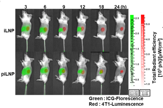
D-Luciferin purchased from MedChemExpress. Usage Cited in: ACS Nano. 2024 Dec 10;18(49):33366-33380. [Abstract]
4T1-luc tumor-bearing BALB/c mice were intravenously (i.v.) injected with 100 mg/kg of iLNPs and 100 mg/kg piLNPs. D-Luciferin sodium (150 mg/kg) was administered to mice 5 min before fluorescence measurement, and ICG fluorescence was measured using an in vivo imaging system (IVIS) at each indicated time.
-
J Nanobiotechnology
AMFR-mediated ER-phagy regulation and therapeutic targeting in osteosarcoma: a multifunctional nanoplatform strategy. [Abstract]2025 Nov 18;23(1):717. PMID: 41250206 -
J Nanobiotechnology
Targeting Dicer reprograms tumor-associated macrophages to promote anti-tumoral immunity in colorectal cancer liver metastasis. [Abstract]2025 Jun 6;23(1):421. PMID: 40481520 -
J Nanobiotechnology
5-FU@HFn combined with decitabine induces pyroptosis and enhances antitumor immunotherapy for chronic myeloid leukemia. [Abstract]2025 Mar 28;23(1):252. PMID: 40148810 -
J Nanobiotechnology
Combination of ferroptosis and pyroptosis dual induction by triptolide nano-MOFs for immunotherapy of Melanoma. [Abstract]2023 Oct 19;21(1):383. PMID: 37858186
D-Luciferin purchased from MedChemExpress. Usage Cited in: J Nanobiotechnology. 2023 Oct 19;21(1):383. [Abstract]
On day 15 after intravenous injection of B16F10-luc cells into C57BL/6 mice to establish a melanoma lung metastasis model, D-Luciferin potassium (150 mg/kg) is intrabitoneally injected into the mice, and lung tissue is collected from the mice for bioluminescence imaging.
-
Acta Pharm Sin B
Converting bacteria into autologous tumor vaccine via surface biomineralization of calcium carbonate for enhanced immunotherapy. [Abstract]2023 Dec;13(12):5074-5090. PMID: 38045045
D-Luciferin purchased from MedChemExpress. Usage Cited in: Acta Pharm Sin B. 2023 Dec;13(12):5074-5090. [Abstract]
The bioluminescence images of lungs harvested from mice post different treatments. On day 13 after intravenous injection of B16F10-luc cells to establish a lung metastasis model of melanoma in mice, D-luciferin potassium (150 mg/kg) is intraperitoneally injected into the mice.
-
Acta Pharm Sin B
Concurrent silencing of TBCE and drug delivery to overcome platinum-based resistance in liver cancer. [Abstract]2023 Mar;13(3):967-981. PMID: 36970197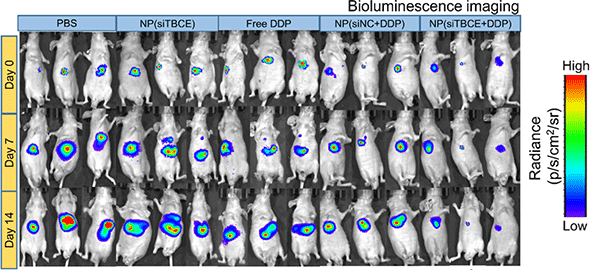
D-Luciferin purchased from MedChemExpress. Usage Cited in: Acta Pharm Sin B. 2023 Mar;13(3):967-981. [Abstract]
Tumor growth is monitored by bioluminescence imaging. Prior to imaging, D-luciferin substrate is injected intraperitoneally into the mice (150 mg/kg per mouse).
-
J Clin Invest
Targeting RANKL-independent osteoclastogenesis overcomes denosumab resistance in models of ER+ breast cancer bone metastasis. [Abstract]2026 May 15;136(10):e199285. PMID: 42138086 -
J Exp Clin Cancer Res
MMP28 recruits M2-type tumor-associated macrophages through MAPK/JNK signaling pathway-dependent cytokine secretion to promote the malignant progression of pancreatic cancer. [Abstract]2025 Feb 19;44(1):60. PMID: 39972459 -
Adv Sci (Weinh)
pH/ROS-Responsive Injectable Hydrogel Co-Loaded with B7-H3 Blocker and NETs Suppressor Boosts OSCC Synergistic Immunotherapy. [Abstract]2026 Apr;13(20):e15431. PMID: 41684277 -
Adv Sci (Weinh)
Chronic ER Stress Triggers Cell-Surface Chaperones as the Therapeutic Targets of CAR Cells in Acute Myeloid Leukemia. [Abstract]2025 Oct 23:e11573. PMID: 41126722 -
Adv Sci (Weinh)
A Novel tRF, HCETSR, Derived From tRNA-Glu/TTC, Inhibits HCC Malignancy by Regulating the SPBTN1-catenin Complex Axis. [Abstract]2025 Apr;12(13):e2415229. PMID: 39921434 -
-
Sci Adv
Synergistic antioxidant and gene supplementation for high-efficacy retinitis pigmentosa therapy. [Abstract]2026 May 15;12(20):eaed0154. PMID: 42127191 -
Sci Adv
Macrophage-targeted Mms6 mRNA-lipid nanoparticles promote locomotor functional recovery after traumatic spinal cord injury in mice. [Abstract]2025 Mar 28;11(13):eads2295. PMID: 40138430
D-Luciferin purchased from MedChemExpress. Usage Cited in: Sci Adv. 2025 Mar 28;11(13):eads2295. [Abstract]
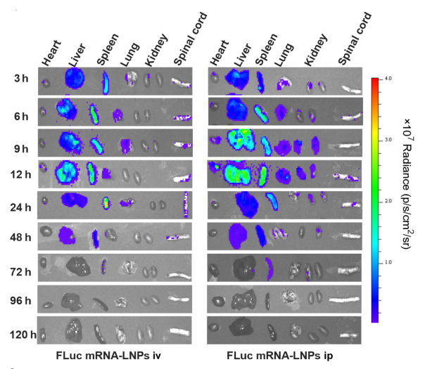
Briefly, d-luciferin sodium (150 mg/kg in PBS) was injected intraperitoneally into the mice. Fifteen minutes later, mice were euthanized, and the heart, liver, spleen, lungs, kidneys, and spinal cord were collected. Bioluminescence images (yellow-green light) in these tissues were analyzed using Living Image Software (PerkinElmer). Representative in vivo imaging system images of major organs in SCI mice after being administered with firefly luciferase (FLuc) mRNA-LNPs (0.5 mg mRNA/kg).
-
Sci Adv
Epinephrine promotes breast cancer metastasis through a ubiquitin-specific peptidase 22-mediated lipolysis circuit. [Abstract]2024 Aug 16;10(33):eado1533. PMID: 39151008
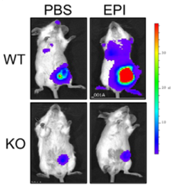
D-Luciferin purchased from MedChemExpress. Usage Cited in: Sci Adv. 2024 Aug 16;10(33):eado1533. [Abstract]
Monitoring of primary tumor growth and the occurrence of lung metastasis was conducted using bioluminescence imaging after intraperitoneal injection of D-luciferin sodium (150 mg/kg).
-
Plant Commun
The TATA-box binding protein-associated factor TAF12b facilitates the degradation of type-B response regulators to negatively regulate cytokinin signaling. [Abstract]2024 Dec 9;5(12):101076. PMID: 39228128 -
Plant Commun
Clathrin Light Chains negatively regulate plant immunity by hijacking the autophagy pathway. [Abstract]2024 Aug 12;5(8):100937. PMID: 38693694 -
Biomaterials
Radiation-triggerable bioreactors enable bioenergetic reprograming of cancer stem cell plasticity via targeted arginine metabolism disruption for augmented radio-immunotherapy. [Abstract]2025 Nov:322:123391. PMID: 40344881 -
Plant Cell
The E3 ubiquitin ligases SlXERICO1 and 3 regulate thermotolerance in Solanum lycopersicum. [Abstract]2026 May 11:koag137. PMID: 42116660 -
Plant Cell
RALF33-FERONIA signaling orchestrates postwounding root-tip regeneration via TPR4-ERF115 dynamics. [Abstract]2025 Jun 4;37(6):koaf098. PMID: 40323783 -
Carbohydr Polym
Characterization and biological activities of a high-yield galactosylated heparan sulfate from Bursatella leachii egg strings. [Abstract]2026 Mar 1:375:124760. PMID: 41475732 -
Asian J Pharm Sci
Non-cytotoxic nanoparticles re-educating macrophages achieving both innate and adaptive immune responses for tumor therapy. [Abstract]2022 Jul;17(4):557-570. PMID: 36101893 -
J Control Release
Local delivery of IL-12 mRNA and indoximod prodrug potentiates antitumor immunity by increasing T cell effector function. [Abstract]2025 Sep 10:385:113970. PMID: 40541741 -
Pharmacol Res
Fig-derived exosome-like nanoparticles attenuating bone metastasis of breast cancer through establishing an anti-tumor microenvironment. [Abstract]2026 Feb:224:108088. PMID: 41500295 -
Pharmacol Res
Oligosaccharides from fucosylated glycosaminoglycan prevent breast cancer metastasis in mice by inhibiting heparanase activity and angiogenesis. [Abstract]2021 Apr:166:105527. PMID: 33667689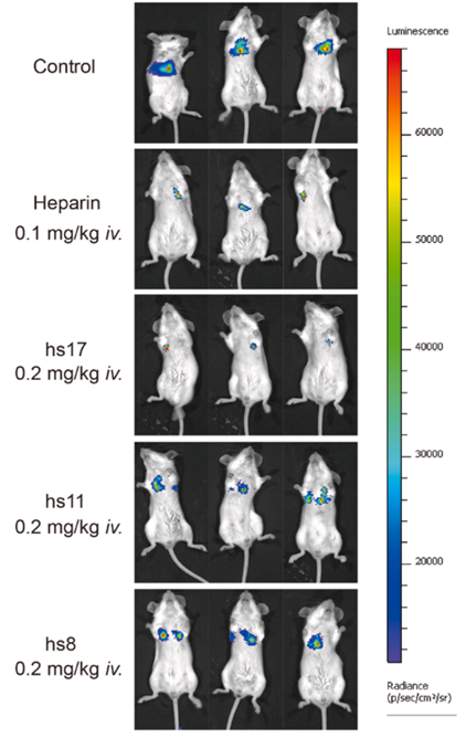
D-Luciferin purchased from MedChemExpress. Usage Cited in: Pharmacol Res. 2021 Apr:166:105527. [Abstract]
After cell inoculation for 7 days, mice were injected intraperitoneally with D-luciferin substrate at 150 mg/kg and anesthetized with 10% chloral hydrate (5 mL/kg).
-
Cell Death Dis
Dual inhibition of HDAC and tyrosine kinase signaling pathways with CUDC-907 attenuates TGFβ1 induced lung and tumor fibrosis. [Abstract]2020 Sep 17;11(9):765. PMID: 32943605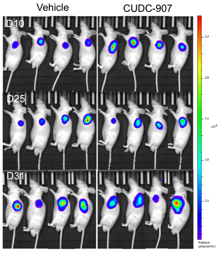
D-Luciferin purchased from MedChemExpress. Usage Cited in: Cell Death Dis. 2020 Sep 17;11(9):765. [Abstract]
At Day 10, 25, and 31, mice were anesthetized with isoflurane and then injected with 75 mg/kg D-Luciferin solution for imaging.
-
-
Cancer Lett
FABP4 in macrophages facilitates obesity-associated pancreatic cancer progression via the NLRP3/IL-1β axis. [Abstract]2023 Oct 28:575:216403. PMID: 37741433 -
J Immunother Cancer
CXCL16-driven CD4+ T cells orchestrate immunosurveillance against MHC-I-deficient hepatocellular tumors. [Abstract]2026 May 12;14(5):e012347. PMID: 42120180 -
J Immunother Cancer
METTL3 promotes an immunosuppressive microenvironment in bladder cancer via m6A-dependent CXCL5/CCL5 regulation. [Abstract]2025 Apr 15;13(4):e011108. PMID: 40234090 -
Mol Ther
Nicotinamide riboside enhances adoptive T cell therapy by promoting memory differentiation and metabolic fitness. [Abstract]2026 Jun 2:S1525-0016(26)00470-3. PMID: 42231576 -
Phytomedicine
Jianpi Huayu decoction exerts antitumor effects in pancreatic cancer via SCD1-mediated lipid metabolism remodeling/ferroptosis axis. [Abstract]2025 Nov 25:148:157389. PMID: 41101075 -
Phytomedicine
Shenling Baizhu Powder potentiates immunotherapy response: putative roles of gut microbial remodeling and fatty acid metabolism modulation. [Abstract]2025 Dec:149:157559. PMID: 41270389 -
Adv Healthc Mater
Ultrasonic Cavitation-Manipulated Macrophages Hitchhiking Augments Delivery of Nanocarriers for Active and Efficient Cancer Therapy. [Abstract]2025 Aug;14(22):e2405061. PMID: 40545970 -
J Hazard Mater
2024 Dec 5:480:136351. PMID: 39488976 -
Acta Biomater
Mechanically heterogeneous nanofibrous hydrogel drives Piezo1-mediated cell aggregation and collective migration. [Abstract]2026 Jul:218:244-260. PMID: 42270027 -
Acta Biomater
Bioorthogonal SERS-bioluminescence dual-modal imaging for real-time tracking of triple-negative breast cancer metastasis. [Abstract]2025 May 1:197:431-443. PMID: 40101869 -
Acta Pharmacol Sin
Okicamelliaside targets the N-terminal chaperone pocket of HSP90 disrupts the chaperone protein interaction of HSP90-CDC37 and exerts antitumor activity. [Abstract]2022 Apr;43(4):1046-1058. PMID: 34326484 -
J Transl Med
Mito-LND and (E)-Akt inhibitor-IV: novel compounds inducing endoplasmic reticulum stress and ROS accumulation against hepatocellular carcinoma. [Abstract]2024 Aug 28;22(1):792. PMID: 39198815 -
Sci China Life Sci
2022 Feb;65(2):341-361. PMID: 34047913 -
-
Hortic Res
A novel leucine-rich repeat receptor-like kinase MRK1 regulates resistance to multiple stresses in tomato. [Abstract]2022 Jan 20:9:uhab088. PMID: 35048129 -
Cell Chem Biol
Temporal proteomics reveal specific cell cycle oncoprotein downregulation by p97/VCP inhibition. [Abstract]2022 Mar 17;29(3):517-529.e5. PMID: 34847375 -
Int J Nanomedicine
Intraperitoneal Co-Delivery of Claudin18.2×41BB and EpCAM×CD3 Bispecific Antibodies via mRNA-LNPs Synergistically Suppresses Gastric Cancer Peritoneal Metastasis Through T Cell Co-Activation. [Abstract]2026 Mar 20:21:577606. PMID: 41884275 -
New Phytol
2025 Dec 7. PMID: 41355201 -
Int J Biol Macromol
circSCYL2 antagonizes hnRNPA2B1-mediated m6A-chromatin crosstalk to promote ferroptosis and inhibit metastasis of breast cancer to brain. [Abstract]2025 Dec 15:149699. PMID: 41407226 -
Int J Biol Macromol
Exosomal microRNA-20b-5p contributes to cytarabine resistance in acute myeloid leukemia via the microtubule-associated serine/threonine kinase-like-phosphatidylinositol 3-kinase-protein kinase B signaling axis. [Abstract]2025 Dec;333(Pt 2):148750. PMID: 41197693 -
New Phytol
SlWRI5a and SlHY5 co-activate SlFatM-mediated fatty acid biosynthesis during arbuscular mycorrhizal symbiosis in tomato. [Abstract]2025 Nov 3. PMID: 41185456 -
New Phytol
SlMPK1- and SlMPK2-mediated SlBBX17 phosphorylation positively regulates CBF-dependent cold tolerance in tomato. [Abstract]2023 Sep;239(5):1887-1902. PMID: 37322592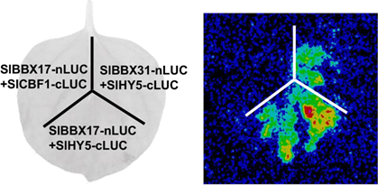
D-Luciferin purchased from MedChemExpress. Usage Cited in: New Phytol. 2023 Sep;239(5):1887-1902. [Abstract]
The full-length coding sequences (CDS) of target genes are cloned into either the pCAMBIA-nLuc or pCAMBIA-cLuc vectors. The resulting recombinant constructs are transformed into Agrobacterium tumefaciens strain GV3101, which is then infiltrated into Nicotiana benthamiana leaves. After 2 days, the infiltrated leaves are painted with 1 mM D-Luciferin potassium.
-
New Phytol
Herbivore-induced Ca2+ signals trigger a jasmonate burst by activating ERF16-mediated expression in tomato. [Abstract]2022 Dec;236(5):1796-1808. PMID: 36052744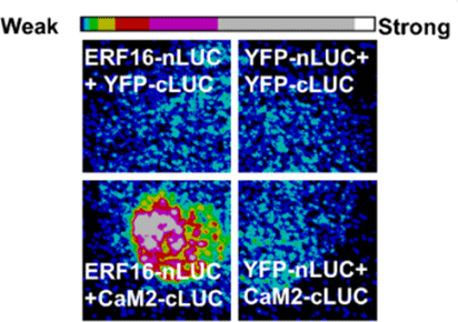
D-Luciferin purchased from MedChemExpress. Usage Cited in: New Phytol. 2022 Dec;236(5):1796-1808. [Abstract]
ERF16-nLUC and CaM2-cLUC vectors are cotransfected in N. benthamiana leaves. 10 min after the abaxial sides of leaves are sprayed with 1 mM Luciferin.
-
Plant Physiol
Fungal effector CgCAP1 suppresses the MdMVQ4-MdWRKY100-MdChi5 immune module during Colletotrichum gloeosporioides infection. [Abstract]2026 Jun 18:kiag396. PMID: 42315118 -
Plant Physiol
C2-domain abscisic acid-related proteins regulate the dynamics of a plasma membrane H+-ATPase in response to alkali stress. [Abstract]2024 Dec 2;196(4):2784-2794. PMID: 39217410 -
Plant Physiol
CALMODULIN6 negatively regulates cold tolerance by attenuating ICE1-dependent stress responses in tomato. [Abstract]2023 Oct 26;193(3):2105-2121. PMID: 37565524 -
Plant Physiol
The protein kinase CPK28 phosphorylates ascorbate peroxidase and enhances thermotolerance in tomato. [Abstract]2021 Jun 11;186(2):1302-1317. PMID: 33711164 -
Cell Mol Gastroenterol Hepatol
METTL3/METTL14 Transactivation and m6A-Dependent TGF-β1 Translation in Activated Kupffer Cells. [Abstract]2021;12(3):839-856. PMID: 33992834
D-Luciferin purchased from MedChemExpress. Usage Cited in: Cell Mol Gastroenterol Hepatol. 2021;12(3):839-856. [Abstract]
HEK293T cells were seeded into a 6-well plate and transfected with in vitro synthesized mRNA containing the luciferase gene by Lipofectamine 2000. Luciferase substrate D-Luciferin sodium salt (1 mM) was added into the culture medium immediately after transfection. Luciferase activity was detected on the GloMax 96 Microplate Luminometer.
-
Free Radic Biol Med
The non-metabolic role of MTHFD2 in regulating mitochondrial fission-dependent mitophagy via stabilizing TOP2A mRNA in glioblastoma. [Abstract]2026 Mar 16:246:93-106. PMID: 41534569 -
ACS Appl Mater Interfaces
Nanoparticle-Based Mitochondrial Disruptor Drives Tumor-Selective Pyroptosis for Effective Colorectal Cancer Immunotherapy. [Abstract]2026 Jun 24;18(24):33542-33558. PMID: 42273718 -
ACS Appl Mater Interfaces
Gastrointestinal Device-Mediated Delivery of mRNA-Lipid Nanoparticles Achieves Distinct Expression and Biodistribution in Mice and Pigs. [Abstract]2024 Dec 11;16(49):67192-67202. PMID: 39621822 -
Cell Rep
The TNRC6B/circTNRC6B axis drives lymphovascular invasion and metastasis through an intragenic self-degradation loop. [Abstract]2026 Jun 25;45(7):117606. PMID: 42360875 -
J Med Chem
Prioritization of Eleven-Nineteen-Leukemia Inhibitors as Orally Available Drug Candidates for Acute Myeloid Leukemia. [Abstract]2024 Nov 28;67(22):20100-20117. PMID: 39530508
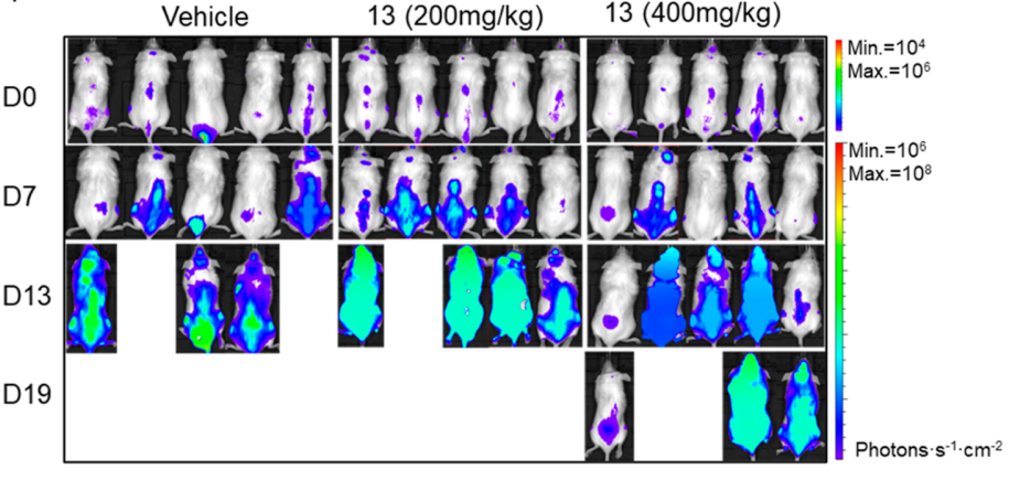
D-Luciferin purchased from MedChemExpress. Usage Cited in: J Med Chem. 2024 Nov 28;67(22):20100-20117. [Abstract]
The tumor model was established by tail vein injection of luciferase-labeled MOLM-13-Luc cells. After allowing 6 days for tumor growth post-inoculation, daily oral gavage treatment was initiated (designated as day 0 of treatment) at doses of 200 or 400 mg/kg. Subsequently, bioluminescence imaging was performed via intraperitoneal injection of 150 mg/kg D-Luciferin potassium.
-
Int J Oncol
Establishment of a novel human cell line retaining the characteristics of the original pancreatic adenocarcinoma, and evaluation of MEK as a therapeutic target. [Abstract]2020 Mar;56(3):761-771. PMID: 32124956 -
Plant Cell Environ
SlIAA23-SlARF6 module controls arbuscular mycorrhizal symbiosis by regulating strigolactone biosynthesis in tomato. [Abstract]2023 Jun;46(6):1921-1934. PMID: 36891914 -
Plant Cell Environ
PIF8-WRKY42-mediated salicylic acid synthesis modulates red light induced powdery mildew resistance in oriental melon. [Abstract]2023 May;46(5):1726-1742. PMID: 36759948 -
J Agric Food Chem
Transcription Repressor SsGATA2 Regulates Broad-Spectrum Resistance to Fungicides and Pathogenicity in Sclerotinia sclerotiorum. [Abstract]2025 Jul 9;73(27):16787-16803. PMID: 40558024 -
Mol Ther Nucleic Acids
Myomerger-derived peptide enhances skeletal muscle tropism and reduces liver transduction of lipid nanoparticles for gene delivery. [Abstract]2025 Nov 27;37(1):102785. PMID: 41458876 -
J Mater Chem B
GalNAc-modified five-component lipid nanoparticles for liver-targeted delivery of p65 siRNA to alleviate cytokine storm in liver injury via NF-κB targeting. [Abstract]2026 May 15. PMID: 42149053 -
Breast Cancer Res
ALDH3A2 targets arachidonic acid to promote cell metastasis in TNBC via AMPK/m-TOR signaling pathway. [Abstract]2025 Dec 18;27(1):217. PMID: 41413590 -
Biosensors (Basel)
The Construction and Application of a New Screening Method for Phosphodiesterase Inhibitors. [Abstract]2024 May 16;14(5):252. PMID: 38785726 -
Colloids Surf B Biointerfaces
Tumor exosome-based drug delivery system targeting ferroptosis and apoptosis for glioblastoma therapy. [Abstract]2026 Jan:257:115180. PMID: 41072330 -
-
Cell Oncol (Dordr)
2026 Apr 20;49(3):77. PMID: 42009994 -
Int Immunopharmacol
Identification of Siglec-15 as a novel target for CAR-T cell therapy in acute myeloid leukemia. [Abstract]2026 Mar 1:172:116170. PMID: 41558291 -
Int Immunopharmacol
Complement C1S is a potential prognostic biomarker and associated with M2 macrophage infiltration in gliomas: From bioinformatics to comprehensive experimental validation. [Abstract]2024 Nov 7;143(Pt 3):113573. PMID: 39515040 -
mBio
A Ralstonia solanacearum effector regulates plant cell death by disrupting the homeostasis of the BPA1-ACD11 complex. [Abstract]2025 Apr 9;16(4):e0366524. PMID: 39998214
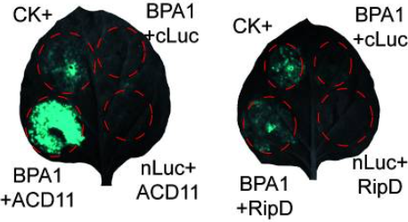
D-Luciferin purchased from MedChemExpress. Usage Cited in: mBio. 2025 Apr 9;16(4):e0366524. [Abstract]
Luciferase complementation imaging assay (LCA) of the interactions between BPA1 and either RipD or ACD11 in N. benthamiana. The interaction of Ca1 (AT3G01500) and Ca4 (AT1G70410) was used as a positive control, and n-Luc or c-Luc was used as a negative control. The interacting target proteins are fused to either the N-terminal or C-terminal fragment of luciferase, and the recombinant constructs are transformed into Agrobacterium tumefaciens GV3101. Forty hours after agroinfiltration,
-
Sci Rep
Tie1 derived from cervical cancer promotes the invasion and metastasis by Ang1 mediating Tie2/PI3K/Akt signaling axis and angiogenesis. [Abstract]2025 Dec 17;15(1):43979. PMID: 41407748 -
Sci Rep
The regulatory effect of CoL10A1 to the intracranial vascular invasion and cell proliferation in breast cancer via EMT pathway. [Abstract]2025 Apr 1;15(1):11040. PMID: 40169690 -
Neoplasia
ZC3H15 suppression ameliorates bone cancer pain through inhibiting neuronal oxidative stress and microglial inflammation. [Abstract]2025 Mar:61:101123. PMID: 39908779 -
Cell Signal
ROCK1 drives colorectal cancer liver metastasis via PRKCH-mediated bioenergetic reprogramming. [Abstract]2025 Oct 18:112174. PMID: 41115563 -
iScience
Down-regulation of MFNG promotes the metastatic potential of lung adenocarcinoma by regulating TGF-β/Smad signaling. [Abstract]2025 Sep 24;28(11):113647. PMID: 41210989 -
Nanomedicine
Generation of fluorescent and bioluminescent induced pluripotent stem cells with their application in tracking organoid development. [Abstract]2025 Oct:69:102849. PMID: 40789431 -
Antiviral Res
A rapid and versatile reverse genetic approach and visualization animal models for emerging zoonotic pseudorabies virus. [Abstract]2024 Dec:232:106036. PMID: 39522887 -
Front Biosci (Landmark Ed)
Argon Alleviates Bone Cancer Pain by Mitigating Neuronal Ferroptosis via the TLR2-COX-2 Pathway. [Abstract]2025 Sep 25;30(9):43761. PMID: 41074445 -
BMC Cancer
Immune characteristics analysis and construction of a four-gene prognostic signature for lung adenocarcinoma based on estrogen reactivity. [Abstract]2023 Oct 31;23(1):1047. PMID: 37907850 -
J Cell Commun Signal
CD2-CD58 axis orchestrates cytotoxic T lymphocyte function and metabolic crosstalk in breast cancer brain metastasis. [Abstract]2025 Aug 24;19(3):e70040. PMID: 40859981 -
Vet Res
CRISPR/Cas9-based generation of a recombinant double-reporter pseudorabies virus and its characterization in vitro and in vivo. [Abstract]2021 Jun 26;52(1):95. PMID: 34174954 -
Pathol Res Pract
Chromatin architecture protein HMGA2 promotes glioma malignancy via novel mechanism of IGFBP3 transcription inhibition. [Abstract]2025 Aug:272:156051. PMID: 40450974 -
Parasit Vectors
Clonorchis sinensis granulin promotes malignant transformation of human intrahepatic biliary epithelial cells through interaction with M2 macrophages via regulation of STAT3 phosphorylation and the MEK/ERK pathway. [Abstract]2023 Apr 24;16(1):139. PMID: 37095578 -
Arch Biochem Biophys
tsRNA-10547 generated by m1A modification-mediated tRNA shearing promotes colorectal cancer metastasis via suppressing CHRNA9. [Abstract]2026 May 15:110866. PMID: 42142645 -
Bioorg Med Chem
2025 Nov 19:133:118489. PMID: 41317695 -
Immun Inflamm Dis
B7-H3 promotes nasopharyngeal carcinoma progression by regulating CD8+ T cell exhaustion. [Abstract]2024 Sep;12(9):e70005. PMID: 39267471 -
J Leukoc Biol
Ginsenoside Rh2 reverses cyclophosphamide-induced immune deficiency by regulating fatty acid metabolism. [Abstract]2019 Nov;106(5):1089-1100. PMID: 31211478 -
Gastro Hep Adv
Hepatic Aquaporin 10 Expression Is Downregulated by Activated NFκB Signaling in Human Obstructive Cholestasis. [Abstract]2022 Nov 8;2(3):412-423. PMID: 39132646 -
-
J Gastrointest Oncol
Inhibition of hepatocellular carcinoma progression by artesunate via modulation of the TLR4/MyD88/NF-κB signaling pathway. [Abstract]2025 Apr 30;16(2):599-614. PMID: 40386608 -
J Vis Exp
2025 Oct 17:(224). PMID: 41182951 -
-
-
-
-
-
-
-
-
-
Biomed Pharmacother
FMNL1 down-regulation suppresses bone metastasis through reducing TGF-β1 expression in non-small cell lung cancer (NSCLC). [Abstract]2019 Sep:117:109126. PMID: 31387165


Lösungsmittel & Löslichkeit
DMSO : 125 mg/mL (445.90 mM; Need ultrasonic; Hygroscopic DMSO has a significant impact on the solubility of product, please use newly opened DMSO)
H2O : 9.09 mg/mL (32.43 mM; ultrasonic and adjust pH to 9 with 1M NaOH)
H2O : < 0.1 mg/mL (insoluble)
Please refer to the solubility information to select the appropriate solvent. Once prepared, please aliquot and store the solution to prevent product inactivation from repeated freeze-thaw cycles.
Storage method and period of stock solution: -80°C, 6 months; -20°C, 1 month (protect from light). When stored at -80°C, please use it within 6 months. When stored at -20°C, please use it within 1 month.
Konzentration (Stammlösung) × Volumen (Stammlösung) = Konzentration (Ziellösung) × Volumen (Ziellösung)

Protokoll
Mice[2]
In vivo BLI is performed using a cooled charge-coupled device camera system (IVIS Imaging System 100) 3, 5, 7, 10, 12, 14, 19, 21, 24, and 28 days after the inoculation of HCT116-Luc cells. Mice are injected with 75 mg/kg D-luciferin in 100 μL of phosphate-buffered saline subcutaneously near the scapula and were placed in the light-tight chamber of the imaging system. Beginning 5 min after injection, dorsal luminescent images with an exposure time of 1 s are acquired sequentially at a rate of one image per min until 20 min after D-luciferin injection. Data acquisition is continued until 40 min postinjection on days 3 or 5 and until 25 min on day 7, because of the prolonged time course of light emission. Binning is 4 and the field of view is 15 cm.
MedChemExpress (MCE) has not independently confirmed the accuracy of these methods. They are for reference only.


Reinheit & Dokumentation
-
Data Sheet (278 KB)
-
SDS (394 KB)
- English - EN (394 KB)
- Français - FR (394 KB)
- Deutsch - DE (394 KB)
- Norwegian - NO (394 KB)
- Español - ES (394 KB)
- Swedish - SV (394 KB)
- Italian - IT (394 KB)
- Korean - KR (394 KB)
- Portuguese - PT (394 KB)
-
Handling Instructions (2659 KB)
Verweise
[2]. Rajesh Shinde, et al. Luciferin derivatives for enhanced in vitro and in vivo bioluminescence assays. Biochemistry. 2006 Sep 19;45(37):11103-12. [Content Brief]
[3]. Inoue Y, et al. Timing of imaging after d-luciferin injection affects the longitudinal assessment of tumor growthusing in vivo bioluminescence imaging. Int J Biomed Imaging. 2010;2010:471408. [Content Brief]


Complete Stock Solution Preparation Table
Please refer to the solubility information to select the appropriate solvent. Once prepared, please aliquot and store the solution to prevent product inactivation from repeated freeze-thaw cycles.
Storage method and period of stock solution: -80°C, 6 months; -20°C, 1 month (protect from light). When stored at -80°C, please use it within 6 months. When stored at -20°C, please use it within 1 month.
| Optional Solvent | Concentration Solvent Mass | 1 mg | 5 mg | 10 mg | 25 mg |
|---|---|---|---|---|---|
| H2O / DMSO | 1 mM | 3.5672 mL | 17.8361 mL | 35.6722 mL | 89.1806 mL |
| 5 mM | 0.7134 mL | 3.5672 mL | 7.1344 mL | 17.8361 mL | |
| 10 mM | 0.3567 mL | 1.7836 mL | 3.5672 mL | 8.9181 mL | |
| 15 mM | 0.2378 mL | 1.1891 mL | 2.3781 mL | 5.9454 mL | |
| 20 mM | 0.1784 mL | 0.8918 mL | 1.7836 mL | 4.4590 mL | |
| 25 mM | 0.1427 mL | 0.7134 mL | 1.4269 mL | 3.5672 mL | |
| 30 mM | 0.1189 mL | 0.5945 mL | 1.1891 mL | 2.9727 mL | |
| DMSO | 40 mM | 0.0892 mL | 0.4459 mL | 0.8918 mL | 2.2295 mL |
| 50 mM | 0.0713 mL | 0.3567 mL | 0.7134 mL | 1.7836 mL | |
| 60 mM | 0.0595 mL | 0.2973 mL | 0.5945 mL | 1.4863 mL | |
| 80 mM | 0.0446 mL | 0.2230 mL | 0.4459 mL | 1.1148 mL | |
| 100 mM | 0.0357 mL | 0.1784 mL | 0.3567 mL | 0.8918 mL |


 Powered by Bioz