


Nile Red
Based on 129 publication(s) in Google Scholar
Nile red (Nile blue oxazone) is a lipophilic stain. Nile red has environment-sensitive fluorescence. Nile red is intensely fluorescent in a lipid-rich environment while it has minimal fluorescence in aqueous media. Nile red is an excellent vital stain for the detection of intracellular lipid droplets by fluorescence microscopy and flow cytof uorometry. Nile red stains intracellular lipid droplets red. The fluorescence wavelength is 559/635 nm.
商品は「研究用試薬」です。人や動物の医療用・臨床診断用・食品用の製品ではありません。
研究用途以外に使用した場合、当社は一切の責任を負いかねます。
- 純度: 99.74%
- CAS 番号: 7385-67-3
- 分子式: C20H18N2O2
- 分子量:318.37
-
保管条件:
4°C, protect from light
* In solvent : -80°C, 2 years; -20°C, 1 year (protect from light)

To place orders, for customer services and technical support, please contact: MedChemExpress USA
Tel: 609-228-6898 E-mail: [email protected] [email protected]


-
 生物活性
生物活性
-
化学情報
-
溶剤 & 溶解度
- 純度とドキュメンテーション
- 参考文献
-
 Help & FAQs


MedChemExpress(MCE)の使用を引用している文献 Nile Red
More- Bioact Mater. 2026 Apr 29;64:34-55. [Abstract]
- J Extracell Vesicles. 2024 Jan;13(1):e12401. [Abstract]
- Nat Neurosci. 2023 Apr;26(4):542-554. [Abstract]
- Autophagy. 2026 Jul 1:1-14. [Abstract]
- Nat Commun. 2026 Feb 12;17(1):2681. [Abstract]
- Nat Commun. 2025 Aug 26;16(1):7945. [Abstract]
- Nat Commun. 2025 Apr 3;16(1):3194. [Abstract]
- Nat Commun. 2023 Aug 28;14(1):5242. [Abstract]
- Nat Commun. 2022 Oct 5;13(1):5871. [Abstract]
- Nat Commun. 2020 Jan 13;11(1):240. [Abstract]
- Int J Oral Sci. 2025 Feb 1;17(1):6. [Abstract]
- J Adv Res. 2026 Apr 6:S2090-1232(26)00298-5. [Abstract]
- J Adv Res. 2025 Oct 10:S2090-1232(25)00764-7. [Abstract]
- J Adv Res. 2024 Apr 14:S2090-1232(24)00155-3. [Abstract]
- Redox Biol. 2023 Dec:68:102959. [Abstract]
- Nucleic Acids Res. 2022 Jun 24;50(12):6953-6967. [Abstract]
- Theranostics. 2022 May 26;12(10):4513-4535. [Abstract]
- Theranostics. 2021 Jan 1;11(5):2149-2169. [Abstract]
- Acta Pharm Sin B. 2025 Dec;15(12):6382-6398. [Abstract]
- Acta Pharm Sin B. 2025 May;15(5):2703-2722. [Abstract]
- J Clin Invest. 2024 Oct 22;134(24):e182217. [Abstract]
- Adv Sci (Weinh). 2025 Nov 19:e13711. [Abstract]
- Adv Sci (Weinh). 2025 Jun 20:e03233. [Abstract]
- Adv Sci (Weinh). 2025 Jul;12(28):e04532. [Abstract]
- Sci Adv. 2026 May 29;12(22):eaea6467. [Abstract]
- Chem Eng J. 2024 Sep 1.
- J Control Release. 2026 May 10:393:114759. [Abstract]
- J Control Release. 2024 Oct 24:376:553-565. [Abstract]
- J Control Release. 2024 Aug 20:374:242-253. [Abstract]
- J Control Release. 2021 Apr 10:332:194-209. [Abstract]
- Cell Death Dis. 2025 May 16;16(1):391. [Abstract]
- Cell Death Dis. 2025 Feb 17;16(1):104. [Abstract]
- Pharmacol Res. 2023 Feb:188:106666. [Abstract]
- Cancer Lett. 2026 Sep 28:656:218655. [Abstract]
- J Neuroinflammation. 2025 Oct 2;22(1):220. [Abstract]
- J Neuroinflammation. 2023 Nov 15;20(1):264. [Abstract]
- Adv Healthc Mater. 2025 Sep 30:e01619. [Abstract]
- Nano Today. 2025 Apr.
- Nano Today. 2024 Dec.
- J Clean Prod. 2024 Jun 10, 457, 142487.
- Nano Today. 47 (2022) 101675
- J Hazard Mater. 2025 Jul 9:496:139198. [Abstract]
- J Hazard Mater. 2024 Dec 5:480:136030. [Abstract]
- Part Fibre Toxicol. 2024 Mar 7;21(1):13. [Abstract]
- Food Chem. 2026 Apr 6:12:100401. [Abstract]
- Cell Death Discov. 2026 Apr 13;12(1):227. [Abstract]
- Acta Pharmacol Sin. 2026 Feb 27. [Abstract]
- Acta Biomater. 2025 Mar 1:194:305-322. [Abstract]
- J Transl Med. 2024 Aug 22;22(1):782. [Abstract]
- J Transl Med. 2022 May 14;20(1):222. [Abstract]
- Oncogene. 2026 Jun;45(22):2155-2170. [Abstract]
- Int J Biol Macromol. 2025 Dec 9:149590. [Abstract]
- Int J Mol Med. 2024 Aug;54(2):69. [Abstract]
- JACC Basic Transl Sci. 2025 Jul;10(7):101222. [Abstract]
- EMBO J. 2022 Aug 16;41(16):e110439. [Abstract]
- Anim Nutr. 2025 Jul 12.
- Environ Res. 2024 May 15:249:118402. [Abstract]
- Phytother Res. 2025 Aug 6. [Abstract]
- Free Radic Biol Med. 2025 Apr 28:235:150-161. [Abstract]
- Clin Transl Med. 2023 Jan;13(1):e1158. [Abstract]
- ACS Appl Mater Interfaces. 2024 Sep 18;16(37):48969-48981. [Abstract]
- Cell Rep. 2026 Jun 11;45(6):117550. [Abstract]
- Cell Prolif. 2024 Jun 12:e13687. [Abstract]
- Cell Prolif. 2021 Nov;54(11):e13134. [Abstract]
- Br J Pharmacol. 2025 Nov 11. [Abstract]
- Br J Pharmacol. 2025 Oct 3. [Abstract]
- Small Sci. 2025 Dec 12.
- Cancer Cell Int. 2025 Jan 27;25(1):25. [Abstract]
- J Ethnopharmacol. 2024 Sep 3:118777. [Abstract]
- J Agric Food Chem. 2025 Dec 29. [Abstract]
- Adv Intell Syst. 2025 Jan 28.
- J Agric Food Chem. 2024 Sep 4;72(35):19323-19332. [Abstract]
- Cell Mol Life Sci. 2026 May 22. [Abstract]
- Cell Mol Life Sci. 2025 Jul 3;82(1):270. [Abstract]
- Life Sci. 2018 Jul 1:204:55-64. [Abstract]
- Food Funct. 2025 Apr 22. [Abstract]
- Food Funct. 2024 Sep 30;15(19):9863-9879. [Abstract]
- Food Funct. 2021 Mar 7;12(5):2171-2188. [Abstract]
- Biosensors (Basel). 2026 May 1;16(5):261. [Abstract]
- Diabetes Obes Metab. 2025 Jun 17. [Abstract]
- RSC Adv. 2021 Nov 2;11(56):35331-35341. [Abstract]
- Oncogenesis. 2026 Mar 17;15(1):16. [Abstract]
- Mater Adv. 2025 May 7.
- Polymers. 2024 Aug 23;16(17):2394. [Abstract]
- Eur J Pharmacol. 2026 Mar 28:1019:178698. [Abstract]
- Cell Biol Toxicol. 2025 Feb 12;41(1):44. [Abstract]
- Eur J Pharmacol. 2023 Jan 5:938:175428. [Abstract]
- Int Immunopharmacol. 2025 Sep 6:165:115445. [Abstract]
- ACS Appl Bio Mater. 2024 May 20;7(5):2899-2910. [Abstract]
- Int J Mol Sci. 2021 May 31;22(11):5951. [Abstract]
- mBio. 2025 Apr 16:e0060225. [Abstract]
- mBio. 2024 May 16:e0067924. [Abstract]
- J Nutr Biochem. 2023 Sep:119:109408. [Abstract]
- ACS Omega. 2022 Dec 23;8(1):1710-1722. [Abstract]
- Molecules. 2023 Mar 31;28(7):3121. [Abstract]
- Eur J Pharm Biopharm. 2026 Jul:224:115081. [Abstract]
- Eur J Pharm Biopharm. 2023 Sep:190:24-34. [Abstract]
- Am J Pathol. 2025 Nov 7:S0002-9440(25)00410-9. [Abstract]
- Sci Rep. 2024 Dec 28;14(1):31310. [Abstract]
- Med Oncol. 2025 Jul 14;42(8):332. [Abstract]
- J Inflamm Res. 2025 Apr 25:18:5573-5586. [Abstract]
- iScience. 2025 Apr 3;28(5):112344. [Abstract]
- Brain Res Bull. 2022 Dec:191:93-106. [Abstract]
- Biochim Biophys Acta Mol Cell Res. 2026 Mar;1873(3):120107. [Abstract]
- Antiviral Res. 2023 Mar:211:105542. [Abstract]
- Pest Manag Sci. 2025 Apr;81(4):1923-1933. [Abstract]
- J Cell Physiol. 2024 Jan;239(1):180-192. [Abstract]
- Endocrinology. 2023 Jan 9;164(3):bqac215. [Abstract]
- Insect Sci. 2026 Apr 15. [Abstract]
- Int J Biochem Cell Biol. 2025 Jun 9:186:106820. [Abstract]
- Int J Biochem Cell Biol. 2024 May 9:172:106585. [Abstract]
- Arch Biochem Biophys. 2023 Aug:744:109689. [Abstract]
- Insect Mol Biol. 2024 Dec;33(6):792-805. [Abstract]
- Exp Eye Res. 2026 May:266:110932. [Abstract]
- Mol Pharmacol. 2025 May 16;107(7):100047. [Abstract]
- ACS Food Sci Technol. 2023 Nov 17.
- Theriogenology. 2023 Mar 15:199:11-18. [Abstract]
- Discov Oncol. 2023 May 12;14(1):67. [Abstract]
- Cytotechnology. 2025 Jun;77(3):105. [Abstract]
- bioRxiv. 2026 Mar 4.
- Res Sq. 2026 Jan 19.
- Patent. US20250154193A1.
- J Mol Liq. 2025 Apr 30.
- Research Square Preprint. 2024 Nov 17.
- bioRxiv. 2024 July 05.
- bioRxiv. 2024 Jul 30:2024.07.29.605485. [Abstract]
- SSRN. 2024 May 30.
- SSRN. 2023 Dec 2.
- Research Square Print. October 20th, 2022.
 Customer Validation & Images

-
IF
-
IF
-
Cell Imaging/Staining
-
IF
-
IF


生物活性
|
Cell Line
|
Type | Value | Description | References |
|---|---|---|---|---|
| L6 | IC50 |
65.9 μM
Compound: 7
|
Cytotoxicity against rat L6 cells
Cytotoxicity against rat L6 cells
|
[PMID: 24900219] |
Guide (The following is our recommended solution. This solution is merely a guideline and should be modified according to your specific needs.)
1. Preparation of storage solution and working solution
1.1 Preparation of storage solution
Prepare 1 mM storage solution using DMSO.
1.2 Preparation of working solution
Dilute the storage solution with pre-heated serum-free cell culture medium or PBS to a final concentration of 200-1000 nM.
Note: Please adjust the concentration of Nile Red working solution according to the actual situation, and prepare it as needed.
2. Cell staining
2.1 Suspension cells: Centrifuge to collect cells, add PBS for two washes, each for 5 minutes.
Adherent cells: Discard the culture medium, add trypsin to digest the cells. Centrifuge and discard the supernatant, then add PBS for two washes, each for 5 minutes.
2.2 Add 1 mL of Nile Red working solution, incubate at room temperature for 5-10 minutes.
2.3 400 g, 4℃ centrifuge for 3-4 minutes, discard the supernatant.
2.4 Add PBS for two washes of the cells, each for 5 minutes.
2.5 Resuspend the cells in 1 mL of serum-free medium or PBS, and observe using a fluorescence microscope.
MedChemExpress (MCE) has not independently confirmed the accuracy of these methods. They are for reference only.
MedChemExpress (MCE) has not independently confirmed the accuracy of these methods. They are for reference only.
598
543


化学情報
-
CAS 番号 7385-67-3
-
性状 Solid
-
分子量 318.37
-
分子式 C20H18N2O2
-
Color Green to dark green
-
SMILES
O=C1C2=CC=CC=C2C3=NC4=CC=C(N(CC)CC)C=C4OC3=C1
-
別名
ナイルレッド; Phenoxazone 9; Nile Blue A oxazone; ナイルブルーA オキサゾン
-
輸送条件
Room temperature in continental US; may vary elsewhere.
-
保管条件
4°C, protect from light
* In solvent : -80°C, 2 years; -20°C, 1 year (protect from light)


Publications (129)
-
Journal Impact Factor
-
Most Recent
-
Bioact Mater
Fe3+-based nanovesicle mediated transferrin hijacking for glioblastoma stem cell tumoricidal treatment and postoperative recurrence inhibition. [Abstract]2026 Apr 29;64:34-55. PMID: 42093838 -
J Extracell Vesicles
Extracellular vesicles from organoid-derived human retinal progenitor cells prevent lipid overload-induced retinal pigment epithelium injury by regulating fatty acid metabolism. [Abstract]2024 Jan;13(1):e12401. PMID: 38151470 -
Nat Neurosci
2023 Apr;26(4):542-554. PMID: 36941428 -
Autophagy
Genome-wide CRISPR screen identifies ELFN2 as a key regulator of host autophagy and lipid metabolism important for Toxoplasma gondii proliferation. [Abstract]2026 Jul 1:1-14. PMID: 42329082 -
Nat Commun
2026 Feb 12;17(1):2681. PMID: 41680151 -
Nat Commun
Nascent liver proteome reveals enzymes and transcription regulators under physiological and alcohol exposure conditions. [Abstract]2025 Aug 26;16(1):7945. PMID: 40858584 -
Nat Commun
Intrapleural pressure-controlled piezo-catalytic nanozyme for the inhibition of malignant pleural effusion. [Abstract]2025 Apr 3;16(1):3194. PMID: 40180981 -
Nat Commun
CircRREB1 mediates lipid metabolism related senescent phenotypes in chondrocytes through FASN post-translational modifications. [Abstract]2023 Aug 28;14(1):5242. PMID: 37640697 -
Nat Commun
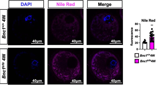
BNC1 deficiency-triggered ferroptosis through the NF2-YAP pathway induces primary ovarian insufficiency. [Abstract]2022 Oct 5;13(1):5871. PMID: 36198708
Nile Red purchased from MedChemExpress. Usage Cited in: Nat Commun. 2022 Oct 5;13(1):5871. [Abstract]
Isolated oocytes are fixed with 4% paraformaldehyde for 1 hour at room temperature and washed 3 times with 1% BSA in PBS. Then, they are stained with 10 µg/ml Nile red at 4 °C overnight.
-
Nat Commun
2020 Jan 13;11(1):240. PMID: 31932588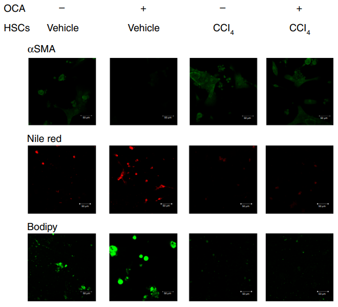
Nile Red purchased from MedChemExpress. Usage Cited in: Nat Commun. 2020 Jan 13;11(1):240. [Abstract]
αSMA, Bodipy and Nile Red staining of HSCs, data are representative of n = 3 biological independent samples. Scale bar, 50 μm.
-
Int J Oral Sci
Strontium-Alix interaction enhances exosomal miRNA selectively loading in synovial MSCs for temporomandibular joint osteoarthritis treatment. [Abstract]2025 Feb 1;17(1):6. PMID: 39890774 -
J Adv Res
LPCAT3 deficiency Drives phospholipid remodeling and mitochondrial oxidative dysfunction to accelerate MASH-HCC development. [Abstract]2026 Apr 6:S2090-1232(26)00298-5. PMID: 41951050 -
J Adv Res
MK8722 alleviates osteoarthritis by activating Sesn2 and transcriptionally upregulating BNIP3 to promote mitophagy and inhibit chondrocyte ferroptosis. [Abstract]2025 Oct 10:S2090-1232(25)00764-7. PMID: 41077348 -
J Adv Res
Interplay between lipid dysregulation and ferroptosis in chondrocytes and the targeted therapy effect of metformin on osteoarthritis. [Abstract]2024 Apr 14:S2090-1232(24)00155-3. PMID: 38621621 -
Redox Biol
A novel CPT1A covalent inhibitor modulates fatty acid oxidation and CPT1A-VDAC1 axis with therapeutic potential for colorectal cancer. [Abstract]2023 Dec:68:102959. PMID: 37977042 -
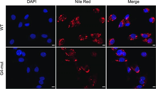
Nucleic Acids Res
DNA G-quadruplex structure participates in regulation of lipid metabolism through acyl-CoA binding protein. [Abstract]2022 Jun 24;50(12):6953-6967. PMID: 35748856
Nile Red purchased from MedChemExpress. Usage Cited in: Nucleic Acids Res. 2022 Jun 24;50(12):6953-6967. [Abstract]
The wild-type or G4-mutant human hepatic adenocarcinoma HepG2 cells are stained with Nile Red reagent to visualize the lipids with an Olympus Fluoview FV1000 confocal microscope.
-
Theranostics
Blockade of phosphotyrosine pathways suggesting SH2 superbinder as a novel therapy for pulmonary fibrosis. [Abstract]2022 May 26;12(10):4513-4535. PMID: 35832075 -
Theranostics
Intravital imaging of interactions between iNKT and kupffer cells to clear free lipids during steatohepatitis. [Abstract]2021 Jan 1;11(5):2149-2169. PMID: 33500717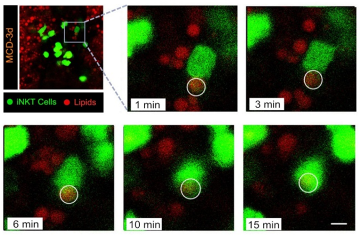
Nile Red purchased from MedChemExpress. Usage Cited in: Theranostics. 2021 Jan 1;11(5):2149-2169. [Abstract]
Injected 5 μg of Nile Red lipid dye into the tail vein of mice to label triglycerides in the liver prior to intravital imaging.
-
Acta Pharm Sin B
2025 Dec;15(12):6382-6398. PMID: 41477334 -
Acta Pharm Sin B
A cisplatin prodrug-based self-assembling ozone delivery nanosystem sensitizes radiotherapy in triple-negative breast cancer. [Abstract]2025 May;15(5):2703-2722. PMID: 40487662 -
J Clin Invest
MOGAT3-Mediated DAG Accumulation Drives Acquired Resistance to Anti-BRAF/EGFR Therapy in BRAFV600E-Mutant Metastatic Colorectal Cancer. [Abstract]2024 Oct 22;134(24):e182217. PMID: 39436710 -
Adv Sci (Weinh)
Combined Photothermal and mTOR-Targeted Therapy Overcomes Immune Evasion and Enhances Checkpoint Blockade Efficacy in Metastatic Triple-Negative Breast Cancer. [Abstract]2025 Nov 19:e13711. PMID: 41255280 -
Adv Sci (Weinh)
NPR1 Promotes Lipid Droplet Lipolysis to Enhance Mitochondrial Oxidative Phosphorylation and Fuel Gastric Cancer Metastasis. [Abstract]2025 Jun 20:e03233. PMID: 40539884 -
Adv Sci (Weinh)
A Feedback Loop Between Fatty Acid Metabolism and Epigenetics in Clear Cell Renal Carcinoma. [Abstract]2025 Jul;12(28):e04532. PMID: 40391655 -
Sci Adv
TPM1 drives cytoskeleton-immunometabolism coupling and LGALS9/CD45-mediated neuroinflammatory propagation in retinitis pigmentosa. [Abstract]2026 May 29;12(22):eaea6467. PMID: 42202020 -
-
J Control Release
Liposomal particokinetics and intratumoral microdistribution: A quantitative reassessment of the enhanced permeability and retention effect. [Abstract]2026 May 10:393:114759. PMID: 41763268 -
J Control Release
Size-dependent translocation and lymphatic transportation of polymeric nanocarriers post intraperitoneal administration. [Abstract]2024 Oct 24:376:553-565. PMID: 39427777 -
J Control Release
2024 Aug 20:374:242-253. PMID: 39153723 -
J Control Release
Combination of MAPK inhibition with photothermal therapy synergistically augments the anti-tumor efficacy of immune checkpoint blockade. [Abstract]2021 Apr 10:332:194-209. PMID: 33631225 -
Cell Death Dis
MARCH8 suppresses hepatocellular carcinoma by promoting SREBP1 degradation and modulating fatty acid de novo synthesis. [Abstract]2025 May 16;16(1):391. PMID: 40379644 -
Cell Death Dis
BRD1 deficiency affects SREBF1-related lipid metabolism through regulating H3K9ac/H3K9me3 transition to inhibit HCC progression. [Abstract]2025 Feb 17;16(1):104. PMID: 39962068 -
Pharmacol Res
PXR triggers YAP-TEAD binding and Sirt2-driven YAP deacetylation and polyubiquitination to promote liver enlargement and regeneration in mice. [Abstract]2023 Feb:188:106666. PMID: 36657504 -
Cancer Lett
Targeting C/EBPβ/SCD1/C16:1 loop-driven macrophage M2 polarization by Bruceine D improves anti-PD-1 immunotherapy in colorectal cancer. [Abstract]2026 Sep 28:656:218655. PMID: 42250753 -
J Neuroinflammation
Salt-sensitive hypertension promotes neuronal mitochondrial stress and neurodegenerative alterations via neuro-vascular metabolic reprogramming and local RAS signaling. [Abstract]2025 Oct 2;22(1):220. PMID: 41039568 -
J Neuroinflammation
Glyceryl triacetate promotes blood-brain barrier recovery after ischemic stroke through lipogenesis-mediated IL-33 in mice. [Abstract]2023 Nov 15;20(1):264. PMID: 37968698 -
Adv Healthc Mater
Trans-Organ Early Intervention for Myocardial Infarction Based on Bone Marrow-Homing Biomimetic Nanomedicine. [Abstract]2025 Sep 30:e01619. PMID: 41025748 -
-
-
-
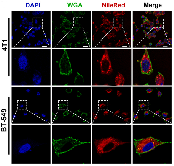
Nile Red purchased from MedChemExpress. Usage Cited in: Nano Today. 47 (2022) 101675
Subcellular biodistribution of NileRed-labeled PFD Liposome nano-particles in 4T1 and BT-549 cells was visualized by confocal laser microscopy. Blue, DAPI; Green, WGA 488; Red, NileRed-labeled PFD Liposome.
-
J Hazard Mater
2025 Jul 9:496:139198. PMID: 40652855 -
J Hazard Mater
Trophic transfer induced gut inflammation, dysbiosis, and inflammatory pathways in zebrafish via Artemia franciscana: A differential analysis of nanoplastic toxicity. [Abstract]2024 Dec 5:480:136030. PMID: 39362123 -
Part Fibre Toxicol
Exposure to high dose of polystyrene nanoplastics causes trophoblast cell apoptosis and induces miscarriage. [Abstract]2024 Mar 7;21(1):13. PMID: 38454452 -
Food Chem
Multi-omics reveals molecular basis of beef quality differences between Qinchuan and Nanyang cattle. [Abstract]2026 Apr 6:12:100401. PMID: 42011267 -
Cell Death Discov
Oncoprotein CYB561, acting in IRE1-XBP1-SREBF1 and FAK-ERK pathway, promotes breast cancer lipogenesis and progression. [Abstract]2026 Apr 13;12(1):227. PMID: 41974658 -
Acta Pharmacol Sin
2026 Feb 27. PMID: 41760780 -
Acta Biomater
Promotion of triple negative breast cancer immunotherapy by combining bioactive radicals with immune checkpoint blockade. [Abstract]2025 Mar 1:194:305-322. PMID: 39805523 -
J Transl Med
Chemically synthesized osteocalcin alleviates NAFLD via the AMPK-FOXO1/BCL6-CD36 pathway. [Abstract]2024 Aug 22;22(1):782. PMID: 39175012 -
J Transl Med
SIRT1 prevents cigarette smoking-induced lung fibroblasts activation by regulating mitochondrial oxidative stress and lipid metabolism. [Abstract]2022 May 14;20(1):222. PMID: 35568871
Nile Red purchased from MedChemExpress. Usage Cited in: J Transl Med. 2022 May 14;20(1):222. [Abstract]
Living cells are incubated with Nile Red (1 μM) for 15 min at 37 °C in dark. Then, cells are washed with HBSS/Ca/Mg and analyzed by fluorescence microscopy.
-
Oncogene
Targeting YRDC blocks codon-biased FABP7 translation and lipid droplet formation to overcome chemoresistance in glioblastoma. [Abstract]2026 Jun;45(22):2155-2170. PMID: 42014887 -
Int J Biol Macromol
Targeting aldose reductase in pulmonary fibrosis: Blocking de novo fatty acid synthesis halts pro-fibrotic M2 polarization. [Abstract]2025 Dec 9:149590. PMID: 41380895 -
Int J Mol Med
Therapeutic impacts of GNE‑477‑loaded H2O2 stimulus‑responsive dodecanoic acid‑phenylborate ester‑dextran polymeric micelles on osteosarcoma. [Abstract]2024 Aug;54(2):69. PMID: 38940336 -
JACC Basic Transl Sci
FNDC4 Prevents Aging-Related Cardiac Dysfunction: By Restoring AMPKα/PPARα-Dependent Mitochondrial Function. [Abstract]2025 Jul;10(7):101222. PMID: 40464727 -
EMBO J
PGE2 -EP3 axis promotes brown adipose tissue formation through stabilization of WTAP RNA methyltransferase. [Abstract]2022 Aug 16;41(16):e110439. PMID: 35781818
Nile Red purchased from MedChemExpress. Usage Cited in: EMBO J. 2022 Aug 16;41(16):e110439. [Abstract]
Representative pictures of Nile red-stained human BAs differentiated from hESC-derived brown progenitors.
-
-
Environ Res
Microcystin-RR promote lipid accumulation through CD36 mediated signal pathway and fatty acid uptake in HepG2 cells. [Abstract]2024 May 15:249:118402. PMID: 38309560 -
Phytother Res
Oridonin Ameliorates Metabolic-Dysfunction-Associated Fatty Liver Disease by Inhibiting PANoptosis via Modulating SIRT2/NLRP3 Pathway. [Abstract]2025 Aug 6. PMID: 40765411 -
Free Radic Biol Med
Role of ferroptosis mediated by abnormal membrane structure in DEHP-induced reproductive injury. [Abstract]2025 Apr 28:235:150-161. PMID: 40306442 -
Clin Transl Med
CircZSWIM6 mediates dysregulation of ECM and energy homeostasis in ageing chondrocytes through RPS14 post-translational modification. [Abstract]2023 Jan;13(1):e1158. PMID: 36604982 -
ACS Appl Mater Interfaces
Percutaneous Delivery of Hederacoside C-Loaded Nanoliposome Gel Alleviates Psoriasiform Skin Inflammation through the CCL17/Treg Axis. [Abstract]2024 Sep 18;16(37):48969-48981. PMID: 39233638 -
Cell Rep
High glucose impairs cognitive function by inducing lipid droplet accumulation through lactylation of HSD17B10 at K105. [Abstract]2026 Jun 11;45(6):117550. PMID: 42275213 -
Cell Prolif
Procyanidin B2 improves developmental capacity of bovine oocytes via promoting PPARγ/UCP1-mediated uncoupling lipid catabolism during in vitro maturation. [Abstract]2024 Jun 12:e13687. PMID: 38864666 -
Cell Prolif
D-mannose alleviates osteoarthritis progression by inhibiting chondrocyte ferroptosis in a HIF-2α-dependent manner. [Abstract]2021 Nov;54(11):e13134. PMID: 34561933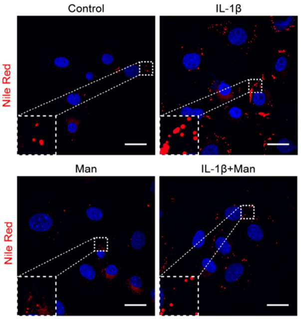
Nile Red purchased from MedChemExpress. Usage Cited in: Cell Prolif. 2021 Nov;54(11):e13134. [Abstract]
For lipid droplet staining, fixed cells are stained for 30 minutes with 0.1 µg/mL Nile Red (MCE), and the nucleus is counterstained using DAPI.
-
Br J Pharmacol
Imperatorin ameliorates metabolic dysfunction-associated fatty liver disease through modulating Suv39h1/Fabps/Cept1 signalling pathway. [Abstract]2025 Nov 11. PMID: 41218845 -
Br J Pharmacol
Acetaminophen aggravates valproate-induced hepatic lipid accumulation and apoptosis by facilitating valproate retention. [Abstract]2025 Oct 3. PMID: 41041829 -
-
Cancer Cell Int
DLAT is involved in ovarian cancer progression by modulating lipid metabolism through the JAK2/STAT5A/SREBP1 signaling pathway. [Abstract]2025 Jan 27;25(1):25. PMID: 39871246 -
J Ethnopharmacol
Chemometric-based analysis and bioassay guided identification of potent compounds with intestinal motility promoting effects from Dalitong Granules. [Abstract]2024 Sep 3:118777. PMID: 39236779 -
J Agric Food Chem
Genistein Ameliorates Rifampicin-Undermined Hepatic Cholesterol Efflux via the CH25H-LXRα-ABCA Pathway. [Abstract]2025 Dec 29. PMID: 41459645 -
-
J Agric Food Chem
Azadirachtin Induces Fat Body Apoptosis by Suppressing Caspase- 8 in the Fall Armyworm, Spodoptera frugiperda. [Abstract]2024 Sep 4;72(35):19323-19332. PMID: 39174876 -
Cell Mol Life Sci
Obesity-induced oleic acid metabolic dysregulation may exacerbate osteoarthritis through the degradation of SOX9. [Abstract]2026 May 22. PMID: 42171703 -
Cell Mol Life Sci
Aquaglyceroporin-7 ameliorates sorafenib resistance and immune evasion in hepatocellular carcinoma through inhibition of lipid accumulation. [Abstract]2025 Jul 3;82(1):270. PMID: 40610622 -
Life Sci
Polyacetylene glycoside attenuates ischemic kidney injury by co-inhibiting inflammation, mitochondria dysfunction and lipotoxicity. [Abstract]2018 Jul 1:204:55-64. PMID: 29733848 -
Food Funct
Crocin extends lifespan by mitigating oxidative stress and regulating lipid metabolism through the DAF-16/FOXO pathway. [Abstract]2025 Apr 22. PMID: 40260541 -
Food Funct
2024 Sep 30;15(19):9863-9879. PMID: 39246047 -
Food Funct
Plant sterol ester of α-linolenic acid ameliorates high-fat diet-induced nonalcoholic fatty liver disease in mice: association with regulating mitochondrial dysfunction and oxidative stress via activating AMPK signaling. [Abstract]2021 Mar 7;12(5):2171-2188. PMID: 33566044 -
Biosensors (Basel)
Imaging Study of MnO2-Based Nanomotors Modulating HIF-1α/Lipid Droplet Biogenesis and Activating the cGAS-STING Pathway. [Abstract]2026 May 1;16(5):261. PMID: 42187457 -
Diabetes Obes Metab
AdipoRon attenuates steatosis, inflammation and fibrosis in murine diet-induced NASH via inhibiting ER stress. [Abstract]2025 Jun 17. PMID: 40528684 -
RSC Adv
BSA-MnO2-SAL multifunctional nanoparticle-mediated M1 macrophages polarization for glioblastoma therapy. [Abstract]2021 Nov 2;11(56):35331-35341. PMID: 35493189 -
Oncogenesis
IFFO1 inhibits breast cancer by coordinating mitochondrial fission and fatty acid synthesis via the LaminA/C-PGC1α axis. [Abstract]2026 Mar 17;15(1):16. PMID: 41844580 -
-
Polymers
Composite Mineralized Collagen/Polycaprolactone Scaffold-Loaded Microsphere System with Dual Osteogenesis and Antibacterial Functions. [Abstract]2024 Aug 23;16(17):2394. PMID: 39274026 -
Eur J Pharmacol
Phellopterin from angelica dahurica is a natural antagonist of glucocorticoid receptors regulating lipid and cholesterol metabolism. [Abstract]2026 Mar 28:1019:178698. PMID: 41765273 -
Cell Biol Toxicol
Enhancing cardiac repair post-myocardial infarction: a study on GATM/Gel hydrogel therapeutics. [Abstract]2025 Feb 12;41(1):44. PMID: 39937362 -
Eur J Pharmacol
A novel therapy for hepatic cholestasis treatment-the combination of rosiglitazone and fenofibrate. [Abstract]2023 Jan 5:938:175428. PMID: 36436592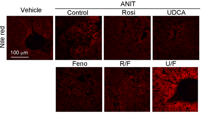
Nile Red purchased from MedChemExpress. Usage Cited in: Eur J Pharmacol. 2023 Jan 5:938:175428. [Abstract]
Hepatic lipid and FFA content were determined by Nile red staining of liver frozen sections.
-
Int Immunopharmacol
RNF180 suppressed aggressiveness by degrading NOTCH1, TRIM24 and FOXC1, and chemoresistance by degrading ACC1 and ACLY in colorectal cancer. [Abstract]2025 Sep 6:165:115445. PMID: 40915184 -
ACS Appl Bio Mater
Ionic Liquid Pretreatment Enhances Skin Penetration of 5-Aminolevulinic Acid: A Promising Scheme for Photodynamic Therapy for Acne Vulgaris. [Abstract]2024 May 20;7(5):2899-2910. PMID: 38607995 -
Int J Mol Sci
Serratia symbiotica Enhances Fatty Acid Metabolism of Pea Aphid to Promote Host Development. [Abstract]2021 May 31;22(11):5951. PMID: 34073039
Nile Red purchased from MedChemExpress. Usage Cited in: Int J Mol Sci. 2021 May 31;22(11):5951. [Abstract]
For lipid staining, fat bodies are submerged in Nile red solution at a final working concentration of 10 μg/mL in an acetone/water (1:9) mixture and visualized using a fluorescent microscope at Ex543/Em626 nm.
-
mBio
AUP1 and UBE2G2 complex targets STING signaling and regulates virus-induced innate immunity. [Abstract]2025 Apr 16:e0060225. PMID: 40237449 -
mBio
Phage protein Gp11 blocks Staphylococcus aureus cell division by inhibiting peptidoglycan biosynthesis. [Abstract]2024 May 16:e0067924. PMID: 38752726 -
J Nutr Biochem
Through regulation of the SIRT1 pathway plant sterol ester of α-linolenic acid inhibits pyroptosis thereby attenuating the development of NASH in mice. [Abstract]2023 Sep:119:109408. PMID: 37336331 -
ACS Omega
2022 Dec 23;8(1):1710-1722. PMID: 36643556 -
Molecules
Uncarboxylated Osteocalcin Decreases SCD1 by Activating AMPK to Alleviate Hepatocyte Lipid Accumulation. [Abstract]2023 Mar 31;28(7):3121. PMID: 37049884 -
Eur J Pharm Biopharm
Spatiotemporally controlled systemic delivery reshapes the in vivo fate of a cationic antimicrobial peptide for lung-selective exposure and an improved therapeutic index. [Abstract]2026 Jul:224:115081. PMID: 42044857 -
Eur J Pharm Biopharm
2023 Sep:190:24-34. PMID: 37433416 -
Am J Pathol
Nonalcoholic Steatohepatitis Exacerbates Inflammatory Bone Destruction and Macrophage NLR Family Pyrin Domain-Containing 3 Inflammasome Activation in Ligature-Induced Periodontitis. [Abstract]2025 Nov 7:S0002-9440(25)00410-9. PMID: 41205805 -
Sci Rep
VPS28 regulates triglyceride synthesis via ubiquitination in bovine mammary epithelial cells. [Abstract]2024 Dec 28;14(1):31310. PMID: 39732879 -
Med Oncol
Enhanced LDL uptake and PPARα signaling support OSCC cell survival under glutamine deprivation. [Abstract]2025 Jul 14;42(8):332. PMID: 40660028 -
J Inflamm Res
Calycosin-7-Glucoside Alleviates Atherosclerosis by Inhibiting Ox-LDL-Induced Foam Cell Formation and Inflammatory Response in THP-1-Derived Macrophages via ATF-1 Activation Through the p38/MAPK Pathway. [Abstract]2025 Apr 25:18:5573-5586. PMID: 40303003 -
iScience
2025 Apr 3;28(5):112344. PMID: 40276762 -
Brain Res Bull
Movement disorder caused by FRRS1L deficiency may be associated with morphological and functional disorders in Purkinje cells. [Abstract]2022 Dec:191:93-106. PMID: 36330921 -
Biochim Biophys Acta Mol Cell Res
USP49 regulates lipid metabolism in hepatocellular carcinoma by stabilizing RACK1 to promote tumor proliferation and migration. [Abstract]2026 Mar;1873(3):120107. PMID: 41519241 -
Antiviral Res
Identification of desoxyrhapontigenin as a novel antiviral agent against congenital Zika virus infection. [Abstract]2023 Mar:211:105542. PMID: 36646387 -
Pest Manag Sci
Knockout BR-C induces premature expression of E93 thus triggering adult differentiation under larval morphology. [Abstract]2025 Apr;81(4):1923-1933. PMID: 39641237 -
J Cell Physiol
NAMPT regulates mitochondria function and lipid metabolism during porcine oocyte maturation. [Abstract]2024 Jan;239(1):180-192. PMID: 37992208 -
Endocrinology
STRA6 promotes thyroid carcinoma progression via activation of the ILK/AKT/mTOR axis in cells and female nude mice. [Abstract]2023 Jan 9;164(3):bqac215. PMID: 36592123 -
Insect Sci
Single-minded regulates larval energy homeostasis in the fall armyworm Spodoptera frugiperda. [Abstract]2026 Apr 15. PMID: 41982118 -
Int J Biochem Cell Biol
Cofilin is a key regulator of oxidative stress-induced intercellular tunneling nanotubes formation. [Abstract]2025 Jun 9:186:106820. PMID: 40499810 -
Int J Biochem Cell Biol
Tamoxifen upregulates the peroxisomal β-oxidation enzyme Enoyl CoA hydratase and 3-hydroxyacyl CoA hydratase ameliorating hepatic lipid accumulation in mice. [Abstract]2024 May 9:172:106585. PMID: 38734232 -
Arch Biochem Biophys
Exogenous galanin alleviates hepatic steatosis by promoting autophagy via the AMPK-mTOR pathway. [Abstract]2023 Aug:744:109689. PMID: 37429535 -
Insect Mol Biol
Juvenile hormone-induced microRNA miR-iab-8 regulates lipid homeostasis and metamorphosis in Drosophila melanogaster. [Abstract]2024 Dec;33(6):792-805. PMID: 39005109 -
Exp Eye Res
SREBP1-driven SCD1 protects retinal pigment epithelium from oxidative damage by activating the NRF2/GPX4 axis. [Abstract]2026 May:266:110932. PMID: 41720386 -
Mol Pharmacol
Fenofibrate promotes erucic acid metabolism by peroxisome enzyme EHHADH activation alleviating high-fat diet-induced steatotic liver disease. [Abstract]2025 May 16;107(7):100047. PMID: 40516250 -
-
Theriogenology
Leonurine improves bovine oocyte maturation and subsequent embryonic development by reducing oxidative stress and improving mitochondrial function. [Abstract]2023 Mar 15:199:11-18. PMID: 36680865 -
Discov Oncol
Myc derived circRNA promotes triple-negative breast cancer progression via reprogramming fatty acid metabolism. [Abstract]2023 May 12;14(1):67. PMID: 37173608 -
Cytotechnology
Catalpol inhibits Hedgehog signaling pathway to suppress proliferation and promote lipid accumulation in rat meibomian gland epithelial cells. [Abstract]2025 Jun;77(3):105. PMID: 40406032 -
-
-
-
-
-
-
bioRxiv
2024 Jul 30:2024.07.29.605485. PMID: 39131374 -
-
-


溶剤 & 溶解度
DMSO : 2 mg/mL (6.28 mM; ultrasonic and warming and heat to 60°C; Hygroscopic DMSO has a significant impact on the solubility of product, please use newly opened DMSO)
Ethanol : 1 mg/mL (3.14 mM; ultrasonic and warming and heat to 60°C)
Please refer to the solubility information to select the appropriate solvent. Once prepared, please aliquot and store the solution to prevent product inactivation from repeated freeze-thaw cycles.
Storage method and period of stock solution: -80°C, 2 years; -20°C, 1 year (protect from light). When stored at -80°C, please use it within 2 years. When stored at -20°C, please use it within 1 year.
濃度 (開始) × 体積 (開始) = 濃度 (終了) × 体積 (終了)


純度とドキュメンテーション
-
データシート (277 KB)
-
SDS (393 KB)
- English - EN (393 KB)
- Français - FR (393 KB)
- Deutsch - DE (393 KB)
- Norwegian - NO (393 KB)
- Español - ES (393 KB)
- Swedish - SV (393 KB)
- Italian - IT (393 KB)
- Korean - KR (393 KB)
- Portuguese - PT (393 KB)
-
取扱説明書 (2659 KB)
参考文献
[1]. Greenspan P, et al. Nile red: a selective fluorescent stain for intracellular lipid droplets. J Cell Biol. 1985 Mar;100(3):965-73. [Content Brief]
[2]. Gibrán S Alemán-Nava, et al. How to use Nile Red, a selective fluorescent stain for microalgal neutral lipids. J Microbiol Methods. 2016 Sep;128:74-79. [Content Brief]
[3]. Wilber Escorcia, et al. Quantification of Lipid Abundance and Evaluation of Lipid Distribution in Caenorhabditis elegans by Nile Red and Oil Red O Staining. J Vis Exp. 2018 Mar 5;(133):57352. [Content Brief]
[4]. Elizabeth C Pino, et al. Biochemical and high throughput microscopic assessment of fat mass in Caenorhabditis elegans. J Vis Exp. 2013 Mar 30;(73):50180. [Content Brief]


Complete Stock Solution Preparation Table
Please refer to the solubility information to select the appropriate solvent. Once prepared, please aliquot and store the solution to prevent product inactivation from repeated freeze-thaw cycles.
Storage method and period of stock solution: -80°C, 2 years; -20°C, 1 year (protect from light). When stored at -80°C, please use it within 2 years. When stored at -20°C, please use it within 1 year.
| Optional Solvent | Concentration Solvent Mass | 1 mg | 5 mg | 10 mg | 25 mg |
|---|---|---|---|---|---|
| Ethanol / DMSO | 1 mM | 3.1410 mL | 15.7050 mL | 31.4100 mL | 78.5250 mL |
| DMSO | 5 mM | 0.6282 mL | 3.1410 mL | 6.2820 mL | 15.7050 mL |


 Powered by Bioz