



FTI-277
Based on 7 publication(s) in Google Scholar
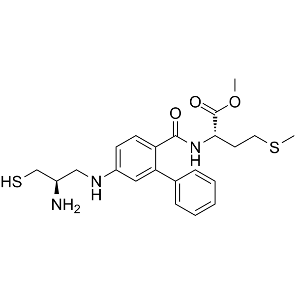
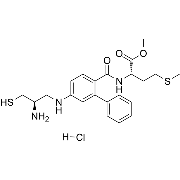
FTI-277 is a farnesyltransferase (FTase) inhibitor. FTI-277 inhibits Ras farnesylation, blocks the phosphorylation of downstream ERK1/2 and mTOR, and reduces membrane-bound active N-ras protein. FTI-277 activates caspase 3, upregulates Bim expression, induces cell apoptosis, suppresses regulatory T cell expansion, enhances macrophage phagocytosis, and improves bacterial clearance. FTI-277 activates the PI3K/Akt signaling pathway, inhibits osteoblast differentiation, and reduces the proliferation ability of neuroblastoma cells. FTI-277 can be used in research related to head and neck squamous cell carcinoma, neuroblastoma, sepsis, and vascular calcification.
Nur für Forschungszwecke. Wir verkaufen nicht an Patienten.
- CAS. Nr.: 170006-73-2
- Formel: C22H29N3O3S2
- Molecular Weight:447.61
-
Speicherung:
Please store the product under the recommended conditions in the Certificate of Analysis.

To place orders, for customer services and technical support, please contact: MedChemExpress USA
Tel: 609-228-6898 E-mail: [email protected] [email protected]


-
 Biologische Aktivität
-
Chemical Information
- Protokoll
- Reinheit & Dokumentation
- Verweise
-
 Help & FAQs


-
HY-10502
TipifarnibFTase Inhibitor -
HY-16273A
L-778123 hydrochlorideFarnesyl Transferase Inhibitor -
HY-16111
BMS-214662Farnesyl Transferase Inhibitor -
HY-15872A
FTI-277 hydrochlorideFTase Inhibitor -
HY-16373
CP-609754Farnesyltransferase Inhibitor -
HY-10502A
Tipifarnib (S enantiomer) -

HY-10502R
Tipifarnib (Standard)Reference Standard -
HY-15136
LonafarnibFarnesyltransferase Inhibitor -
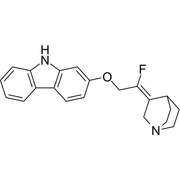
HY-100313
YM-53601 free baseSqualene Synthetase Inhibitor -
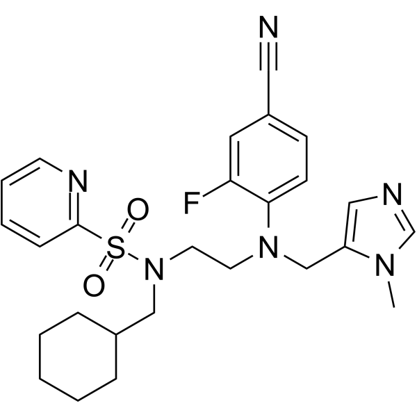
HY-128350
FGTI-2734Farnesyl/Geranylgeranyl Transferase Inhibitor -
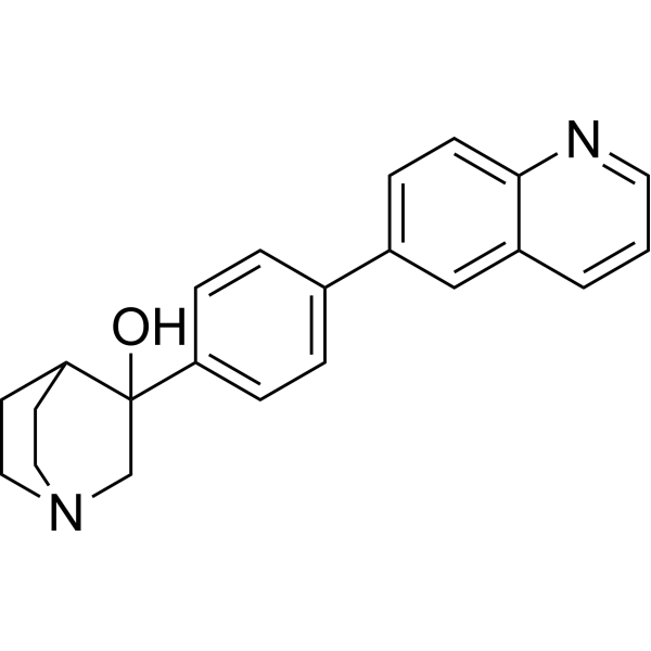
HY-100299
RPR107393 free baseSqualene Synthase Inhibitor -
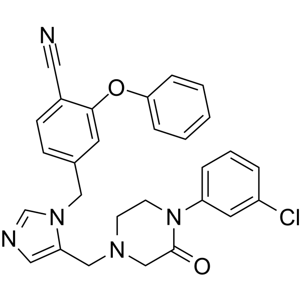
HY-U00327
Prenyl-IN-1
Publications Citing Use of MedChemExpress (MCE) FTI-277
More- Adv Sci (Weinh). 2024 Aug;11(30):e2307751. [Abstract]
- Mol Cell Proteomics. 2023 Aug;22(8):100593. [Abstract]
- J Cell Mol Med. 2024 Dec;28(24):e70273. [Abstract]
- Fish Shellfish Immunol. 2019 Jun:89:281-289. [Abstract]
- J Virol. 2026 Apr 21;100(4):e0209725. [Abstract]
- FEBS Open Bio. 2025 Aug 8. [Abstract]
- Oncotarget. 2017 Nov 22;8(65):109135-109150. [Abstract]
 Customer Validation & Images

-
WB
Alle Caspase Isoform-spezifische Produkte anzeigen
More

Biologische Aktivität
Beschreibung
IC50 & Target
[1]|
ERK1 |
ERK2 |
Caspase 3 |
Cellular Effect
|
Cell Line

|
Type | Value | Description | References |
|---|---|---|---|---|
| NIH3T3 | ED50 |
>25 μM
Compound: FTI-277
|
Effective dose against murine 3T3 fibroblasts cells
Effective dose against murine 3T3 fibroblasts cells
|
[PMID: 14711313] |
| NIH3T3 | ED50 |
0.2 μM
Compound: 2
|
Inhibition of Ha-Ras processing in NIH3T3 Ras transformed cells.
Inhibition of Ha-Ras processing in NIH3T3 Ras transformed cells.
|
[PMID: 9784104] |
In Vitro
FTI-277 (1-40 μM; 24-72 h) decreases the viability of HEp-2 head and neck squamous cell carcinoma cells in a concentration-dependent manner, with a 58.5% reduction in viability at the highest tested concentration (40 μM) after 72 h of treatment[1].
FTI-277 (0.5-10 μM; 24-72 h) decreases the viability of HSC-3 head and neck squamous cell carcinoma cells in a concentration-dependent manner, with a 77.0% reduction in viability at the highest tested concentration (10 μM) after 72 h of treatment, and exhibits greater potency against HSC-3 cells than HEp-2 cells[1].
FTI-277 (40 μM; 36 h) significantly increases caspase-3 activity in HEp-2 head and neck squamous cell carcinoma cells[1].
FTI-277 (5 μM; 36 h) significantly increases caspase-3 activity in HSC-3 head and neck squamous cell carcinoma cells, demonstrating greater potency than against HEp-2 cells[1].
FTI-277 (40 μM; 36 h) significantly increases the percentage of annexin V-positive HEp-2 head and neck squamous cell carcinoma cells, indicating induction of apoptosis[1].
FTI-277 (5 μM; 36 h) significantly increases the percentage of annexin V-positive HSC-3 head and neck squamous cell carcinoma cells, indicating induction of apoptosis with greater potency than against HEp-2 cells[1].
FTI-277 (40 μM; 48 h) suppresses Ras membrane localization, reduces phosphorylation of ERK1/2 and mTOR, and increases Bim protein expression in HEp-2 head and neck squamous cell carcinoma cells[1].
FTI-277 (5 μM; 48 h) suppresses Ras membrane localization, reduces phosphorylation of ERK1/2 and mTOR, and increases Bim protein expression in HSC-3 head and neck squamous cell carcinoma cells, demonstrating greater potency than against HEp-2 cells[1].
FTI-277 (5 μM; 72 h) significantly decreases the viability of NW7 v-H-Ras-transfected NIH3T3 cells but has no significant effect on NV20 empty vector-transfected NIH3T3 cells, indicating selective activity against Ras-overexpressing cells[1].
FTI-277 (1-20 μM; 8-10 days) inhibits osteogenic differentiation and βGP-induced mineralization in primary bovine vascular smooth muscle cells, with significant effects observed at concentrations ≥10 μM, and even when added up to 6 days post-βGP initiation[3].
FTI-277 (10 μM; 77 h) enhances Akt phosphorylation in primary bovine vascular smooth muscle cells when followed by serum starvation and short-term serum stimulation[3].
FTI-277 (10-20 μM; 9-10 days) maintains Akt phosphorylation during βGP-induced mineralization in primary bovine vascular smooth muscle cells, and this Akt activation is required for FTI-277's inhibitory effect on mineralization[3].
FTI-277 (10 μM) inhibits phosphate-induced apoptosis in human coronary artery vascular smooth muscle cells, and this effect depends on Akt activation[3].
FTI-277 (10 μM; 96 h) reduces farnesylated N-Ras protein levels by approximately 50% in IMR-5 neuroblastoma cells[4].
FTI-277 (10 μM; 96 h) reduces basal MAP-kinase activity by ~47% and blunts BDNF-induced MAP-kinase activation to 35% of control levels in IMR-5 neuroblastoma cells[4].
FTI-277 (10 μM; 96 h) reduces basal N-myc expression to 70% of controls and blunts BDNF-induced N-myc expression to 53% of control-stimulated levels in IMR-5 neuroblastoma cells[4].
FTI-277 (10 μM; 96 h) significantly reduces cell proliferation across complete, serum-free, and BDNF-supplemented media, and abolishes BDNF-induced proliferation in IMR-5 neuroblastoma cells[4].
MedChemExpress (MCE) has not independently confirmed the accuracy of these methods. They are for reference only.
-
Cell Line:HEp-2 head and neck squamous cell carcinoma cells
-
Concentration:1 μM, 5 μM, 10 μM, 20 μM, 40 μM
-
Incubation Time:24 h, 48 h, 72 h
-
Result:Decreased HEp-2 cell viability in a concentration-dependent manner.
Reduced cell viability to 97.9% with 1 μM, 98.9% with 5 μM, 89.1% with 10 μM, 78.3% with 20 μM, and 41.5% with 40 μM after 72 h treatment.
Induced statistically significant viability reductions with 20 μM and 40 μM at 48 h, and with 10 μM, 20 μM, and 40 μM at 72 h relative to controls.
-
Cell Line:HSC-3 head and neck squamous cell carcinoma cells
-
Concentration:0.5 μM, 1 μM, 2.5 μM, 5 μM, 10 μM
-
Incubation Time:24 h, 48 h, 72 h
-
Result:Decreased HSC-3 cell viability in a concentration-dependent manner, with greater potency than in HEp-2 cells.
Reduced cell viability to 89.7% with 0.5 μM, 69.1% with 1 μM, 52.9% with 2.5 μM, 33.4% with 5 μM, and 23.0% with 10 μM after 72 h treatment.
Induced statistically significant viability reductions with all tested concentrations at 24, 48, and 72 h relative to controls.
-
Cell Line:HEp-2 head and neck squamous cell carcinoma cells
-
Concentration:40 μM
-
Incubation Time:36 h
-
Result:Increased the percentage of annexin V-positive HEp-2 cells to approximately 42%, relative to control cells with approximately 7% annexin V-positive cells.
Induced statistically significant elevation of annexin V-positive cells, indicating apoptosis induction.
-
Cell Line:HSC-3 head and neck squamous cell carcinoma cells
-
Concentration:5 μM
-
Incubation Time:36 h
-
Result:Increased the percentage of annexin V-positive HSC-3 cells to approximately 47%, relative to control cells with approximately 9% annexin V-positive cells.
Induced statistically significant elevation of annexin V-positive cells, indicating apoptosis induction with greater potency than against HEp-2 cells.
-
Cell Line:HEp-2 head and neck squamous cell carcinoma cells
-
Concentration:40 μM
-
Incubation Time:48 h
-
Result:Inhibited Ras membrane localization and increased Ras cytoplasmic localization in HEp-2 cells.
Decreased the expression of phosphorylated ERK1/2 and phosphorylated mTOR.
Increased the expression of Bim protein, relative to control cells.
-
Cell Line:HSC-3 head and neck squamous cell carcinoma cells
-
Concentration:5 μM
-
Incubation Time:48 h
-
Result:Inhibited Ras membrane localization and increased Ras cytoplasmic localization in HSC-3 cells.
Decreased the expression of phosphorylated ERK1/2 and phosphorylated mTOR.
Increased the expression of Bim protein, relative to control cells.
Demonstrated greater potency than against HEp-2 cells.
-
Cell Line:NV20 empty vector-transfected NIH3T3 cells, NW7 v-H-Ras-transfected NIH3T3 cells
-
Concentration:5 μM
-
Incubation Time:72 h
-
Result:Had no significant effect on the viability of NV20 empty vector-transfected NIH3T3 cells.
Significantly reduced the viability of NW7 v-H-Ras-transfected NIH3T3 cells to approximately 35%, relative to untreated NW7 cells.
Exhibited selective activity against Ras-overexpressing cells.
-
Cell Line:IMR-5 neuroblastoma cells
-
Concentration:10 μM
-
Incubation Time:96 h
-
Result:Reduced farnesylated N-Ras protein levels by 47%.\nReduced basal MAP-kinase activity by 47% compared to untreated controls.
Blunted BDNF-induced MAP-kinase activation to 35% of control levels.
-
Cell Line:IMR-5 neuroblastoma cells
-
Concentration:10 μM
-
Incubation Time:96 h
-
Result:Reduced basal N-myc expression to 70% of untreated control levels.
Blunted BDNF-induced N-myc expression to 53% of control-stimulated levels.
-
Cell Line:IMR-5 neuroblastoma cells
-
Concentration:10 μM
-
Incubation Time:96 h
-
Result:Decreased cell numbers by 10% in complete medium over 48 hours, compared to an ~80% increase in untreated controls.
Resulted in significantly lower cell numbers than controls in serum-free medium.
Abolished BDNF-induced 22% proliferation increase seen in control cells in serum-free medium.
Reduced cell proliferation significantly across all three media conditions.
In Vivo
MedChemExpress (MCE) has not independently confirmed the accuracy of these methods. They are for reference only.
-
Animal Model:C57BL/6 (male, 7 weeks of age, sepsis induced by cecal ligation and puncture)[2]
-
Dosage:25 mg/kg
-
Administration:i.p.; single dose
-
Result:Increased survival rate of septic mice to 67% (10/15 survived to 7 days) compared to 7% (1/15) in vehicle-treated group.
Significantly reduced bacterial colony-forming units in blood and peritoneal cavity at 16 hours post-CLP.
Almost completely blocked sepsis-induced increase in serum HMGB1 concentration at 16 hours post-CLP.
Reduced percentage of TUNEL-positive apoptotic cells in spleen from ~7% to ~2% and in thymus from ~9% to ~1% at 16 hours post-CLP.
Reversed sepsis-induced increase in splenic farnesylated protein levels and farnesyltransferase activity to sham-equivalent levels at 16 hours post-CLP.
Partially reversed sepsis-induced increase in splenic CD4+Foxp3+ regulatory T cells from ~22% to ~15% of total CD4+ cells without altering total CD4+ splenocyte counts.
Restored IFN-γ secretion from ~30 pg/mL to ~100 pg/mL and splenocyte proliferative response to sham-equivalent levels, and reversed sepsis-induced decrease in IFN-γ to IL-4 ratio.
Reduced sepsis-induced increases in PD-L1 and PD-1 expression on splenic CD4+ T cells and macrophages.
Increased phagocytotic activity of peritoneal F4/80+ macrophages, with percentage of fluorescent microsphere-positive cells rising from ~45% to ~65% and mean fluorescence intensity per cell rising from ~220 to ~300.
- Western Blot
- RNA extraction experimental
- Annexin V plus membrane-impermeant dye apoptosis staining
- Research Protocol for Cancer Immunology
- Cell Viability Determination by MTT Colorimetric Assay
- Kinase activity and phosphorylation assays
- Phagocytosis Functional Assay
- Detection of Apoptosis
- TUNEL staining for apoptotic DNA fragmentation
- LPS-Induced Endotoxemia/Systemic Inflammation
- Protocol for Pharmacokinetic Study
- Apoptosis Solutions
- Protocol for Kinase activity and phosphorylation assays


Chemical Information
-
CAS. Nr. 170006-73-2
-
Molecular Weight 447.61
-
Formel C22H29N3O3S2
-
SMILES
CSCC[C@@H](C(OC)=O)NC(C1=CC=C(NC[C@@H](N)CS)C=C1C2=CC=CC=C2)=O
-
Versand
Room temperature in continental US; may vary elsewhere.
-
Speicherung
Please store the product under the recommended conditions in the Certificate of Analysis.


Publications (7)
-
Journal Impact Factor
-
Most Recent
-
Adv Sci (Weinh)
The Compromised Fanconi Anemia Pathway in Prelamin A-Expressing Cells Contributes to Replication Stress-Induced Genomic Instability. [Abstract]2024 Aug;11(30):e2307751. PMID: 38894550 -
Mol Cell Proteomics
Prenylated PALM2 Promotes the Migration of Esophageal Squamous Cell Carcinoma Cells through Activating Ezrin. [Abstract]2023 Aug;22(8):100593. PMID: 37328063 -
J Cell Mol Med
Geranylgeranyl Pyrophosphate Promotes Profibrotic Factors and Collagen-Specific Chaperone HSP47 in Fibroblasts. [Abstract]2024 Dec;28(24):e70273. PMID: 39716037 -
Fish Shellfish Immunol
Involvement of H-Ras in the adaptive immunity of Nile tilapia by regulating lymphocyte activation. [Abstract]2019 Jun:89:281-289. PMID: 30953781 -
J Virol
PEX19 restricts porcine deltacoronavirus replication through farnesylation-dependent and -independent mechanisms. [Abstract]2026 Apr 21;100(4):e0209725. PMID: 41874194 -
FEBS Open Bio
Statins induce monocytic differentiation in acute myeloid leukemia cells through the KLF4/DPYSL2A axis. [Abstract]2025 Aug 8. PMID: 40781787 -
Oncotarget
2017 Nov 22;8(65):109135-109150. PMID: 29312596
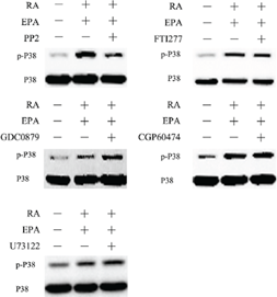
FTI-277 purchased from MedChemExpress. Usage Cited in: Oncotarget. 2017 Nov 22;8(65):109135-109150. [Abstract]
MCF-7 cells are pretreated with the indicated chemical inhibitors for 30min, followed by 15 min treatment with RA (20 μM) + EPA (80 μM).Cell extracts are prepared and subjected to western blotting analysis.


Reinheit & Dokumentation
Verweise
[1]. Tateishi K, et al. FTI-277 and GGTI-289 induce apoptosis via inhibition of the Ras/ERK and Ras/mTOR pathway in head and neck carcinoma HEp-2 and HSC-3 cells. J BUON. 2021;26(2):606-612. [Content Brief]
[2]. Yang W, et al. Farnesyltransferase inhibitor FTI-277 reduces mortality of septic mice along with improved bacterial clearance. J Pharmacol Exp Ther. 2011;339(3):832-841. [Content Brief]
[3]. Ponnusamy A, et al. FTI-277 inhibits smooth muscle cell calcification by up-regulating PI3K/Akt signaling and inhibiting apoptosis. PLoS One. 2018;13(4):e0196232. Published 2018 Apr 24. [Content Brief]
[4]. Girgert R, et al. Farnesyltransferase inhibitor FTI-277 prevents autocrine growth stimulation of neuroblastoma by BDNF. J Cancer Res Clin Oncol. 2003;129(4):227-233. [Content Brief]

Calculators
Konzentration (Stammlösung) × Volumen (Stammlösung) = Konzentration (Ziellösung) × Volumen (Ziellösung)


FTI-277 Related Classifications
HY-15872 Related Classifications
 Powered by Bioz