



Aβ-IN-6
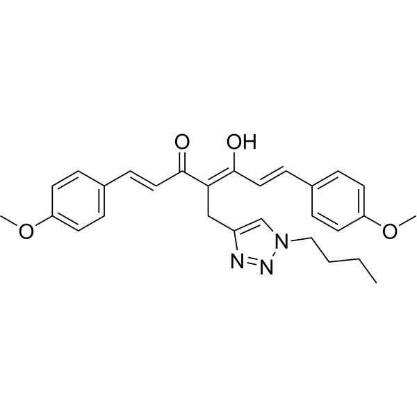
Aβ-IN-6 reduces pro-inflammatory cytokine release from microglia cells. Aβ-IN-6 significantly induces Nrf2 nuclear translocation and hamperes Aβ oligomers formation. Aβ-IN-6 exerts a consistent neuroprotective effect by modulating the redox-sensitive signalling pathways in vivo oxidative stress model. Aβ-IN-6 is an orally active and has antiinflammatory, Antioxidant and Anti-oligomeric activity. Aβ-IN-6 has the potential for Alzheimer's disease (AD) research.
For research use only. We do not sell to patients.
- CAS No.: 3043752-01-5
- Formula: C28H31N3O4
- Molecular Weight:473.56
-
Storage:
Please store the product under the recommended conditions in the Certificate of Analysis.

To place orders, for customer services and technical support, please contact: MedChemExpress USA
Tel: 609-228-6898 E-mail: [email protected] [email protected]


-
 Biological Activity
-
Chemical Information
- Protocols
- Purity & Documentation
- References
-
 Help & FAQs




Biological Activity
Description
In Vitro
Aβ-IN-6 (compound 4; 1-20 μM; 24 h) markedly reduces microglia viability starting from the concentration of 5 μM[1].
Aβ-IN-6 (1.25-40 μM; 24 h) causes significant cytotoxicity at concentrations higher than 2.5 μM in SH-SY5Y neuroblastoma cells[1].
Aβ-IN-6 (2.5 μM; 3 h) significantly induces Nrf2 nuclear translocation[1].
Aβ-IN-6 (2.5 μM) markedly suppresses the LPS-induced increase of mRNA levels of the two cytokines and NLRP3[1].
Aβ-IN-6 (1, 2.5 μM; protreamt for 1 h then stimulated with LPS for 24 h) significantly decreases LPS treatment induced the release of TNF-α and IL-1β[1].
Aβ-IN-6 (2.5 μM; for 24 h) before tert-butyl hydroperoxide (t-BuOOH; 50 μM for 30 min) reduces ROS formation with inhibition of around 18%[1].
MedChemExpress (MCE) has not independently confirmed the accuracy of these methods. They are for reference only. Further protocols information, click here.
-
Cell Line:Microglia
-
Concentration:1-20 μM
-
Incubation Time:24 h
-
Result:Markedly reduced microglia viability starting from the concentration of 5 μM.
-
Cell Line:SH-SY5Y neuroblastoma cells
-
Concentration:1.25, 2.5, 5, 10, 20, 40 μM
-
Incubation Time:24 h
-
Result:Recorded significant cytotoxicity at concentrations higher than 2.5 μM.
-
Cell Line:SH-SY5Y cells
-
Concentration:2.5 μM 3 h Significantly induced Nrf2 nuclear translocation. SH-SY5Y cells 2.5 μM
-
Incubation Time:3 h
-
Result:Significantly induced Nrf2 nuclear translocation.
-
Cell Line:Microglia
-
Concentration:2.5 μM
-
Incubation Time:Pretreated for 1 h and then stimulated with 100 ng/mL LPS for 6 h
-
Result:Markedly suppressed the LPS-induced increase of mRNA levels of the two cytokines and NLRP3, confirming the anti-inflammatory properties.
In Vivo
MedChemExpress (MCE) has not independently confirmed the accuracy of these methods. They are for reference only.
-
Animal Model:Spastin Drosophila model[1]
-
Dosage:10 μM
-
Administration:Added to standard food (dissolved in DMSO)
-
Result:Efficiently restored the increased ROS level in larval muscles and brains under neurodegenerative conditions (D-spastin loss of function model) to that observed for the control.
Significantly ameliorated the phenotype associated with spastin reduction.


Chemical Information
-
CAS No. 3043752-01-5
-
Molecular Weight 473.56
-
Formula C28H31N3O4
-
SMILES
CCCCN1C=C(N=N1)C/C(C(/C=C/C2=CC=C(C=C2)OC)=O)=C(\C=C\C3=CC=C(C=C3)OC)O
-
Shipping
Room temperature in continental US; may vary elsewhere.
-
Storage
Please store the product under the recommended conditions in the Certificate of Analysis.

Protocols
-
Cytoplasmic-Nuclear Fractionated Protein Extraction
Cytoplasmic-nuclear fractionated protein extraction separates soluble cytoplasmic proteins from nuclear-enriched proteins by mild plasma-membrane permeabilization, differential centrifugation, washing of nuclei, and extraction of nuclear proteins for downstream immunoblotting or related molecular analysis. The readout is the relative abundance of a protein in cytoplasmic and nuclear fractions, commonly assessed by western blotting together with compartment markers such as tubulin or pyruvate kinase for cytoplasm and lamin, nucleoporin, hnRNP, H2AX, or Lamin B for nuclear fractions.
-
ROS/oxidative-stress fluorescent staining
ROS/oxidative-stress fluorescent staining uses cell-permeant fluorogenic probes that become fluorescent after oxidation inside cells or tissues; commonly used examples include DCFH-DA/DCFDA for broad cellular oxidant detection, DHE for superoxide-related signal detection, MitoSOX for mitochondrial superoxide-related signal detection, and CellROX probes for oxidative-stress-associated fluorescence readouts. The assay detects probe oxidation rather than a single ROS species unless the probe and analysis method have been chemically validated for that species. DCFH-DA enters cells, is deacetylated by intracellular esterases to DCFH, and produces fluorescent DCF after oxidation, so the readout is used as an operational measure of total cellular oxidative stress rather than a species-specific ROS measurement. DHE and MitoSOX can report superoxide-related oxidation, but red fluorescence alone can include non-specific ethidium-like oxidation products; HPLC or optimized spectral approaches are
-
Research Protocol for Inflammation-related Diseases
The NLRP3 inflammasome is a cytosolic innate immune signaling platform that integrates priming signals and danger-signal activation to promote caspase-1 activation, maturation of IL-1β and IL-18, and gasdermin D-mediated pyroptotic cell death. The core experimental logic is to determine whether inflammatory disease phenotypes are driven by increased NLRP3 expression, ASC-containing inflammasome assembly, caspase-1 cleavage, GSDMD cleavage, and extracellular release of IL-1β/IL-18 rather than by nonspecific cell injury alone. The pathway is strongly linked to inflammation-related disease phenotypes because monosodium urate crystals activate NALP3/NLRP3 inflammasome signaling in gout-like crystal inflammation, cholesterol crystals activate NLRP3 inflammasomes in atherogenesis models, and DSS-induced intestinal inflammation has been reported to involve NLRP3 inflammasome activity. However, experimental colitis studies also show context-dependent protective effects of NLRP3 inflammasome co
-
Alzheimer’s Disease Modeling
Alzheimer’s Disease (AD) is a neurodegenerative disorder characterized by a progressive decline in cognitive functions and loss of specific types of neurons and synapses. Alzheimer's symptoms can be simulated in mice by injecting drugs (such as Aβ) or genetically modified.


Purity & Documentation
References

Calculators
Concentration (start) × Volume (start) = Concentration (final) × Volume (final)


Aβ-IN-6 Related Classifications
HY-149246 Related Classifications
 Powered by Bioz