



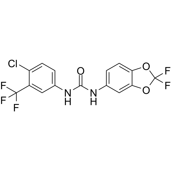
PK150

Based on 1 Customer Validation
PK150, an analogue of Sorafenib, shows oral bioavailability and antibacterial activity against several pathogenic strains at submicromolar concentrations. PK150 inhibits Gram-positive Methicillin-sensitive S. aureus (MSSA), Methicillin-resistant S. aureus (MRSA), Vancomycin intermediate S. aureus (VISA) with MICs of 0.3, 0.3-1, 0.3 μM, respectively.
Nur für Forschungszwecke. Wir verkaufen nicht an Patienten.
- Reinheit : 99.63%
- CAS. Nr.: 2165324-62-7
- Formel: C15H8ClF5N2O3
- Molecular Weight:394.68
-
Speicherung:Powder -20°C, 3 years , 4°C, 2 years ; In solvent -80°C, 6 months , -20°C, 1 month

To place orders, for customer services and technical support, please contact: MedChemExpress USA
Tel: 609-228-6898 E-mail: [email protected] [email protected]


-
 Biologische Aktivität
-
Chemical Information
-
Lösungsmittel & Löslichkeit
- Protokoll
- Reinheit & Dokumentation
- Verweise
-
 Help & FAQs




Biologische Aktivität
Beschreibung
IC50 & Target
MIC: 0.3 µM (MSSA), 0.3-1 µM (MRSA), 0.3 µM (VISA)[1]
Cellular Effect
|
Cell Line

|
Type | Value | Description | References |
|---|---|---|---|---|
| HeLa | IC50 |
9.02 μM
Compound: PK150
|
Cytotoxicity against human HeLa cells by SRB assay
Cytotoxicity against human HeLa cells by SRB assay
|
[PMID: 36067930] |
| HepG2 | IC50 |
5.68 μM
Compound: PK150
|
Cytotoxicity against human HepG2 cells by SRB assay
Cytotoxicity against human HepG2 cells by SRB assay
|
[PMID: 36067930] |
In Vivo
PK150 (10 and 20mg/kg orally; or 10mg/kg intravenously) shows no obvious signs of toxicity in mice. Higher i.v. dosing of 20mg/kg results in severe toxic effects and is thus avoided for subsequent therapeutic models[1].
MedChemExpress (MCE) has not independently confirmed the accuracy of these methods. They are for reference only.
-
Animal Model:Pathogen-free 9-week old female C57BL/6J mice[1]
-
Dosage:20 mg/kg
-
Administration:Administered p.o.
-
Result:Bacterial loads in the liver and heart were both significantly reduced by approximately 100-fold.
-
Animal Model:Outbred male CD-1 mice, 4 weeks old[1]
-
Dosage:10 and 20 mg/kg (Pharmacokinetic Analysis)
-
Administration:Administered by intragastric gavage at 10 and 20 mg/kg or intravenously at 10 mg/kg
-
Result:Oral bioavailability was approximately 63% and the mean residence time was slightly enhanced via this administration route.
T1/2=11.69±1.5, 9.67±0.2, and 9.37±0.5 hours for 10 mg/kg i.v., 10 mg/kg p.o., and 20 mg/kg p.o., respectively.


Chemical Information
-
CAS. Nr. 2165324-62-7
-
Appearance Solid
-
Molecular Weight 394.68
-
Formel C15H8ClF5N2O3
-
Color White to off-white
-
SMILES
O=C(NC1=CC=C(OC(F)(F)O2)C2=C1)NC3=CC=C(Cl)C(C(F)(F)F)=C3
-
Versand
Room temperature in continental US; may vary elsewhere.
-
Speicherung
Powder -20°C 3 years 4°C 2 years In solvent -80°C 6 months -20°C 1 month


Lösungsmittel & Löslichkeit
In Vitro:
DMSO : 250 mg/mL (633.42 mM; Need ultrasonic; Hygroscopic DMSO has a significant impact on the solubility of product, please use newly opened DMSO)
Please refer to the solubility information to select the appropriate solvent. Once prepared, please aliquot and store the solution to prevent product inactivation from repeated freeze-thaw cycles.
Storage method and period of stock solution: -80°C, 6 months; -20°C, 1 month. When stored at -80°C, please use it within 6 months. When stored at -20°C, please use it within 1 month.
Please refer to the solubility information to select the appropriate solvent. Once prepared, please aliquot and store the solution to prevent product inactivation from repeated freeze-thaw cycles.
Storage method and period of stock solution: -80°C, 6 months; -20°C, 1 month. When stored at -80°C, please use it within 6 months. When stored at -20°C, please use it within 1 month.
Konzentration (Stammlösung) × Volumen (Stammlösung) = Konzentration (Ziellösung) × Volumen (Ziellösung)
In Vivo:
Select the appropriate dissolution method based on your experimental animal and administration route.
- For the following dissolution methods, please ensure to first prepare a clear stock solution using an In Vitro approach and then sequentially add co-solvents:
- To ensure reliable experimental results, the clarified stock solution can be appropriately stored based on storage conditions. As for the working solution for In Vivo experiments, it is recommended to prepare freshly and use it on the same day.
- The percentages shown for the solvents indicate their volumetric ratio in the final prepared solution. If precipitation or phase separation occurs during preparation, heat and/or sonication can be used to aid dissolution.
Add each solvent one by one: 10% DMSO 40% PEG300 5% Tween-80 45% Saline
Solubility: ≥ 2.08 mg/mL (5.27 mM); Clear solution
This protocol yields a clear solution of ≥ 2.08 mg/mL (saturation unknown).
Taking 1 mL working solution as an example, add 100 μL DMSO stock solution (20.8 mg/mL) to 400 μL PEG300, and mix evenly; then add 50 μL Tween-80 and mix evenly; then add 450 μL Saline to adjust the volume to 1 mL.
Preparation of Saline: Dissolve 0.9 g sodium chloride in ddH₂O and dilute to 100 mL to obtain a clear Saline solution.
In Vivo Dissolution Calculator
Please enter the basic information of animal experiments:
-
-
-
-
Recommended: Prepare an additional quantity of animals to account for potential losses during experiments.
Please enter your animal formula composition:
-
%DMSO +
Recommended: Keep the proportion of DMSO in working solution below 2% if your animal is weak.
-
%+
-
+%Tween-80 + +
-
%Saline +
The co-solvents required include: DMSO, . All of co-solvents are available by MedChemExpress (MCE). , Tween 80. All of co-solvents are available by MedChemExpress (MCE).
Working solution concentration: 0.22 mg/mL
Method for preparing stock solution: mg drug dissolved in μL DMSO. Stock solution concentration: mg/mL.
1. Take μL DMSO stock solution;
2. Add μL .
μL , mix evenly;
3. Then add μL Tween 80, mix evenly;
4. Then add μL
Please ensure that the stock solution in the first step is dissolved to a clear state, and add co-solvents in sequence. You can use ultrasonic heating (ultrasonic cleaner, recommended frequency 20-40 kHz), vortexing, etc. to assist dissolution.


Reinheit & Dokumentation
-
Data Sheet (272 KB)
-
SDS (396 KB)
- English - EN (396 KB)
- Français - FR (396 KB)
- Deutsch - DE (396 KB)
- Norwegian - NO (396 KB)
- Español - ES (396 KB)
- Swedish - SV (396 KB)
- Italian - IT (396 KB)
- Korean - KR (396 KB)
- Portuguese - PT (396 KB)
-
Handling Instructions (2659 KB)
Verweise


Complete Stock Solution Preparation Table
Please refer to the solubility information to select the appropriate solvent. Once prepared, please aliquot and store the solution to prevent product inactivation from repeated freeze-thaw cycles.
Storage method and period of stock solution: -80°C, 6 months; -20°C, 1 month. When stored at -80°C, please use it within 6 months. When stored at -20°C, please use it within 1 month.
| Optional Solvent | Concentration Solvent Mass | 1 mg | 5 mg | 10 mg | 25 mg |
|---|---|---|---|---|---|
| DMSO | 1 mM | 2.5337 mL | 12.6685 mL | 25.3370 mL | 63.3425 mL |
| 5 mM | 0.5067 mL | 2.5337 mL | 5.0674 mL | 12.6685 mL | |
| 10 mM | 0.2534 mL | 1.2668 mL | 2.5337 mL | 6.3342 mL | |
| 15 mM | 0.1689 mL | 0.8446 mL | 1.6891 mL | 4.2228 mL | |
| 20 mM | 0.1267 mL | 0.6334 mL | 1.2668 mL | 3.1671 mL | |
| 25 mM | 0.1013 mL | 0.5067 mL | 1.0135 mL | 2.5337 mL | |
| 30 mM | 0.0845 mL | 0.4223 mL | 0.8446 mL | 2.1114 mL | |
| 40 mM | 0.0633 mL | 0.3167 mL | 0.6334 mL | 1.5836 mL | |
| 50 mM | 0.0507 mL | 0.2534 mL | 0.5067 mL | 1.2668 mL | |
| 60 mM | 0.0422 mL | 0.2111 mL | 0.4223 mL | 1.0557 mL | |
| 80 mM | 0.0317 mL | 0.1584 mL | 0.3167 mL | 0.7918 mL | |
| 100 mM | 0.0253 mL | 0.1267 mL | 0.2534 mL | 0.6334 mL |


PK150 Related Classifications
HY-133119 Related Classifications
 Powered by Bioz