



EphA7 Protein, Human (HEK293, His)

Based on 1 Customer Validation
The EphA7 protein is a receptor tyrosine kinase that promiscuously binds to the GPI-anchored ephrin-A ligand to initiate bidirectional signaling. It participates in forward and reverse signaling pathways, uses EFNA5 as a functional ligand, regulates brain development and affects cell adhesion and repulsion. EphA7 Protein, Human (HEK293, His) is the recombinant human-derived EphA7 protein, expressed by HEK293 , with C-6*His labeled tag.
- Species: Human
- Source: HEK293
-
Storage:Stored at -20°C for 2 years from date of receipt. After reconstitution, it is stable at 4°C for 1 week or -20°C for longer (with carrier protein). It is recommended to freeze aliquots at -20°C or -80°C for extended storage.


To place orders, for customer services and technical support, please contact: MedChemExpress USA
Tel: 609-228-6898 E-mail: [email protected] [email protected]


-
 Biological Activity
-
Technical Parameters
-
Product Properties
- Documentation
- Help & FAQs




Biological Activity
Description
The EphA7 protein is a receptor tyrosine kinase that promiscuously binds to the GPI-anchored ephrin-A ligand to initiate bidirectional signaling. It participates in forward and reverse signaling pathways, uses EFNA5 as a functional ligand, regulates brain development and affects cell adhesion and repulsion. EphA7 Protein, Human (HEK293, His) is the recombinant human-derived EphA7 protein, expressed by HEK293 , with C-6*His labeled tag.
Background
The EphA7 protein, a receptor tyrosine kinase, engages in promiscuous binding to GPI-anchored ephrin-A family ligands on adjacent cells, initiating contact-dependent bidirectional signaling. The downstream pathway originating from the receptor is termed forward signaling, while the pathway downstream of the ephrin ligand is referred to as reverse signaling. Among the GPI-anchored ephrin-A ligands, EFNA5 serves as a cognate/functional ligand for EPHA7, regulating brain development and modulating cell-cell adhesion and repulsion. EphA7 exhibits repellent activity on axons, playing a crucial role in guiding corticothalamic axons and ensuring the proper topographic mapping of retinal axons to the colliculus. Additionally, EphA7 may contribute to brain development through a caspase (CASP3)-dependent proapoptotic activity. Forward signaling through EphA7 may result in the activation of components of the ERK signaling pathway, including MAP2K1, MAP2K2, MAPK1, and MAPK3, which are phosphorylated upon EphA7 activation.
Verified Bioactivity
1.This product does not contain protein kinase domain.
2.Measured by its binding ability in a functional ELISA. Immobilized Human EphA7 at 2 μg/mL (100 μL/well) on the plate can bind Biotinylated Mouse EphrinA4. The ED50 for this effect is 1.0-10.0 ng/mL.
MCE Validation Data
-
Purity - SDS-PAGE
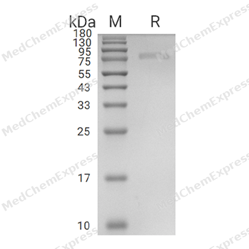
Purity - SDS-PAGE
-
Bioactivity - ELISA
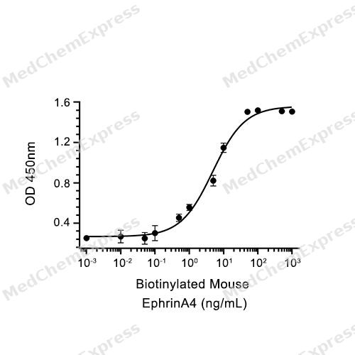
Bioactivity - ELISA


Technical Parameters
-
Species Human
-
Source HEK293
-
Tag C-6*His
-
Accession
Q15375-1 (Q28-I556)
-
Molecular Construction
-
N-term
-
EphA7 (Q28-I556)
Accession # Q15375-1 -
6*His
-
C-term
-
-
Protein Length
Extracellular Domain
-
Synonyms
EPHA7; EK11; EPH Receptor A7; Receptor Protein-Tyrosine Kinase HEK11; Ephrin Type-A Receptor 7; Tyrosine-Protein Kinase Receptor EHK-3; EPH Homology Kinase 3; Receptor Protein-Tyrosine Kinase; Hek11; Alternative Protein EPHA7; EPH-Like Kinase 11; Eph Homo
-
AA Sequence
QAAKEVLLLDSKAQQTELEWISSPPNGWEEISGLDENYTPIRTYQVCQVMEPNQNNWLRTNWISKGNAQRIFVELKFTLRDCNSLPGVLGTCKETFNLYYYETDYDTGRNIRENLYVKIDTIAADESFTQGDLGERKMKLNTEVREIGPLSKKGFYLAFQDVGACIALVSVKVYYKKCWSIIENLAIFPDTVTGSEFSSLVEVRGTCVSSAEEEAENAPRMHCSAEGEWLVPIGKCICKAGYQQKGDTCEPCGRGFYKSSSQDLQCSRCPTHSFSDKEGSSRCECEDGYYRAPSDPPYVACTRPPSAPQNLIFNINQTTVSLEWSPPADNGGRNDVTYRILCKRCSWEQGECVPCGSNIGYMPQQTGLEDNYVTVMDLLAHANYTFEVEAVNGVSDLSRSQRLFAAVSITTGQAAPSQVSGVMKERVLQRSVELSWQEPEHPNGVITEYEIKYYEKDQRERTYSTVKTKSTSASINNLKPGTVYVFQIRAFTAAGYGNYSPRLDVATLEEATGKMFEATAVSSEQNPVI
-
Predicted Molecular Mass
60.2 kDa
-
Molecular Weight
Approximately 72-77 kDa, based on SDS-PAGE under reducing conditions.
-
Glycosylation
Yes
-
Purity
≥ 95%, as determined by reducing SDS-PAGE.


Product Properties
Lyophilized powder.
Lyophilized from a 0.22 μm filtered solution of 20 mM PB, 150 mM NaCl, pH 7.4.
<1 EU/μg, determined by LAL method.
It is not recommended to reconstitute to a concentration less than 100 μg/mL in ddH2O. For long term storage it is recommended to add a carrier protein (0.1% BSA, 5% HSA, 10% FBS or 5% Trehalose).
Stored at -20°C for 2 years from date of receipt. After reconstitution, it is stable at 4°C for 1 week or -20°C for longer (with carrier protein). It is recommended to freeze aliquots at -20°C or -80°C for extended storage.
Room temperature in continental US; may vary elsewhere.


Documentation
-
Data Sheet (239 KB)
-
SDS (251 KB)
- English - EN (251 KB)
- Français - FR (251 KB)
- Deutsch - DE (251 KB)
- Norwegian - NO (251 KB)
- Español - ES (251 KB)
- Swedish - SV (251 KB)
- Italian - IT (251 KB)
- Korean - KR (251 KB)
- Portuguese - PT (251 KB)
-
Handling Instructions (2659 KB)

Calculators
Concentration (start) × Volume (start) = Concentration (final) × Volume (final)


 Powered by Bioz

