1. Resources
  2. Articles
  3. Efferocytosis: the terminal station for dying cells
Efferocytosis: the terminal station for dying cells
The human body produces billions of dead cells every day. Removing old cells and giving space for new ones can keep tissues or organs maintain vitality and homeostasis. If this clearance process does not work well, it may lead to adverse consequences. For example, one of the causes of systemic lupus erythematosus is the accumulation of apoptotic cells to induce secondary cell necrosis, then releasing damage-associated molecular patterns, thereby causing a wide range of inflammatory reactions. Therefore, the removal of dead cells is important for homeostasis.
Efferocytosis is the process in which phagocytes remove programmed dead cells. Programmed cells death includes apoptosis, pyroptosis, autophagy, necrosis and so on. In most human tissues, efferocytosis is performed by professional phagocytes (macrophages and dendritic cells) or non-professional phagocytes (epithelial cells and fibrocytes) near the dying cells[1]. Efferocytosis prevents secondary necrosis of dying cells from releasing harmful cell contents (such as oxides and proteases) that may cause inflammation. So how do phagocytes recognize dying cells and get rid of them? Here, we will introduce three stages of efferocytosis: Find, Eat, Digest.
Efferocytosis Trilogy

• Find the hidden dying cells

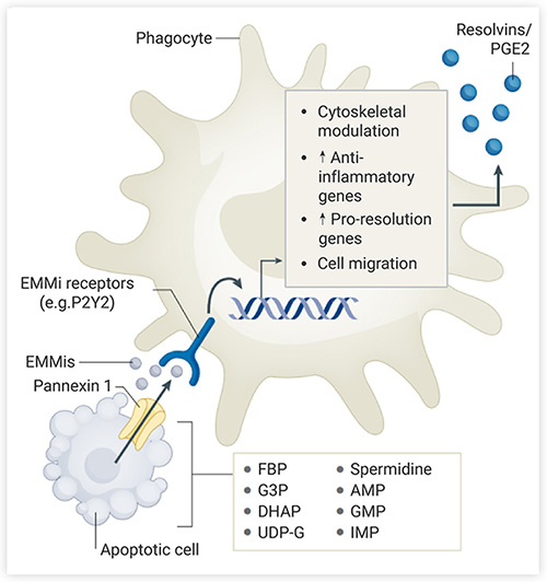
Dying cells release soluble media, which contain "Find me" signals for phagocytic cells to recognize, including nucleotides (ATP, UTP), membrane lipids (sphingosine 1-phosphate), and chemokines (CX3CL1). These signals not only attract the phagocytes, but also prepare the phagocytes for "fight", such as the upregulation of the phagocytic receptor and the enhancement of digestive mechanism[1].

Apoptotic cells release the nucleotides "Find me" signals through the pannexin channel (activated by the cleavage of caspase 3/7 during apoptosis). The released ATP induces phagocyte migration via the purine receptor P2Y. Besides, nucleotides regulate the immune mechanism of macrophages. For instance, ATP or AMP of apoptotic cells is converted to adenosine, which inhibits inflammation through adenosine receptors and increases the expression of anti-inflammatory and pro-resolution genes (including Nr4a1 and thrombospondin).

Non-apoptotic cells possess the damaged lipid membrane, thus inflammatory signals are released directly outside the cell. Pathogen-infected cells release pathogen-associated molecular pattern (PAMP) signals that bind to pattern recognition receptors (PRR) on or in phagocytes to influence macrophage function and immune activation. In addition, non-apoptotic cells also release damage-associated molecular patterns (DAMP) that trigger inflammatory responses and act as chemokines for macrophages.

Fig 1. Phagocytes recognize apoptotic cells
Fig 1. Phagocytes recognize apoptotic cells [1]
Apoptotic cells release “Find me” signals (nucleotides, membrane lipids, chemokines, etc.), which bind to corresponding receptors on phagocytes to induce phagocyte migration, up-regulate anti-inflammatory/pro-anti-inflammatory gene expression, and regulate the cytoskeleton.

• Eat the recognized dying cells

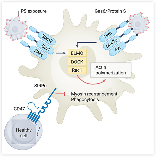
After phagocytes migrate to the vicinity of dying cells, surface receptors recognize the “Eat me” signals of dying cells and bind to them. "Eat me" signals include phosphatidylserine (PS), calreticulin, oxidized low-density lipoprotein, and so on. PS is the most efficient and evolutionally-conserved signaling molecule. A common feature of all dying cells is the loss of phospholipid asymmetry in the plasma membrane, where PS is exposed outside the cell and promotes the phagocytosis for dead cells. During apoptosis, PS located on the medial side of the cell are flipped from the medial side to the surface at an early stage. PS receptors of phagocytes can directly or indirectly recognize PS exposed on the surface. After binding to PS, one of the PS receptors, BAI1, initiates intracellular signaling via the ELMO1-DOCK complex to induce RAC1-mediated actin cytoskeletal rearrangement (Fig. 2) in preparation for "eating" apoptotic cells.

Unlike dying cells, healthy cells expose "don't eat me" signals on their surfaces, including CD47 and CD24. CD47 on living cells binds to SIRP-α on macrophages, resulting in tyrosine phosphorylation of the cytoplasmic domain of SIRP-α. The recruited SHP1/2 is then inhibited by non-muscle myosin IIA to inhibit phagocytosis. These signals prevent healthy cells from being cleared by phagocytes.

Fig 2. Phagocytes bind to apoptotic cells
Fig 2. Phagocytes bind to apoptotic cells[1]
Phagocytes recognize PS on the apoptotic cell surface by direct ways (BAI1, TIM1) or indirect ways (AXL, MerTK), then activate the expression of ELMO and DOCK proteins, and in turn activate GTPase RAC1, enhancing actin remodeling and phagocytosis cup formation. CD47 on the surface of healthy cells binds to SIRP-α and inhibits phagocytic cup by inhibiting non-muscle myosin IIA.

• Digest the eaten dead cells

1) The phagocytosis of dying cells

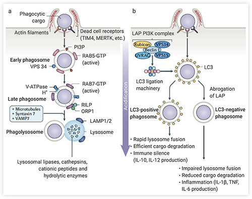
After binding with dying cells, phagocytes initiate actin remodeling, and the plasma membrane invagination and local overflow form phagosomes. Phagocytes then engulf the dying cells through endocytosis. Phagosomes are formed after endocytosis, and the phagosome membrane undergoes various biochemical changes, which are controlled by the Rab GTPase protein family. Mature phagosomes fuse directly with lysosomes by forming a Ca2+-dependent SNARE complex consisting of VAMP7 and Syntaxin 7 (Fig. 3a)[2]. Lysosomes contain a large number of proteases, nucleases and lipases that digest apoptotic cells in phagosomes.

There is another phagocytosis pathway--the LC3 (microtubule-associated protein 1A/1B-light chain 3) associated phagocytosis (LAP) pathway (Fig. 3b). After phagocytosis of dying cells, the LAP PI3K complex is recruited to the LAP-associated phagosomes (LAPosome) containing dying cells[2]. This complex is essential for the maintenance of LAPosome and activate the LC3 binding. LC3 binding promotes the fusion of LAPosome with lysosomes, improving the efficiency of clearance of dead cells and maintaining immune silencing.

Fig. 3 Phagocytosis for dying cells
Fig. 3 Phagocytosis for dying cells[2]
a. Lysosome degradation pathway: VPS34 was recruited into RAB5-positive early phagosomes and catalyzed phosphatidylinositol to form PI3P (all of which were necessary for phagosome maturation). After the early phagosomes matured into late phagosomes, they finally merged with lysosomes. b. LAP pathway: The LAP PI3K complex (consisting of Beclin 1, VPS34, VPS15, UVRAG, and Rubicon) activates LC3 binding to LAPosome, promotes LAPosome maturation, and lysosome fusion.

2) Digestion of dying cells

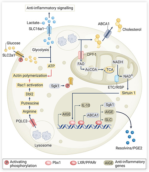
In most cases, dead cells outnumber phagocytes, so the phagocytes need to clear more than one cell at a time. Dying cells carry membranes, cholesterol, proteins, nucleic acids, and so on, which need to be metabolized. This is a huge burden for phagocytes to control their own volume and surface area and to rapidly change their immune metabolic environment. Apoptotic cells themselves can also change the immune metabolism of phagocytes (Fig. 4):

(a) PS on the surface of apoptotic cells can activate or up-regulate LXR and ABCA1 after binding with receptors, which can promote the metabolism and excretion of cholesterol;

(b) Fatty acid oxidation in mitochondria of apoptotic cells can promote the expression of anti-inflammatory factor IL-10;

(c) Phagocytes use Arginine from apoptotic cells to drive actin cytoskeleton rearrangement and promote the uptake of apoptotic cells in subsequent rounds;

(d) Increased glycolysis and glucose input also promote ATP production and actin polymerization.

Fig. 4 Apoptotic cells regulate the immune metabolic environment of phagocytes
Fig. 4 Apoptotic cells regulate the immune metabolic environment of phagocytes[1]
Dynamic tracking of efferocytosis

Routine efferocytosis studies are conducted in vitro. For example, the process of efferocytosis can be observed under a microscope after immuno-fluorescence staining of primary human macrophages and apoptotic Jurkat T cells[3]. However, because apoptotic cells are easily eliminated, it is still a great challenge to detect the process of efferocytosis in vivo.

Recently, Raymond et al. developed a gene-encoded fluorescent reporter gene, CharON, to track efferocytosis in live cells during drosophila embryonic development. This study was published in Science.

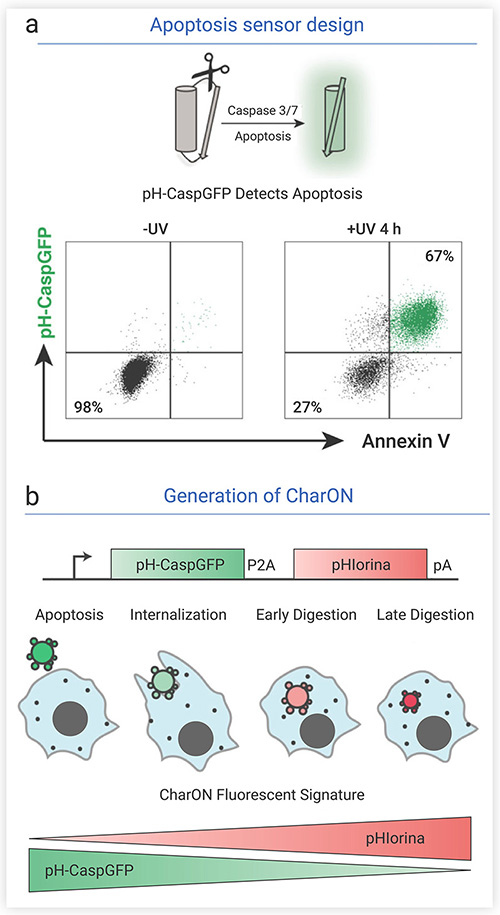
First, the researchers designed sensors that detected apoptosis and phagocytosis. For apoptosis, they designed a special green fluorescent protein (GFP) probe.Caspases 3/7 expressed during apoptosis can cut GFP and make it fluoresce. GFP is easily quenched under acidic lysosomal conditions, but after mutating Q204H gene, GFP can become tolerant to pH. With above modification, they designed a pH-tolerant fluorescent probe called pH-CaspGFP, which accurately reported apoptotic cells. For phagocytosis, they designed a new red fluorescent pH sensor, pHlorina, which increases its fluorescence intensity as the pH decreases. Then, they combined two sensors obtaining the probe “CharON” (Fig 5). CharON transgenic cells first fluoresce green during apoptosis, and after phagocytosis and acidification in lysozyme, the intensity of pHlorina's red fluorescence increases while GFP is gradually quenched. CharON visualizes continuous cellular events.

Fig. 5 Design of pH-CaspGFP and schematic diagram of CharON probe for detection of exocytic process
Fig. 5 Design of pH-CaspGFP and schematic diagram of CharON probe for detection of exocytic process[4]
During cell apoptosis, caspase 3/7 cleaved GFP and showed green fluorescence. After phagocytosis, GFP was gradually quenched in acidic environment, and the intensity of red fluorescence pHlorina increased.

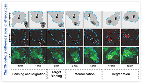
To track the process of efferocytosis, researchers created CharON transgenic drosophila. In drosophila embryo development, there is a wave of apoptosis in the developing central nervous system (CNS). CharON presented a more complete dynamic process of efferocytosis in vivo, including apoptosis, phagocytosis recruitment, Find, Eat and Digest (Fig 6). CharON also observed that the "heavy duty" of clearing apoptotic cells in the CNS of embryonic Drosophila melanogaster is shared by phagocytic glial cells and dispersed ventral blood cells (macrophages). It was also observed that the size of phagosomes in glial cells and macrophages was different, and the fluorescence of apoptotic cells in macrophages was more dense.

Fig. 6 CharON visualized the different stages of efferocytosis in drosophila embryos
Fig. 6 CharON visualized the different stages of efferocytosis in drosophila embryos[4]
0~1 min: apoptotic cells attract macrophages; 2 min: apoptotic cells bind to macrophages; 3-5 min: phagocytosis of macrophages. MΦ (blue outline): GFP-labeled macrophages; A.C. (white outline): CharON transgenic apoptotic cells. Apoptosis induces pH-CaspGFP (green) activation and macrophage migration (green). Apoptotic cell is phagocytosed after target binding to receptor. After digestion, acidification/degradation of apoptotic cells is detected by increasing the pHlorina signal (red)
Conclusion

The process of efferocytosis includes three stages: Find (phagocytes recognize dying cells), Eat (receptors of phagocytes bind targets on dying cells), Digest (phagocytes phagocytose and degrade cells). Efferocytosis involves several signaling pathways, including receptor networks, cell signaling molecules, rapid cytoskeletal rearrangement, apoptotic cell digestion, and immune metabolism pathways. The dysfunction of these pathways may lead to a variety of diseases related to efferocytosis defects (neurodegenerative diseases, retinal degeneration, atherosclerosis, cancer, etc.). Therefore, understanding the mechanism of efferocytosis is important for the treatment of these diseases.