


Obatoclax
Based on 12 publication(s) in Google Scholar
Obatoclax (GX15-070), a BH3 mimetic, is a pan-BCL-2 family proteins inhibitor with a Ki of 220 nM for BCL-2. Obatoclax induces autophagy-dependent cell death and targets cyclin D1 for proteasomal degradation. Obatoclax has anti-cancer and broad-spectrum antiparasitic activity.
For research use only. We do not sell to patients.
- Purity: 98.04%
- CAS No.: 803712-67-6
- Formula: C20H19N3O
- Molecular Weight:317.38
-
Storage:Powder -20°C, 3 years , 4°C, 2 years ; In solvent -80°C, 6 months , -20°C, 1 month

To place orders, for customer services and technical support, please contact: MedChemExpress USA
Tel: 609-228-6898 E-mail: [email protected] [email protected]


-
 Biological Activity
-
Chemical Information
-
Solvent & Solubility
- Purity & Documentation
- References
-
 Help & FAQs
-


Anti-Infection Compound Library
HY-L002
-
Apoptosis Compound Library
HY-L003
-
Anti-Cancer Compound Library
HY-L025
-
Clinical Compound Library
HY-L026
-
CNS-Penetrant Compound Library
HY-L028
-
Autophagy Compound Library
HY-L029
-
Drug Repurposing Compound Library
HY-L035
-
Anti-Blood Cancer Compound Library
HY-L079
-
Antiparasitic Compound library
HY-L082
-
Mitochondria-Targeted Compound Library
HY-L089
-
Rare Diseases Drug Library
HY-L102
-
Protein-protein Interaction Inhibitor Library
HY-L109
-
Anti-Pulmonary Fibrosis Compound Library
HY-L125
-
Cell Death Library
HY-L162
-
Anti-Hematopathy Compound Library
HY-L171
-
Anti-Ovarian Cancer Compound Library
HY-L173
-
Multi-Target Compound Library
HY-L176
-
Mitophagy Compound Library
HY-L180
-
Bioactive Compound Library Max
HY-L181
-
MCE Bioactive Compound Library
HY-L001V
-
Drug Repurposing Compound Library Plus
HY-L035P
-
Clinical Compound Library Plus
HY-L026P
-
Bioactive Compound Library
HY-L001
-
High-Throughput Bioactive Compound Library
HY-L205


Publications Citing Use of MedChemExpress (MCE) Obatoclax
More- Cancer Lett. 2023 Feb 1:554:216028. [Abstract]
- Acta Pharmacol Sin. 2021 Aug;42(8):1298-1310. [Abstract]
- Biomed Pharmacother. 2020 Sep;129:110371. [Abstract]
- Drug Deliv Transl Res. 2025 Oct 29. [Abstract]
- iScience. 2022 Sep 16;25(9):104925. [Abstract]
- Sci Rep. 2019 Sep 24;9(1):13786. [Abstract]
- ACS Infect Dis. 2023 Nov 10;9(11):2105-2118. [Abstract]
- Viruses. 2020 Oct 18;12(10):1178. [Abstract]
- Am J Cancer Res. 2019 Mar 1;9(3):546-561. [Abstract]
- bioRxiv. 2024 Jul 25.
- Norwegian University of Science and Technology. 2021 Oct.
- Universitat Politècnica de València. 2020 Sep 7.
 Customer Validation & Images

-
In Vivo Efficacy Study
-
IF
-
WB
-
Others
-
Cell Proliferation/Viability Assay
All Parasite Isoforms
More

Biological Activity
|
BCL2 200 nM (Ki) |
Mcl-1 1-7 μM (Ki) |
Bcl-xL 1-7 μM (Ki) |
Bcl-W 1-7 μM (Ki) |
Bcl-B 1-7 μM (Ki) |
Obatoclax (GX15-070) inhibits BCL-2, BCL-XL, MCL-1, BCL-w, A1, and BCL-b with Ki values≈1-7 μM[2].
Obatoclax (50-200 nM; 24-72 hours) induces a dose- and time-dependent reduction of cell numbers in all human colorectal cancer cell lines. In particular, the IC50 of cell proliferation at 72 h are 25.85, 40.69, and 40.01 nM for HCT116, HT-29, and LoVo cells, respectively[1].
Obatoclax (400 nM; for 24 hours) induces autophagy in OSCC cells[3].
Obatoclax (50-200 nM; for 24 hours) provokes a dose-dependent increase in the G1-phase cell populations[1].
Obatoclax (25-200 nM; for 24 hours) indicates a marked drop in cyclin D1 levels as low as 50 nM[1].
Obatoclax induces T286 phosphorylation-dependent or -independent cyclin D1 degradation.
in HCT116 and LoVo cells, the steady-state levels of p-Cyclin D (T286) began to decline once exposed to obatoclax (200 nM; 1, 3, 6, 12, 24 hours). Obatoclax inhibits GSK3β but activates p38 MAPK, while barely affecting ERK1/2 activity in HT-29 cells[1].
Obatoclax (50, 100, 150, 200, 250, 300, 350, 400, 450 nM) potently inhibits the clonogenic potential of oral cancer cells[1].
MedChemExpress (MCE) has not independently confirmed the accuracy of these methods. They are for reference only.
-
Cell Line:human colorectal cancer HCT116, HT-29 and LoVo cells
-
Concentration:50, 100, 200 nM
-
Incubation Time:24, 48, and 72 hours
-
Result:Induced a dose- and time-dependent reduction of cell numbers.
-
Cell Line:AW8507 and SCC029B cells
-
Concentration:400 nM
-
Incubation Time:24 hours
-
Result:Induced autophagy in OSCC cells.
-
Cell Line:HCT116 and HT-29 cells
-
Concentration:50, 100, 200 nM
-
Incubation Time:24 hours
-
Result:Provoked a dose-dependent increase in the G1-phase cell populations.
-
Cell Line:HCT116, HT-29 and LoVo cells
-
Concentration:50, 100, 200 nM
-
Incubation Time:24 hours
-
Result:Indicated a marked drop in cyclin D1 levels as low as 50 nM.
MedChemExpress (MCE) has not independently confirmed the accuracy of these methods. They are for reference only.
-
Animal Model:6-8 weeks old female BALB/C nude mice bearing subcutaneous tumors[4]
-
Dosage:1.15, 2.5, 5 mg/kg
-
Administration:Intravenously injected (through lateral tail vein); five consecutive days (i.e. 5 injections)
-
Result:Exhibited potent antitumor activity in xenograft mouse models in a dose-dependent manner.
| NCT Number | Sponsor | Condition | Start Date |
Phase
|
|---|---|---|---|---|
| NCT01329991 | Plexxikon| | 2011-05 | PHASE1 |


Chemical Information
-
CAS No. 803712-67-6
-
Appearance Solid
-
Molecular Weight 317.38
-
Formula C20H19N3O
-
Color Light brown to brown
-
SMILES
COC1=CC(C(N2)=CC3=C2C=CC=C3)=N/C1=C\C4=C(C)C=C(C)N4
-
Synonyms
GX15-070
-
Shipping
Room temperature in continental US; may vary elsewhere.
-
Storage
Powder -20°C 3 years 4°C 2 years In solvent -80°C 6 months -20°C 1 month


Publications (12)
-
Journal Impact Factor
-
Most Recent
-
Cancer Lett
High-content drug screening in zebrafish xenografts reveals high efficacy of dual MCL-1/BCL-XL inhibition against Ewing sarcoma. [Abstract]2023 Feb 1:554:216028. PMID: 36462556 -
Acta Pharmacol Sin
Combinations of proteasome inhibitors with obatoclax are effective for small cell lung cancer. [Abstract]2021 Aug;42(8):1298-1310. PMID: 33139838 -
Biomed Pharmacother
Biomimetic nanoparticle loading obatoclax mesylate for the treatment of non-small-cell lung cancer (NSCLC) through suppressing Bcl-2 signaling. [Abstract]2020 Sep;129:110371. PMID: 32563984
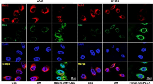
Obatoclax purchased from MedChemExpress. Usage Cited in: Biomed Pharmacother. 2020 Sep;129:110371. [Abstract]
Immunofluorescence staining displays weaker Bcl-2 fluorescence intensity in lung cancer cells incubated with RBCm-Obatoclax Mesylate (OM)/PLGA, accompanied with stronger expression of pro-apoptotic signal Bax in comparison to the Con group.
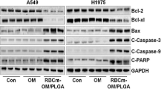
Obatoclax purchased from MedChemExpress. Usage Cited in: Biomed Pharmacother. 2020 Sep;129:110371. [Abstract]
Bcl-2 and Bcl-xl protein expression levels are restrained in A549 and H1975 cells treated with RBCm-OM/PLGA; however, Bax, cleaved Caspase-3, Caspase-9 and PARP are up-regulated following RBCm-OM/PLGA incubation. Also, free Obatoclax Mesylate (OM) does not influence the expression change of all these proteins.
-
Drug Deliv Transl Res
Leveraging quantum chemical properties in transfer learning for predicting blood-brain barrier permeability of drugs. [Abstract]2025 Oct 29. PMID: 41160380 -
iScience
Identification of potent inhibitors of SARS-CoV-2 infection by combined pharmacological evaluation and cellular network prioritization. [Abstract]2022 Sep 16;25(9):104925. PMID: 35992305
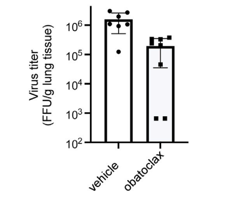
Obatoclax purchased from MedChemExpress. Usage Cited in: iScience. 2022 Sep 16;25(9):104925. [Abstract]
Male and female mice were infected with SARS-CoV-2 virus intranasally, and infection began 6 hours after daily treatment with 3 mg/kg Obatoclax Mesylate. Viral load in the lungs (FFU/g tissue) was measured 4 days post-infection.
-
Sci Rep
2019 Sep 24;9(1):13786. PMID: 31551480
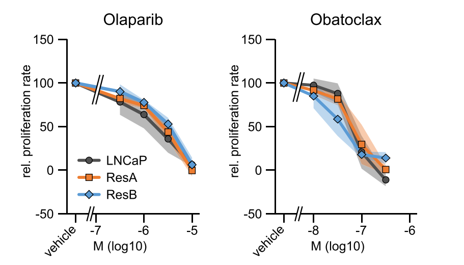
Obatoclax purchased from MedChemExpress. Usage Cited in: Sci Rep. 2019 Sep 24;9(1):13786. [Abstract]
Dose response curves showing the proliferation at increasing concentrations of the PARP inhibitor olaparib and the pan-BCL-2 inhibitor Obatoclax Mesylate in normal growth medium (containing 10 µM enzalutamide for ResA/ResB).
-
ACS Infect Dis
Bcl-2 Antagonist Obatoclax Reactivates Latent HIV-1 via the NF-κB Pathway and Induces Latent Reservoir Cell Apoptosis in Latently Infected Cells. [Abstract]2023 Nov 10;9(11):2105-2118. PMID: 37796279 -
Viruses
2020 Oct 18;12(10):1178. PMID: 33080984
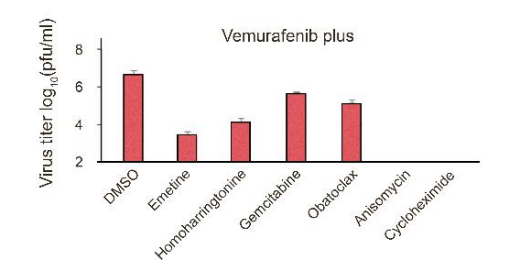
Obatoclax purchased from MedChemExpress. Usage Cited in: Viruses. 2020 Oct 18;12(10):1178. [Abstract]
The effects of 5 µM vemurafenib plus 0.1% DMSO, 0.2 µM emetine, 0.2 µM homoharringtonine, 0.2 µM Obatoclax Mesylate, 1 µL Gemcitabine, 0.1 µL Anisomycin and 1 µL Cycloheximide on viral replication measured by plaque reduction assay.
-
Am J Cancer Res
Targeting sphingosine kinase 2 suppresses cell growth and synergizes with BCL2/BCL-XL inhibitors through NOXA-mediated MCL1 degradation in cholangiocarcinoma. [Abstract]2019 Mar 1;9(3):546-561. PMID: 30949409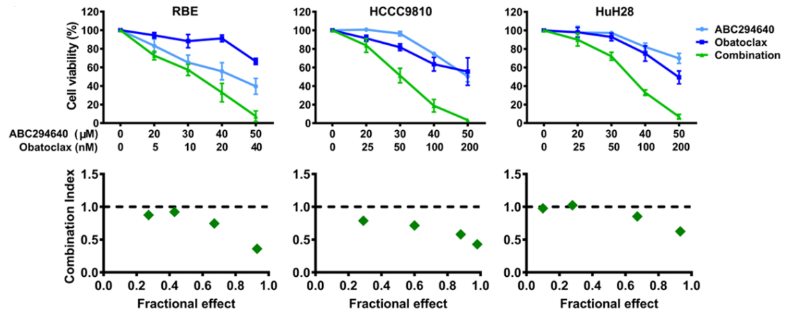
Obatoclax purchased from MedChemExpress. Usage Cited in: Am J Cancer Res. 2019 Mar 1;9(3):546-561. [Abstract]
RBE, HCCC9810, and HuH28 cells were treated with different concentrations of ABC294640 or Obatoclax Mesylate, alone or in combination, for 72 hours, and then cell viability was analyzed using the CCK-8 assay.
-
-
-


Solvent & Solubility
DMSO : 100 mg/mL (315.08 mM; Need ultrasonic; Hygroscopic DMSO has a significant impact on the solubility of product, please use newly opened DMSO)
Please refer to the solubility information to select the appropriate solvent. Once prepared, please aliquot and store the solution to prevent product inactivation from repeated freeze-thaw cycles.
Storage method and period of stock solution: -80°C, 6 months; -20°C, 1 month. When stored at -80°C, please use it within 6 months. When stored at -20°C, please use it within 1 month.
Concentration (start) × Volume (start) = Concentration (final) × Volume (final)
Select the appropriate dissolution method based on your experimental animal and administration route.
- For the following dissolution methods, please ensure to first prepare a clear stock solution using an In Vitro approach and then sequentially add co-solvents:
- To ensure reliable experimental results, the clarified stock solution can be appropriately stored based on storage conditions. As for the working solution for In Vivo experiments, it is recommended to prepare freshly and use it on the same day.
- The percentages shown for the solvents indicate their volumetric ratio in the final prepared solution. If precipitation or phase separation occurs during preparation, heat and/or sonication can be used to aid dissolution.
Add each solvent one by one: 10% DMSO 40% PEG300 5% Tween-80 45% Saline
Solubility: ≥ 2.5 mg/mL (7.88 mM); Clear solution
This protocol yields a clear solution of ≥ 2.5 mg/mL (saturation unknown).
Taking 1 mL working solution as an example, add 100 μL DMSO stock solution (25.0 mg/mL) to 400 μL PEG300, and mix evenly; then add 50 μL Tween-80 and mix evenly; then add 450 μL Saline to adjust the volume to 1 mL.
Preparation of Saline: Dissolve 0.9 g sodium chloride in ddH₂O and dilute to 100 mL to obtain a clear Saline solution.
Please enter the basic information of animal experiments:
-
-
-
-
Recommended: Prepare an additional quantity of animals to account for potential losses during experiments.
Please enter your animal formula composition:
-
%DMSO +
Recommended: Keep the proportion of DMSO in working solution below 2% if your animal is weak.
-
%+
-
+%Tween-80 + +
-
%Saline +
The co-solvents required include: DMSO, . All of co-solvents are available by MedChemExpress (MCE). , Tween 80. All of co-solvents are available by MedChemExpress (MCE).
Working solution concentration: 0.22 mg/mL
Method for preparing stock solution: mg drug dissolved in μL DMSO. Stock solution concentration: mg/mL.
1. Take μL DMSO stock solution;
2. Add μL .
μL , mix evenly;
3. Then add μL Tween 80, mix evenly;
4. Then add μL
Please ensure that the stock solution in the first step is dissolved to a clear state, and add co-solvents in sequence. You can use ultrasonic heating (ultrasonic cleaner, recommended frequency 20-40 kHz), vortexing, etc. to assist dissolution.


Purity & Documentation
-
Data Sheet (284 KB)
-
SDS (396 KB)
- English - EN (396 KB)
- Français - FR (396 KB)
- Deutsch - DE (396 KB)
- Norwegian - NO (396 KB)
- Español - ES (396 KB)
- Swedish - SV (396 KB)
- Italian - IT (396 KB)
- Korean - KR (396 KB)
- Portuguese - PT (396 KB)
-
Handling Instructions (2659 KB)
References
[1]. Or CR, et al. Obatoclax, a Pan-BCL-2 Inhibitor, Targets Cyclin D1 for Degradation to Induce Antiproliferation in Human Colorectal Carcinoma Cells. Int J Mol Sci. 2016 Dec 27;18(1). [Content Brief]
[2]. Nguyen M, et al. Small molecule obatoclax (GX15-070) antagonizes MCL-1 and overcomes MCL-1-mediated resistance to apoptosis. Proc Natl Acad Sci U S A. 2007 Dec 4;104(49):19512-7. Epub 2007 Nov 26. [Content Brief]
[3]. Sulkshane P, et al. BH3 mimetic Obatoclax (GX15-070) mediates mitochondrial stress predominantly via MCL-1 inhibition and induces autophagy-dependent necroptosis in human oral cancer cells. Oncotarget. 2016 Aug 5;8(36):60060-60079. [Content Brief]
[4]. Ehrenkaufer G, et al. Identification of anisomycin, prodigiosin and obatoclax as compounds with broad-spectrum anti-parasitic activity. PLoS Negl Trop Dis. 2020 Mar 20;14(3):e0008150. [Content Brief]


Complete Stock Solution Preparation Table
Please refer to the solubility information to select the appropriate solvent. Once prepared, please aliquot and store the solution to prevent product inactivation from repeated freeze-thaw cycles.
Storage method and period of stock solution: -80°C, 6 months; -20°C, 1 month. When stored at -80°C, please use it within 6 months. When stored at -20°C, please use it within 1 month.
| Optional Solvent | Concentration Solvent Mass | 1 mg | 5 mg | 10 mg | 25 mg |
|---|---|---|---|---|---|
| DMSO | 1 mM | 3.1508 mL | 15.7540 mL | 31.5080 mL | 78.7699 mL |
| 5 mM | 0.6302 mL | 3.1508 mL | 6.3016 mL | 15.7540 mL | |
| 10 mM | 0.3151 mL | 1.5754 mL | 3.1508 mL | 7.8770 mL | |
| 15 mM | 0.2101 mL | 1.0503 mL | 2.1005 mL | 5.2513 mL | |
| 20 mM | 0.1575 mL | 0.7877 mL | 1.5754 mL | 3.9385 mL | |
| 25 mM | 0.1260 mL | 0.6302 mL | 1.2603 mL | 3.1508 mL | |
| 30 mM | 0.1050 mL | 0.5251 mL | 1.0503 mL | 2.6257 mL | |
| 40 mM | 0.0788 mL | 0.3938 mL | 0.7877 mL | 1.9692 mL | |
| 50 mM | 0.0630 mL | 0.3151 mL | 0.6302 mL | 1.5754 mL | |
| 60 mM | 0.0525 mL | 0.2626 mL | 0.5251 mL | 1.3128 mL | |
| 80 mM | 0.0394 mL | 0.1969 mL | 0.3938 mL | 0.9846 mL | |
| 100 mM | 0.0315 mL | 0.1575 mL | 0.3151 mL | 0.7877 mL |


 Powered by Bioz