



Hoechst 33258
Based on 25 publication(s) in Google Scholar
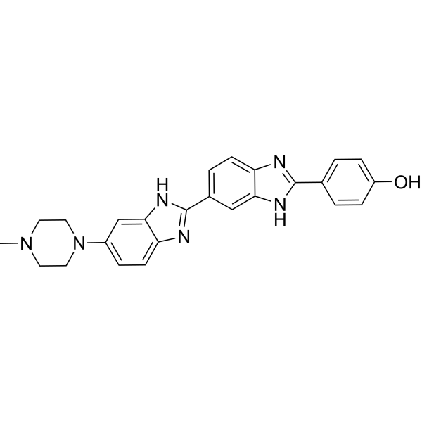
Hoechst 33258 is a blue to blue-green fluorescent live cell dye that can label DNA. Hoechst 33258 can specifically bind to the minor groove of DNA (and tends to bind to A/T-rich DNA), resulting in a significant increase in fluorescence intensity. Hoechst 33258 can cross the cell membrane and cause changes in DNA structure, such as G2/M phase arrest. Hoechst 33258 can bind to live or fixed cells, and the fluorescence intensity increases with increasing solution pH. As a DNA-specific probe, Hoechst 33258 can be used to detect DNA content, analyze cell cycle, etc. The excitation wavelength of Hoechst 33258 is 350-365 nm, and the emission wavelength is 460-490 nm.
Nur für Forschungszwecke. Wir verkaufen nicht an Patienten.
- Reinheit : 98.26%
- CAS. Nr.: 23491-44-3
- Formel: C25H24N6O
- Molecular Weight:424.50
-
Speicherung:
-20°C, protect from light
* In solvent : -80°C, 6 months; -20°C, 1 month (protect from light)

To place orders, for customer services and technical support, please contact: MedChemExpress USA
Tel: 609-228-6898 E-mail: [email protected] [email protected]


-
 Biologische Aktivität
-
Chemical Information
-
Lösungsmittel & Löslichkeit
- Protokoll
- Reinheit & Dokumentation
- Verweise
-
 Help & FAQs


-
HY-D0815
Propidium IodideFluorochrome -
HY-15559
Hoechst 33342DNA Fluorescent Stain -
HY-D0814
DAPI dihydrochlorideFluorescent DNA Stain -
HY-17589A
ChloroquineAntimalarial Agent -
HY-W090090
BODIPY 493/503Neutral Lipid Dye -

HY-DY1006
Propidium Iodide (solution)Fluorochrome -

HY-Y0320
Dimethyl sulfoxide, meets analytical specification of Ch.P.Aprotic Solvent -

HY-13259
MG-132Proteasome Inhibitor -

HY-DY1081
DAPI dihydrochloride (solution)Fluorescent DNA Stain -
HY-Y0873
PEG300Neutral Polymer -
HY-15162
Monomethyl auristatin EAntimitotic Agent -
HY-N1584
HalofuginoneNatural Product
Publications Citing Use of MedChemExpress (MCE) Hoechst 33258
More- Nat Commun. 2020 Feb 5;11(1):719. [Abstract]
- J Nanobiotechnology. 2026 Jul 1.
- Chem Eng J. 2026 Jan 19;529:173215.
- Chem Eng J. 365 (2019) 270-281.
- Phytomedicine. 2025 Jul:142:156632. [Abstract]
- Int J Biol Macromol. 2019 Nov 1:140:709-718. [Abstract]
- Virulence. 2022 Dec;13(1):444-457. [Abstract]
- Food Biosci. 11 December 2021, 101501.
- Int Immunopharmacol. 2023 Jun:119:110136. [Abstract]
- Neurochem Res. 2026 Jun 29;51(4):205.
- Neurochem Res. 2026 Feb 7;51(1):66. [Abstract]
- Acta Biochim Biophys Sin (Shanghai). 2019 Aug 5;51(8):814-825. [Abstract]
- Med Chem Res. 2025 Dec 5.
- Int J Biochem Cell Biol. 2017 Mar:84:75-88. [Abstract]
- J Cell Biochem. 2019 Jun;120(6):9181-9192. [Abstract]
- Discov Oncol. 2025 Feb 18;16(1):199. [Abstract]
- Cancer Manag Res. 2020 Sep 25:12:9097-9111. [Abstract]
- Cancer Manag Res. 2020 Sep 28:12:9197-9209. [Abstract]
- Biochem Biophys Res Commun. 2025 May 5:762:151769. [Abstract]
- Biochem Biophys Res Commun. 2020 Jun 30;527(3):805-810. [Abstract]
- Biochem Cell Biol. 2021 Aug;99(4):403-413. [Abstract]
- Authorea. 2025 Sep 20.
- Linkoping University. 2025.
- Research Square Preprint. 2020 Jun.
- BIOTECHNOLOGY BULLETIN 2018, 34(12):172-178
 Customer Validation & Images

-
Cell Imaging/Staining
-
Cell Imaging/Staining
-
IF
-
Cell Imaging/Staining


Biologische Aktivität
Beschreibung
Cellular Effect
|
Cell Line

|
Type | Value | Description | References |
|---|---|---|---|---|
| A2780 | IC50 |
12 μM
Compound: HOE-33258
|
Antitumor activity of compound for 96-h exposure in A2780 human ovarian cell line
Antitumor activity of compound for 96-h exposure in A2780 human ovarian cell line
|
[PMID: 11170623] |
| A2780cisR | IC50 |
9.5 μM
Compound: HOE-33258
|
Antitumor activity of compound for 96-h exposure in A2780cisR (acquired resistance to cisplatin) human ovarian cell line
Antitumor activity of compound for 96-h exposure in A2780cisR (acquired resistance to cisplatin) human ovarian cell line
|
[PMID: 11170623] |
| A549 | IC50 |
≥31.2 μg/mL
Compound: 2
|
Cytotoxicity against human A549 cells assessed as reduction in cell survival after 24 hrs by resazurin assay
Cytotoxicity against human A549 cells assessed as reduction in cell survival after 24 hrs by resazurin assay
|
[PMID: 27301676] |
| AGS | GI50 |
112 x 10-6 mol/dm3
Compound: Hoechst 33258
|
Cytotoxicity against human AGS cells after 72 hrs by MTT assay
Cytotoxicity against human AGS cells after 72 hrs by MTT assay
|
[PMID: 21227551] |
| BEAS-2B | IC50 |
≥31.2 μg/mL
Compound: 2
|
Cytotoxicity against human BEAS2B cells assessed as reduction in cell survival after 24 hrs by resazurin assay
Cytotoxicity against human BEAS2B cells assessed as reduction in cell survival after 24 hrs by resazurin assay
|
[PMID: 27301676] |
| Caco-2 | GI50 |
84 x 10-6 mol/dm3
Compound: Hoechst 33258
|
Cytotoxicity against human Caco2 cells after 72 hrs by MTT assay
Cytotoxicity against human Caco2 cells after 72 hrs by MTT assay
|
[PMID: 21227551] |
| CH1 | IC50 |
26.5 μM
Compound: HOE-33258
|
Antitumor activity of compound for 96-h exposure in CH1 human ovarian cell line
Antitumor activity of compound for 96-h exposure in CH1 human ovarian cell line
|
[PMID: 11170623] |
| HeLa | GI50 |
115.7 x 10⁻⁶ mol/dm3
Compound: Hoechst 33258
|
Cytotoxicity against human HeLa cells after 72 hrs by MTT assay
Cytotoxicity against human HeLa cells after 72 hrs by MTT assay
|
[PMID: 21227551] |
| HeLa | IC50 |
51.31 μM
Compound: H. 33258, Hoechst 33258
|
Cytotoxicity against human HeLa cells after 72 hrs by MTT assay
Cytotoxicity against human HeLa cells after 72 hrs by MTT assay
|
[PMID: 22959518] |
| HEp-2 | GI50 |
132.5 x 10⁻⁶ mol/dm3
Compound: Hoechst 33258
|
Cytotoxicity against human Hep2 cells after 72 hrs by MTT assay
Cytotoxicity against human Hep2 cells after 72 hrs by MTT assay
|
[PMID: 21227551] |
| HL-60 | IC50 |
32.43 μM
Compound: H. 33258, Hoechst 33258
|
Cytotoxicity against human HL60 cells after 72 hrs by MTT assay
Cytotoxicity against human HL60 cells after 72 hrs by MTT assay
|
[PMID: 22959518] |
| MCF7 | GI50 |
5.7 x 10⁻⁶ mol/dm3
Compound: Hoechst 33258
|
Cytotoxicity against human MCF7 cells after 72 hrs by MTT assay
Cytotoxicity against human MCF7 cells after 72 hrs by MTT assay
|
[PMID: 21227551] |
| MDCK | GI50 |
157.5 x 10⁻⁶ mol/dm3
Compound: Hoechst 33258
|
Cytotoxicity against MDCK cells after 72 hrs by MTT assay
Cytotoxicity against MDCK cells after 72 hrs by MTT assay
|
[PMID: 21227551] |
| MIA PaCa-2 | GI50 |
191.5 x 10⁻⁶ mol/dm3
Compound: Hoechst 33258
|
Cytotoxicity against human MIAPaCa2 cells after 72 hrs by MTT assay
Cytotoxicity against human MIAPaCa2 cells after 72 hrs by MTT assay
|
[PMID: 21227551] |
| NCI-H358 | GI50 |
91.7 x 10⁻⁶ mol/dm3
Compound: Hoechst 33258
|
Cytotoxicity against human NCI-H358 cells after 72 hrs by MTT assay
Cytotoxicity against human NCI-H358 cells after 72 hrs by MTT assay
|
[PMID: 21227551] |
| P388 | IC50 |
1.2 μM
Compound: 4
|
Concentration of the drug to inhibit wild type P388 murine leukemia cell growth in culture
Concentration of the drug to inhibit wild type P388 murine leukemia cell growth in culture
|
[PMID: 7527862] |
| SK-OV-3 | IC50 |
>100 μM
Compound: HOE-33258
|
Antitumor activity of compound for 96-h exposure in SKOV-3 human ovarian cell line
Antitumor activity of compound for 96-h exposure in SKOV-3 human ovarian cell line
|
[PMID: 11170623] |
| U-937 | IC50 |
15.42 μM
Compound: H. 33258, Hoechst 33258
|
Cytotoxicity against human U937 cells after 72 hrs by MTT assay
Cytotoxicity against human U937 cells after 72 hrs by MTT assay
|
[PMID: 22959518] |
In Vitro
Guide (Following is our recommended protocol. This protocol only provides a guideline, and should be modified according to your specific needs).
Hoechst 33258 has two binding modes: Type 1 and Type 2. Type 1 binds at low concentrations, has high fluorescence quantum yield, and has an emission peak at 460 nm; Type 2 binds at higher concentrations, resulting in fluorescence quenching, with an emission peak at around 490 nm[1].
The IC50 values ??of Hoechst 33258 for HeLa, HL60, and U937 cells are 51.31 μM, 32.43 μM, and 15.42 μM, respectively[2].
For the usage of Hoechst 33258, refer to [1]
1 Preparation of Hoechst working solution:
1.1 Preparation of stock solution
Prepare Hoechst 33258 powder into 1-10 mM stock solution, and stir in dark place until completely dissolved.
Note: The dye is light sensitive, so the preparation process needs to be kept away from light.
1.2 Working solution dilution
Before use, dilute the stock solution with PBS or cell culture medium to the working concentration, usually 5-10 μg/mL (cell staining) or 1-50 μM (DNA binding study).
Note: Prepare immediately before use to avoid fluorescence quenching.
2 Adherent cell staining steps:
Cells are seeded in culture plates/dishes and cultured to the logarithmic growth phase. If cells need to be fixed, fix them with 4% paraformaldehyde at room temperature for 10-15 min, and wash twice with PBS for 5 minutes each time.
2.1 Add Hoechst 33258 working solution and incubate at 37°C in the dark for 10-30 min (adjust the time according to the cell type to avoid over-staining).
2.2 For live cell staining, add directly to the culture medium and incubate without fixation; for dead cells, fix first and then stain.
2.3 Wash the cells 2-3 times with PBS or serum-free medium to remove unbound dye and reduce background fluorescence.
3 Suspended cell staining steps:
3.1 Collect cells, centrifuge at 1000 rpm for 5 min, and discard the supernatant.
3.2 Resuspend the cells with PBS, add Hoechst 33258 working solution, and incubate at room temperature in the dark for 15-20 min.
3.3 Resuspend after centrifugation and washing for flow cytometry or fluorescence microscopy detection.
4 Detection channels and tools:
4.1 Excitation wavelength (Ex): 350-365 nm (ultraviolet or near-ultraviolet light).
4.2 Emission wavelength (Em): 460-490 nm (blue to blue-green fluorescence).
4.3 Detection tools:
Fluorescence microscope: equipped with UV excitation filter (such as DAPI channel) to observe the intracellular DNA distribution.
Flow cytometer: Use UV laser (355 nm) and 450/50 nm bandpass filter to detect cell cycle or DNA content.
Microplate reader: When detecting in multi-well plate, select UV excitation and blue emission channels for high-throughput DNA quantification.
5 Precautions:
5.1 The concentration needs to be adjusted according to the cell type and experimental purpose. High concentration (>50 μM) may cause DNA precipitation (e.g., precipitation when D/P>0.4).
5.2 Short-term incubation (<30 min) has low cytotoxicity, and long-term incubation requires verification of cell viability.
MedChemExpress (MCE) has not independently confirmed the accuracy of these methods. They are for reference only.
Emission (Em)
451
Excitation (Ex)
356
Dyeing Example


Chemical Information
-
CAS. Nr. 23491-44-3
-
Appearance Solid
-
Molecular Weight 424.50
-
Formel C25H24N6O
-
Color Light yellow to yellow
-
SMILES
OC1=CC=C(C2=NC3=CC=C(C4=NC5=CC=C(N6CCN(C)CC6)C=C5N4)C=C3N2)C=C1
-
Synonyms
bisBenzimide H 33258; H 33258
-
Versand
Room temperature in continental US; may vary elsewhere.
-
Speicherung
-20°C, protect from light
* In solvent : -80°C, 6 months; -20°C, 1 month (protect from light)


Publications (25)
-
Journal Impact Factor
-
Most Recent
-
Nat Commun
Liver governs adipose remodelling via extracellular vesicles in response to lipid overload. [Abstract]2020 Feb 5;11(1):719. PMID: 32024826 -
-
-
-
Phytomedicine
Curcuminoids WM03 inhibits ovarian cancer cisplatin-resistant cells proliferation and reverses cisplatin resistance by targeting DYRK2. [Abstract]2025 Jul:142:156632. PMID: 40315643 -
Int J Biol Macromol
Protein corona of metal-organic framework nanoparticals: Study on the adsorption behavior of protein and cell interaction. [Abstract]2019 Nov 1:140:709-718. PMID: 31445155 -
Virulence
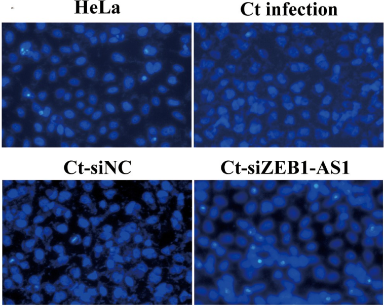
LncRNA ZEB1-AS1/miR-1224-5p / MAP4K4 axis regulates mitochondria-mediated HeLa cell apoptosis in persistent Chlamydia trachomatis infection. [Abstract]2022 Dec;13(1):444-457. PMID: 35266440
Hoechst 33258 purchased from MedChemExpress. Usage Cited in: Virulence. 2022 Dec;13(1):444-457. [Abstract]
The apoptotic rate was detected by Hoechst staining (400×), HeLa cells were treated with siRNA for 24 h and followed by persistent infection.
-
-
Int Immunopharmacol
2023 Jun:119:110136. PMID: 37075668 -
-
Neurochem Res
4-Phenylbutyrate Induces Functional Elongation of the Microglial Process Through Activation of Akt. [Abstract]2026 Feb 7;51(1):66. PMID: 41653373 -
Acta Biochim Biophys Sin (Shanghai)
Downregulation of lncRNA AFAP1-AS1 by oridonin inhibits the epithelial-to-mesenchymal transition and proliferation of pancreatic cancer cells. [Abstract]2019 Aug 5;51(8):814-825. PMID: 31314060 -
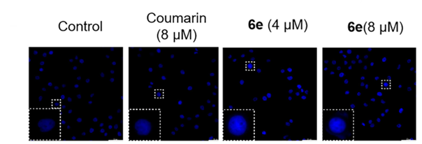
Hoechst 33258 purchased from MedChemExpress. Usage Cited in: Med Chem Res. 2025 Dec 5.
The A549 cells were exposed to coumarin (8 μM) or 6e (0, 4, 8 μM), then stained with 50 mM Hoechst 33258, and examined using fluorescence microscopy.
-
Int J Biochem Cell Biol
Methylene blue increases the amount of HSF1 through promotion of PKA-mediated increase in HSF1-p300 interaction. [Abstract]2017 Mar:84:75-88. PMID: 28104499 -
J Cell Biochem
Notoginsenoside R1 protects oxygen and glucose deprivation-induced injury by upregulation of miR-21 in cardiomyocytes. [Abstract]2019 Jun;120(6):9181-9192. PMID: 30552708 -
Discov Oncol
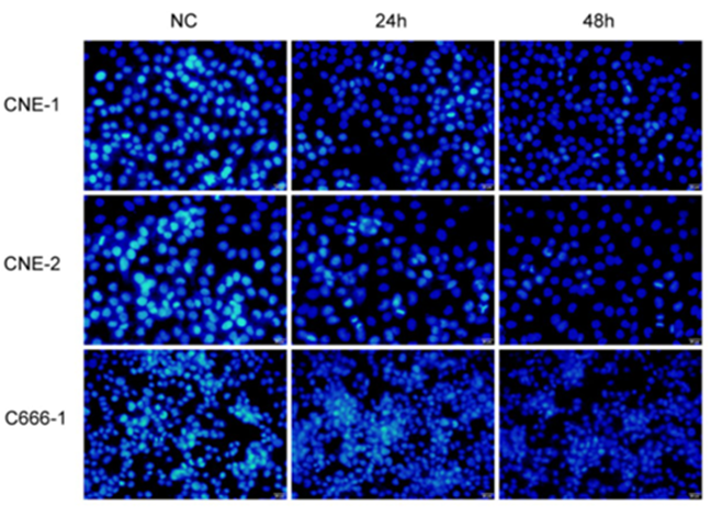
Viral IL-10 can promote the proliferation, migration and invasion of nasopharyngeal carcinoma cells and inhibit their apoptosis. [Abstract]2025 Feb 18;16(1):199. PMID: 39964638
Hoechst 33258 purchased from MedChemExpress. Usage Cited in: Discov Oncol. 2025 Feb 18;16(1):199. [Abstract]
Hoechst 33,258 staining results showing the effect of vIL-10 on the apoptosis of NPC cells.
-
Cancer Manag Res
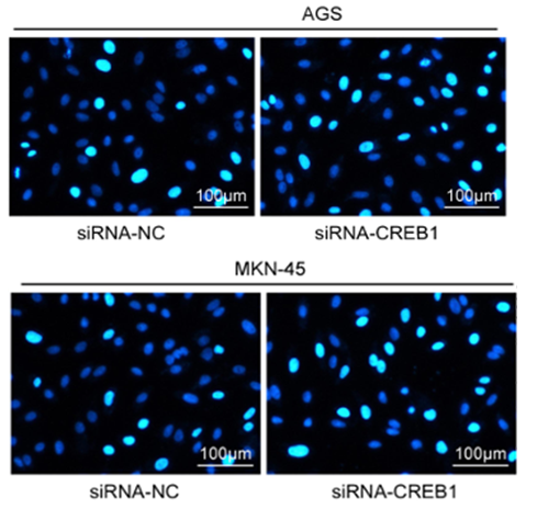
CREB1 Suppresses Transcription of microRNA-186 to Promote Growth, Invasion and Epithelial-Mesenchymal Transition of Gastric Cancer Cells Through the KRT8/HIF-1α Axis. [Abstract]2020 Sep 25:12:9097-9111. PMID: 33061604
Hoechst 33258 purchased from MedChemExpress. Usage Cited in: Cancer Manag Res. 2020 Sep 25:12:9097-9111. [Abstract]
The apoptosis rate of GC cells was assessed by Hoechst 33258 staining (5 μg/mL, 30 min).
-
Cancer Manag Res
microRNA-139-3p Inhibits Malignant Behaviors of Laryngeal Cancer Cells via the KDM5B/SOX2 Axis and the Wnt/β-Catenin Pathway. [Abstract]2020 Sep 28:12:9197-9209. PMID: 33061611 -
Biochem Biophys Res Commun
Tangeretin protects against Aβ1-42-induced toxicity and exploring mitochondria-lysosome interactions in HT22 cells. [Abstract]2025 May 5:762:151769. PMID: 40220719 -
Biochem Biophys Res Commun
Chlamydia trachomatis plasmid-encoded protein pORF5 activates unfolded protein response to induce autophagy via MAPK/ERK signaling pathway. [Abstract]2020 Jun 30;527(3):805-810. PMID: 32446560 -
Biochem Cell Biol
TFAP2A inhibits microRNA-126 expression at the transcriptional level and aggravates ischemic neuronal injury. [Abstract]2021 Aug;99(4):403-413. PMID: 33264079 -
-
-
-
BIOTECHNOLOGY BULLETIN 2018, 34(12):172-178


Lösungsmittel & Löslichkeit
In Vitro:
DMSO : 50 mg/mL (117.79 mM; Need ultrasonic; Hygroscopic DMSO has a significant impact on the solubility of product, please use newly opened DMSO)
Please refer to the solubility information to select the appropriate solvent. Once prepared, please aliquot and store the solution to prevent product inactivation from repeated freeze-thaw cycles.
Storage method and period of stock solution: -80°C, 6 months; -20°C, 1 month (protect from light). When stored at -80°C, please use it within 6 months. When stored at -20°C, please use it within 1 month.
Please refer to the solubility information to select the appropriate solvent. Once prepared, please aliquot and store the solution to prevent product inactivation from repeated freeze-thaw cycles.
Storage method and period of stock solution: -80°C, 6 months; -20°C, 1 month (protect from light). When stored at -80°C, please use it within 6 months. When stored at -20°C, please use it within 1 month.
Konzentration (Stammlösung) × Volumen (Stammlösung) = Konzentration (Ziellösung) × Volumen (Ziellösung)


Reinheit & Dokumentation
-
Data Sheet (279 KB)
-
SDS (394 KB)
- English - EN (394 KB)
- Français - FR (394 KB)
- Deutsch - DE (394 KB)
- Norwegian - NO (394 KB)
- Español - ES (394 KB)
- Swedish - SV (394 KB)
- Italian - IT (394 KB)
- Korean - KR (394 KB)
- Portuguese - PT (394 KB)
-
Handling Instructions (2659 KB)
Verweise
[1]. Stokke T, et al. Multiple binding modes for Hoechst 33258 to DNA. J Histochem Cytochem. 1985 Apr;33(4):333-8. [Content Brief]
[2]. Wang XJ, et al. Newly synthesized bis-benzimidazole derivatives exerting anti-tumor activity through induction of apoptosis and autophagy. Bioorg Med Chem Lett. 2012 Oct 1;22(19):6297-300. [Content Brief]
[3]. Pjura PE, et al. Binding of Hoechst 33258 to the minor groove of B-DNA. J Mol Biol. 1987 Sep 20;197(2):257-71. [Content Brief]


Complete Stock Solution Preparation Table
Please refer to the solubility information to select the appropriate solvent. Once prepared, please aliquot and store the solution to prevent product inactivation from repeated freeze-thaw cycles.
Storage method and period of stock solution: -80°C, 6 months; -20°C, 1 month (protect from light). When stored at -80°C, please use it within 6 months. When stored at -20°C, please use it within 1 month.
| Optional Solvent | Concentration Solvent Mass | 1 mg | 5 mg | 10 mg | 25 mg |
|---|---|---|---|---|---|
| DMSO | 1 mM | 2.3557 mL | 11.7786 mL | 23.5571 mL | 58.8928 mL |
| 5 mM | 0.4711 mL | 2.3557 mL | 4.7114 mL | 11.7786 mL | |
| 10 mM | 0.2356 mL | 1.1779 mL | 2.3557 mL | 5.8893 mL | |
| 15 mM | 0.1570 mL | 0.7852 mL | 1.5705 mL | 3.9262 mL | |
| 20 mM | 0.1178 mL | 0.5889 mL | 1.1779 mL | 2.9446 mL | |
| 25 mM | 0.0942 mL | 0.4711 mL | 0.9423 mL | 2.3557 mL | |
| 30 mM | 0.0785 mL | 0.3926 mL | 0.7852 mL | 1.9631 mL | |
| 40 mM | 0.0589 mL | 0.2945 mL | 0.5889 mL | 1.4723 mL | |
| 50 mM | 0.0471 mL | 0.2356 mL | 0.4711 mL | 1.1779 mL | |
| 60 mM | 0.0393 mL | 0.1963 mL | 0.3926 mL | 0.9815 mL | |
| 80 mM | 0.0294 mL | 0.1472 mL | 0.2945 mL | 0.7362 mL | |
| 100 mM | 0.0236 mL | 0.1178 mL | 0.2356 mL | 0.5889 mL |


Hoechst 33258 Related Classifications
HY-15558 Related Classifications
 Powered by Bioz