



PKN1 Antibody (YA4308)
(Synonyms: DBK; PKN; PAK1; PRK1; PAK-1; PRKCL1; PKN-ALPHA)
Based on 1 Customer Validation
PKN1 Antibody (YA4308) is a Mouse-derived and non-conjugated IgG2b monoclonal antibody, targeting to PKN1.
-
Host:
Mouse
-
Isotype:
IgG
-
Application:
IHC-P, ICC/IF, FC, ELISA
-
Reactivity :
Human
-
Formulation:
Supplied in PBS with 0.05% sodium azide.
-
Conjugation:
Non-conjugated


To place orders, for customer services and technical support, please contact: MedChemExpress USA
Tel: 609-228-6898 E-mail: [email protected] [email protected]





Applications
| Application |
IHC-P

IHC-P: Immunohistochemistry-Paraffin
|
ICC/IF
ICC/IF: Immunocytochemistry/
Immunofluorescence |
FC
FC: Flow Cytometry
|
ELISA
ELISA: Enzyme Linked Immunosorbent Assay
|
|---|---|---|---|---|
| Dilution Ratio | 1:200-1:1000 | 1:200-1:1000 | 1:200-1:400 | 1:10000 |


Product Details
PKN1 Antibody (YA4308) is a Mouse-derived and non-conjugated IgG2b monoclonal antibody, targeting to PKN1.
-
Host Mouse

-
Clonality Monoclonal

-
Species ReactivityHuman
-
Observed Molecular WeightObserved band size: 120 kDa
Note: Due to possible protein modifications or aggregation, the molecular weight should be confirmed by actual measurement, and the predicted value is for reference only.

-
Calculated Molecular Weight Predicted band size: 104 kDa

Purified recombinant fragment of human PKN1 (AA: 442-620) expressed in E. Coli.
affinity purified.
Non-conjugated
Unmodified
IgG


Product Properties
-
Appearance
Solution
-
Formulation
Supplied in PBS with 0.05% sodium azide.
-
Concentration
Batch-dependent, Please check the COA for the concentration of each lot. Check Lot Concentration
-
Storage & Stability
Stored at -20°C for 1 year. Avoid repeated freeze / thaw cycles.
-
Shipping
Shipping with blue ice.


Verification Images
-
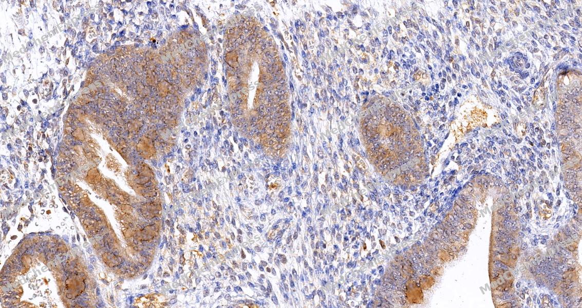 Immunohistochemical analysis of paraffin-embedded human endometrium tissue using PKN1 Antibody (HY-P84611, 1/500). The section was pretreated using heat mediated antigen retrieval with sodium citrate buffer (pH 6.0) for 8 minutes. The tissues were blocked with quick block buffer for 0.5 hours at room temperature, washed with PBS and PBST, and then incubated with the primary antibody overnight at 4℃. The detection was performed using an HRP conjugated compact polymer system. DAB was used as the chromogen. Tissues were counterstained with hematoxylin and mounted with DPX.
 -
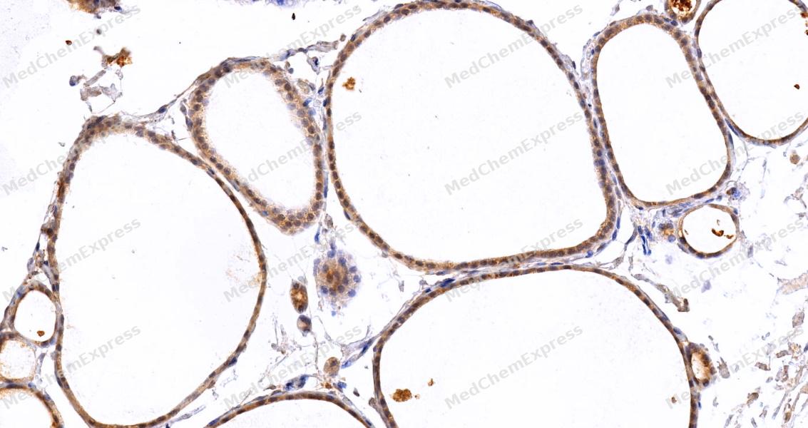 Immunohistochemical analysis of paraffin-embedded human thyroid gland tissue using PKN1 Antibody (HY-P84611, 1/500). The section was pretreated using heat mediated antigen retrieval with sodium citrate buffer (pH 6.0) for 8 minutes. The tissues were blocked with quick block buffer for 0.5 hours at room temperature, washed with PBS and PBST, and then incubated with the primary antibody overnight at 4℃. The detection was performed using an HRP conjugated compact polymer system. DAB was used as the chromogen. Tissues were counterstained with hematoxylin and mounted with DPX.
 -
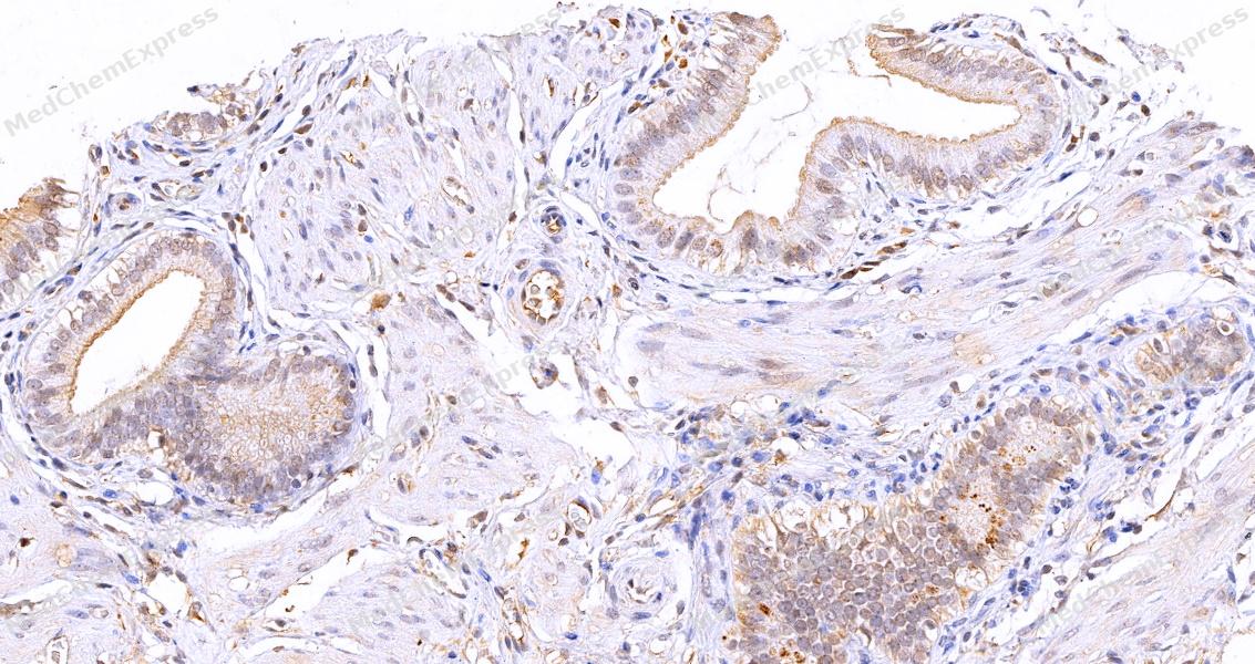 Immunohistochemical analysis of paraffin-embedded human gallbladder tissue using PKN1 Antibody (HY-P84611, 1/500). The section was pretreated using heat mediated antigen retrieval with sodium citrate buffer (pH 6.0) for 8 minutes. The tissues were blocked with quick block buffer for 0.5 hours at room temperature, washed with PBS and PBST, and then incubated with the primary antibody overnight at 4℃. The detection was performed using an HRP conjugated compact polymer system. DAB was used as the chromogen. Tissues were counterstained with hematoxylin and mounted with DPX.
 -
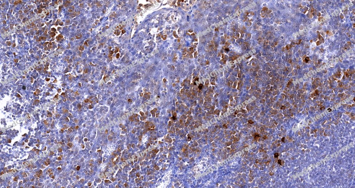 Immunohistochemical analysis of paraffin-embedded human tonsil tissue using PKN1 Antibody (HY-P84611, 1/500). The section was pretreated using heat mediated antigen retrieval with sodium citrate buffer (pH 6.0) for 8 minutes. The tissues were blocked with quick block buffer for 0.5 hours at room temperature, washed with PBS and PBST, and then incubated with the primary antibody overnight at 4℃. The detection was performed using an HRP conjugated compact polymer system. DAB was used as the chromogen. Tissues were counterstained with hematoxylin and mounted with DPX.
 -
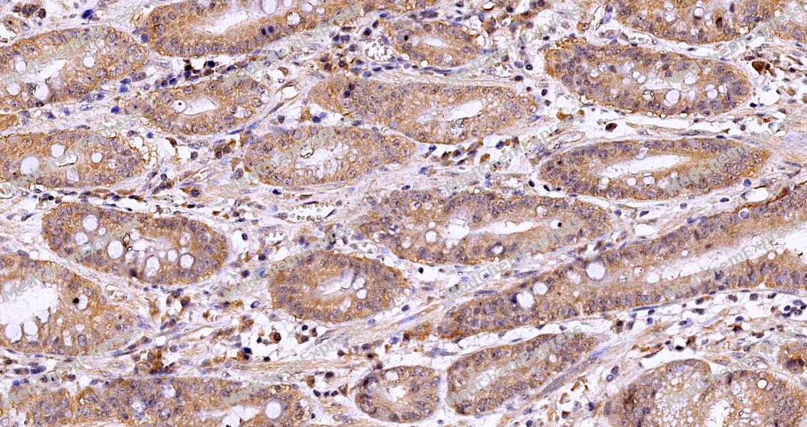 Immunohistochemical analysis of paraffin-embedded human stomach cancer using PKN1 Antibody (HY-P84611, 1/500). The section was pretreated using heat mediated antigen retrieval with sodium citrate buffer (pH 6.0) for 8 minutes. The tissues were blocked with quick block buffer for 0.5 hours at room temperature, washed with PBS and PBST, and then incubated with the primary antibody overnight at 4℃. The detection was performed using an HRP conjugated compact polymer system. DAB was used as the chromogen. Tissues were counterstained with hematoxylin and mounted with DPX.
 -
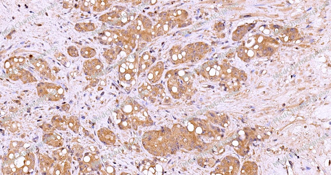 Immunohistochemical analysis of paraffin-embedded human prostate cancer using PKN1 Antibody (HY-P84611, 1/500). The section was pretreated using heat mediated antigen retrieval with sodium citrate buffer (pH 6.0) for 8 minutes. The tissues were blocked with quick block buffer for 0.5 hours at room temperature, washed with PBS and PBST, and then incubated with the primary antibody overnight at 4℃. The detection was performed using an HRP conjugated compact polymer system. DAB was used as the chromogen. Tissues were counterstained with hematoxylin and mounted with DPX.
 -
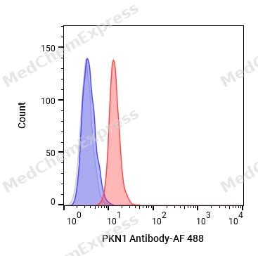 Flow cytometric analysis of 1X10⁶ HeLa cells labeling PKN1 Antibody (HY-P84611, red). Cells were fixed with 4% paraformaldehyde. Then stained with the primary antibody at 1/50 dilution for an hour at 4℃. AF 488-conjugated AffiniPure Goat Anti- Mouse IgG H&L (HY-P8005) was used as the secondary antibody at 1/1,000 dilution for 30 minutes at 4℃. Mouse IgG Isotype Control (HY-P80757, blue) was used as the isotype control, cells without incubation with primary antibody were used as the unlabeled control (black).
 -
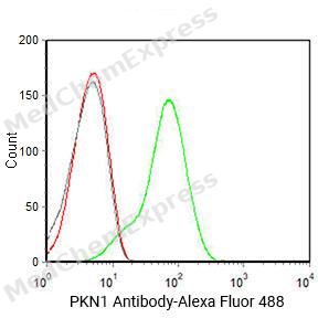 Flow cytometric analysis of 1X10^6 Hela cells labeling PKN1 Antibody(HY-P84611
 -
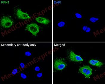 Immunofluorescence analysis of Hela cells labeling PKN1 antibody (HY-P84611) at 1/50 dilution. Cells were fixed in 4% paraformaldehyde for 15 minutes at room temperature


Background
-
Function
PKN1 (protein kinase N1, PRK1) is a member of the protein kinase N family of AGC serine/threonine kinases that functions downstream of Rho family GTPases and phospholipid-dependent signaling to regulate cytoskeletal organization, cell migration, transcriptional control, and cell survival pathways[1][4]. Mechanistically, PKN1 links RhoA-dependent signaling to phosphorylation events that influence intermediate filament dynamics, actin-associated structures, and downstream signaling networks including PI3K/AKT-related processes[1][2]. Through these activities, PKN1 contributes to the regulation of cellular proliferation and differentiation, and genetic or functional perturbation of PKN1 alters signaling responses in multiple experimental systems[2][3]. In disease-relevant contexts, PKN1 has been associated with tumor cell invasion, androgen receptor-dependent transcriptional regulation, and cancer-related signaling pathways, supporting its use as a target for mechanistic studies of oncogenic signaling and cytoskeletal remodeling[4][5]. Compared with related isoforms, PKN1 and PKN2 are broadly expressed across tissues, whereas PKN3 displays a more restricted expression pattern and has been linked more specifically to tumor-associated functions, highlighting important isoform-specific biology within the PKN family[3]. For experimental applications, pharmacological kinase inhibitors and genetic loss-of-function approaches have been used to define PKN1-dependent signaling networks, enabling investigation of isoform-selective functions and evaluation of PKN1 as a potential therapeutic target in cancer and other disease models[2][3].
-
Subcellular Localization
Cytoplasm; Nucleus; Endosome; Cell membrane; Peripheral membrane protein; Cleavage furrow; Midbody
-
Expression
Tissue_specificity:It is widely present in various tissues. It is expressed in the heart, brain, placenta, lungs, skeletal muscle, kidneys, and pancreas. It is also expressed in a variety of tumor cell lines, especially breast tumor cells. -
Subunit
Interacts with ZFAND6 (By similarity). Interacts with AR (PubMed:12514133). Interacts with PRKCB (PubMed:20228790). Interacts (via REM 1 and REM 2 repeats) with RAC1 (PubMed:14514689, PubMed:18006505). Interacts (via REM 1 repeat) with RHOA (PubMed:10619026, PubMed:8571126). Interacts with RHOB (PubMed:9478917). Interacts (via C-terminus) with PDPK1 (PubMed:10792047). Interacts with CCNT2; enhances MYOD1-dependent transcription (PubMed:16331689). Component of a signaling complex containing at least AKAP13, PKN1, MAPK14, ZAK and MAP2K3. Within this complex, AKAP13 interacts directly with PKN1, which in turn recruits MAPK14, MAP2K3 and ZAK (PubMed:21224381)
-
SwissProt ID
-
Synonyms
DBK; PKN; PAK1; PRK1; PAK-1; PRKCL1; PKN-ALPHA


Documentation
-
Data Sheet (262 KB)
-
SDS (251 KB)
- English - EN (251 KB)
- Français - FR (251 KB)
- Deutsch - DE (251 KB)
- Norwegian - NO (251 KB)
- Español - ES (251 KB)
- Swedish - SV (251 KB)
- Italian - IT (251 KB)
- Korean - KR (251 KB)
- Portuguese - PT (251 KB)
-
User Guide for Antibodies (1077 KB)
References
[1]. Uniprotkb.
[2]. Yasui T, et al. Protein kinase N1, a cell inhibitor of Akt kinase, has a central role in quality control of germinal center formation. Proc Natl Acad Sci U S A. 2012 Dec 18;109(51):21022-7. [Content Brief]
[3]. Falk MD, et al. Enzyme kinetics and distinct modulation of the protein kinase N family of kinases by lipid activators and small molecule inhibitors. Biosci Rep. 2014;34(2):e00097. [Content Brief]

 Powered by Bioz
