



RALA Antibody (YA3752)
(Synonyms: RAL)Based on 1 Customer Validation
RALA Antibody (YA3752) is a Mouse-derived and non-conjugated IgG1 monoclonal antibody, targeting to RALA.
-
Host:
Mouse
-
Isotype:
IgG1
-
Application:
WB, IHC-P, ICC/IF, FC, ELISA
-
Reactivity :
Human, Mouse, Rat, Monkey
-
Formulation:
Supplied in PBS with 0.05% sodium azide
-
Conjugation:
Non-conjugated


To place orders, for customer services and technical support, please contact: MedChemExpress USA
Tel: 609-228-6898 E-mail: [email protected] [email protected]





Applications
| Application |
WB

WB: Western Blot
|
IHC-P
IHC-P: Immunohistochemistry-Paraffin
|
ICC/IF
ICC/IF: Immunocytochemistry/
Immunofluorescence |
FC
FC: Flow Cytometry
|
ELISA
ELISA: Enzyme Linked Immunosorbent Assay
|
|---|---|---|---|---|---|
| Dilution Ratio | 1:500-1:2000 | 1:100-1:500 | 1:50-1:500 | 1:200-1:400 | 1:10000 |


Product Details
RALA Antibody (YA3752) is a Mouse-derived and non-conjugated IgG1 monoclonal antibody, targeting to RALA.
-
Host Mouse

-
Clonality Monoclonal

-
Species ReactivityHuman, Mouse, Rat, Monkey
-
Observed Molecular WeightObserved band size: 25 kDa
Note: Due to possible protein modifications or aggregation, the molecular weight should be confirmed by actual measurement, and the predicted value is for reference only.

-
Calculated Molecular Weight Predicted band size: 24 kDa

Purified recombinant fragment of human RALA (AA: 71-203) expressed in E. Coli.
affinity purified.
Non-conjugated
Unmodified
IgG1


Product Properties
-
Appearance
Solution
-
Formulation
Supplied in PBS with 0.05% sodium azide
-
Concentration
Batch-dependent, Please check the COA for the concentration of each lot. Check Lot Concentration
-
Storage & Stability
Stored at -20°C for 1 year. Avoid repeated freeze / thaw cycles.
-
Shipping
Shipping with blue ice.


Verification Images
-
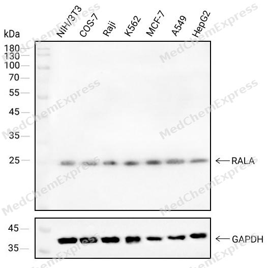 Western blot analysis of extracts from NIH/3T3(lane 1(40μg) ),COS-7(lane 2(40μg) ),Raji(lane 3(40μg) ),K562(lane 4(40μg) ),MCF-7(lane 5(40μg) ),A549(lane 6(40μg) )and HepG2(lane 7(40μg) ) using RALA antibody. Proteins were transferred to a NC membrane and blocked with 5% Skim milk in TBST for 2 hour at room temperature. The primary antibody ( 1/1000) and Loading control antibody (GAPDH,1/1000) was used in 5% Skim milk in TBST at 4°C overnight. Goat Anti-Mouse IgG-HRP Secondary Antibody (1/10,000) was used for 1 hour at room temperature.
 -
 Immunohistochemical analysis of paraffin-embedded human kidney tissue using RALA Antibody (HY-P84055, 1/400). The section was pretreated using heat mediated antigen retrieval with sodium citrate buffer (pH 6.0) for 8 minutes. The tissues were blocked with quick block buffer for 0.5 hours at room temperature, washed with PBS and PBST, and then incubated with the primary antibody overnight at 4℃. The detection was performed using an HRP conjugated compact polymer system. DAB was used as the chromogen. Tissues were counterstained with hematoxylin and mounted with DPX.
 -
 Immunohistochemical analysis of paraffin-embedded human small intestine tissue using RALA Antibody (HY-P84055, 1/400). The section was pretreated using heat mediated antigen retrieval with sodium citrate buffer (pH 6.0) for 8 minutes. The tissues were blocked with quick block buffer for 0.5 hours at room temperature, washed with PBS and PBST, and then incubated with the primary antibody overnight at 4℃. The detection was performed using an HRP conjugated compact polymer system. DAB was used as the chromogen. Tissues were counterstained with hematoxylin and mounted with DPX.
 -
 Immunohistochemical analysis of paraffin-embedded human salivary gland tissue using RALA Antibody (HY-P84055, 1/400). The section was pretreated using heat mediated antigen retrieval with sodium citrate buffer (pH 6.0) for 8 minutes. The tissues were blocked with quick block buffer for 0.5 hours at room temperature, washed with PBS and PBST, and then incubated with the primary antibody overnight at 4℃. The detection was performed using an HRP conjugated compact polymer system. DAB was used as the chromogen. Tissues were counterstained with hematoxylin and mounted with DPX.
 -
 Immunohistochemical analysis of paraffin-embedded human placenta tissue using RALA Antibody (HY-P84055, 1/400). The section was pretreated using heat mediated antigen retrieval with sodium citrate buffer (pH 6.0) for 8 minutes. The tissues were blocked with quick block buffer for 0.5 hours at room temperature, washed with PBS and PBST, and then incubated with the primary antibody overnight at 4℃. The detection was performed using an HRP conjugated compact polymer system. DAB was used as the chromogen. Tissues were counterstained with hematoxylin and mounted with DPX.
 -
 Immunohistochemical analysis of paraffin-embedded human prostate tissue using RALA Antibody (HY-P84055, 1/400). The section was pretreated using heat mediated antigen retrieval with sodium citrate buffer (pH 6.0) for 8 minutes. The tissues were blocked with quick block buffer for 0.5 hours at room temperature, washed with PBS and PBST, and then incubated with the primary antibody overnight at 4℃. The detection was performed using an HRP conjugated compact polymer system. DAB was used as the chromogen. Tissues were counterstained with hematoxylin and mounted with DPX.
 -
 Immunohistochemical analysis of paraffin-embedded human epididymis tissue using RALA Antibody (HY-P84055, 1/400). The section was pretreated using heat mediated antigen retrieval with sodium citrate buffer (pH 6.0) for 8 minutes. The tissues were blocked with quick block buffer for 0.5 hours at room temperature, washed with PBS and PBST, and then incubated with the primary antibody overnight at 4℃. The detection was performed using an HRP conjugated compact polymer system. DAB was used as the chromogen. Tissues were counterstained with hematoxylin and mounted with DPX.
 -
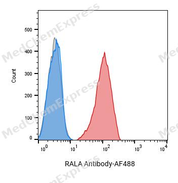 Flow cytometric analysis of 1X10^6 HL-60 cells labeling RALA Antibody(red). Cells were fixed with 4% paraformaldehyde and permeabilised with 0.2% Triton X-100. Then stained with the primary antibody at 1/400 dilution overnight at 4℃. AF 488 Goat Anti-mouse IgG H&L was used as the secondary antibody at 1/1,000 dilution for 45 minutes at room temperature. Mouse IgG Isotype Control (blue) was used as the isotype control, cells without incubation with primary antibody were used as the unlabeled control (black).
 -
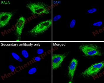 Immunofluorescence analysis of Hela cells labeling RALA antibody at 1/50 dilution. Cells were fixed in 4% paraformaldehyde for 15 minutes at room temperature, permeabilized with 0.1% Triton X-100 in PBS for 15 minutes at room temperature, then blocked with 1% BSA in 10% negative goat serum for 1 hour at room temperature.Cells were then incubated with RALA antibody at 1/50 dilution in 1% BSA in PBST overnight at 4 ℃. AF 488 Goat Anti-mouse IgG H&L (green) was used as the secondary antibody at 1/500 dilution. PBS instead of the primary antibody was used as the secondary antibody only control. The Nuclear counterstain was DAPI (Blue)
 -
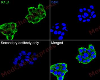 Immunofluorescence analysis of HepG2 cells labeling RALA antibody at 1/50 dilution. Cells were fixed in 4% paraformaldehyde for 15 minutes at room temperature, permeabilized with 0.1% Triton X-100 in PBS for 15 minutes at room temperature, then blocked with 1% BSA in 10% negative goat serum for 1 hour at room temperature.Cells were then incubated with RALA antibody at 1/50 dilution in 1% BSA in PBST overnight at 4 ℃. AF 488 Goat Anti-mouse IgG H&L (green) was used as the secondary antibody at 1/500 dilution. PBS instead of the primary antibody was used as the secondary antibody only control. The Nuclear counterstain was DAPI (Blue)


Background
-
Function
RALA multifunctional GTPase involved in a variety of cellular processes including gene expression, cell migration, cell proliferation, oncogenic transformation and membrane trafficking. Accomplishes its multiple functions by interacting with distinct downstream effectors. Acts as a GTP sensor for GTP-dependent exocytosis of dense core vesicles. The RALA-exocyst complex regulates integrin-dependent membrane raft exocytosis and growth signaling. Key regulator of LPAR1 signaling and competes with GRK2 for binding to LPAR1 thus affecting the signaling properties of the receptor. Required for anchorage-independent proliferation of transformed cells. During mitosis, supports the stabilization and elongation of the intracellular bridge between dividing cells. Cooperates with EXOC2 to recruit other components of the exocyst to the early midbody. During mitosis, also controls mitochondrial fission by recruiting to the mitochondrion RALBP1, which mediates the phosphorylation and activation of DNM1L by the mitotic kinase cyclin B-CDK1[1][2][3][4][5].
-
Subcellular Localization
Cell membrane; Lipid-anchor; Cytoplasmic side; Cleavage furrow; Midbody, Midbody ring; Mitochondrion
-
Expression
Induction:Activated in an LPA-dependent manner by LPAR1 and in an LPA-independent manner by LPAR2 -
Subunit
Interacts (via effector domain) with RALBP1; during mitosis, recruits RALBP1 to the mitochondrion where it promotes DNM1L phosphorylation and mitochondrial fission (PubMed:21822277, PubMed:7673236). Interacts with EXOC2/Sec5 and EXOC8/Exo84; binding to EXOC2 and EXOC8 is mutually exclusive (PubMed:14525976, PubMed:15920473, PubMed:18756269). Interacts with Clostridium exoenzyme C3 (PubMed:15809419, PubMed:16177825). Interacts with RALGPS1 (PubMed:10747847). Interacts with LPAR1 and LPAR2. Interacts with GRK2 in response to LPAR1 activation. RALA and GRK2 binding to LPAR1 is mutually exclusive (PubMed:19306925). Interacts with CDC42 (By similarity)
-
SwissProt ID
-
Synonyms
RAL


Documentation
-
Data Sheet (234 KB)
-
SDS (251 KB)
- English - EN (251 KB)
- Français - FR (251 KB)
- Deutsch - DE (251 KB)
- Norwegian - NO (251 KB)
- Español - ES (251 KB)
- Swedish - SV (251 KB)
- Italian - IT (251 KB)
- Korean - KR (251 KB)
- Portuguese - PT (251 KB)
-
User Guide for Antibodies (1077 KB)

 Powered by Bioz
