



FOXO4-DRI acetate
Based on 1 publication(s) in Google Scholar
FOXO4-DRI acetate is a cell-permeable peptide antagonist that blocks the interaction of FOXO4 and p53. FOXO4-DRI acetate is a senolytic peptide that induces apoptosis of senescent cells.
Nos produits utilisent uniquement pour la recherche. Nous ne vendons pas aux patients.
- Formule: C228H388N86O64.0.145C2H4O2
- Masse moléculaire:5366.77
-
Stockage:
Please store the product under the recommended conditions in the Certificate of Analysis.

To place orders, for customer services and technical support, please contact: MedChemExpress USA
Tel: 609-228-6898 E-mail: [email protected] [email protected]


-
 Activité biologique
-
Chemical Information
-
Solvant et solubilité
- Protocole
- Pureté et documentation
- Références
-
 Help & FAQs


Publications Citing Use of MedChemExpress (MCE) FOXO4-DRI acetate
More

Activité biologique
Description
In Vitro
FOXO4-DRI (25 mM; 3 days) acetate causes nuclear exclusion of active p53 and induces apoptosis in senescent TM3 Leydig cells[1].
FOXO4-DRI (25 μM; 5 days) acetate significantly reduces the senescence level in PDL9 cells[2].
MedChemExpress (MCE) has not independently confirmed the accuracy of these methods. They are for reference only.
-
Cell Line:Senescent Leydig cells
-
Concentration:25 mM
-
Incubation Time:3 days
-
Result:Reduced the viability of senescent as compared to normal TM3 Leydig cells.
-
Cell Line:Senescent Leydig cells
-
Concentration:25 mM
-
Incubation Time:3 days
-
Result:The apoptosis rate increased from 10% to 27%.
-
Cell Line:PDL9 cells
-
Concentration:25 μM
-
Incubation Time:5 days
-
Result:Decreased the protein levels of representative senescent markers, including p16, p21, and p53.
-
Cell Line:PDL9 cells
-
Concentration:25 μM
-
Incubation Time:5 days
-
Result:Enhanced SOX9 expression, and reduced MMP12 and MMP13 expression.
In Vivo
MedChemExpress (MCE) has not independently confirmed the accuracy of these methods. They are for reference only.
-
Animal Model:Naturally aged male C57BL/6 mice (20-24 months old)[1]
-
Dosage:5 mg/kg
-
Administration:Intraperitoneal injection, every other day for three administrations
-
Result:Increased serum testosterone levels. Increased levels of both 3β-HSD and CYP11A1. Decreased interstitial SA-β-gal activity and lowered levels of senescence-associated proteins p53, p21, and p16. Decreased the levels of IL-1β, IL-6 and TGF-β.


Chemical Information
-
Masse moléculaire 5366.77
-
Formule C228H388N86O64.0.145C2H4O2
-
Sequence
D-(Leu-Thr-Leu-Arg-Lys-Glu-Pro-Ala-Ser-Glu-Ile-Ala-Gln-Ser-Ile-Leu-Glu-Ala-Tyr-Ser-Gln-Asn-Gly-Trp-Ala-Asn-Arg-Arg-Ser-Gly-Gly-Lys-Arg-Pro-Pro-Pro-Arg-Arg-Arg-Gln-Arg-Arg-Lys-Lys-Arg-Gly)
-
Sequence Shortening
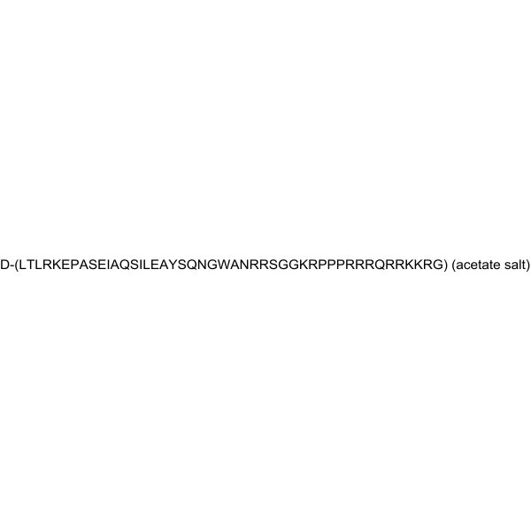
D-(LTLRKEPASEIAQSILEAYSQNGWANRRSGGKRPPPRRRQRRKKRG)
-
Livraison
Room temperature in continental US; may vary elsewhere.
-
Stockage
Please store the product under the recommended conditions in the Certificate of Analysis.


Publications (1)
-
Journal Impact Factor
-
Most Recent


Solvant et solubilité
In Vitro
H2O
Peptide Solubility and Storage Guidelines:
1. Calculate the length of the peptide.
2. Calculate the overall charge of the entire peptide according to the following table:
| Contents | Assign value | |
|---|---|---|
| Acidic amino acid | Asp (D), Glu (E), and the C-terminal -COOH. | -1 |
| Basic amino acid | Arg (R), Lys (K), His (H), and the N-terminal -NH2 | +1 |
| Neutral amino acid | Gly (G), Ala (A), Leu (L), Ile (I), Val (V), Cys (C), Met (M), Thr (T), Ser (S), Phe (F), Tyr (Y), Trp (W), Pro (P), Asn (N), Gln (Q) | 0 |
3. Recommended solution:
| Overall charge of peptide | Details |
|---|---|
| Negative (<0) |
1. Try to dissolve the peptide in water first. 2. If water fails, add NH4OH (<50 μL). 3. If the peptide still does not dissolve, add DMSO (50-100 μL) to solubilize the peptide. |
| Positive (>0) |
1. Try to dissolve the peptide in water first. 2. If water fails, try dissolving the peptide in a 10%-30% acetic acid solution. 3. If the peptide still does not dissolve, try dissolving the peptide in a small amount of DMSO. |
| Zero (=0) |
1. Try to dissolve the peptide in organic solvent (acetonitrile, methanol, etc.) first. 2. For very hydrophobic peptides, try dissolving the peptide in a small amount of DMSO, and then dilute the solution with water to the desired concentration. |


Pureté et documentation
Références
[1]. Zhang C, et al. FOXO4-DRI alleviates age-related testosterone secretion insufficiency by targeting senescent Leydig cells in aged mice. Aging (Albany NY). 2020 Jan 20;12(2):1272-1284. [Content Brief]
[2]. Huang Y, et al. Senolytic Peptide FOXO4-DRI Selectively Removes Senescent Cells From in vitro Expanded Human Chondrocytes. Front Bioeng Biotechnol. 2021 Apr 29;9:677576. [Content Brief]

Calculators
Concentration (start) × Volume (start) = Concentration (final) × Volume (final)


FOXO4-DRI acetate Related Classifications
HY-P4157A Related Classifications
 Powered by Bioz