



IL-1 alpha Protein, Rhesus macaque

Based on 1 Customer Validation
IL-1 alpha, a cytokine present in non-hematopoietic cells, links innate and adaptive immunity. Binding to its receptor IL1R1, IL-1 alpha forms a receptor complex, activating signaling pathways involving MYD88, IRAK1, and IRAK4. It activates NF-kappa-B and MAPK pathways. Released during cell death, IL-1 alpha induces inflammation, senses DNA damage, and interacts with TMED10, IL1R1, and S100A13 for secretion and plasma membrane translocation. IL-1 alpha Protein, Rhesus macaque is the recombinant Rhesus Macaque-derived IL-1 alpha protein, expressed by E. coli , with tag free.
- Species: Rhesus Macaque
- Source: E. coli
-
Storage:Stored at -20°C for 2 years from date of receipt. After reconstitution, it is stable at 4°C for 1 week or -20°C for longer (with carrier protein). It is recommended to freeze aliquots at -20°C or -80°C for extended storage.


To place orders, for customer services and technical support, please contact: MedChemExpress USA
Tel: 609-228-6898 E-mail: [email protected] [email protected]


-
 Biological Activity
-
Technical Parameters
-
Product Properties
- Documentation
- Help & FAQs




Biological Activity
Description
IL-1 alpha, a cytokine present in non-hematopoietic cells, links innate and adaptive immunity. Binding to its receptor IL1R1, IL-1 alpha forms a receptor complex, activating signaling pathways involving MYD88, IRAK1, and IRAK4. It activates NF-kappa-B and MAPK pathways. Released during cell death, IL-1 alpha induces inflammation, senses DNA damage, and interacts with TMED10, IL1R1, and S100A13 for secretion and plasma membrane translocation. IL-1 alpha Protein, Rhesus macaque is the recombinant Rhesus Macaque-derived IL-1 alpha protein, expressed by E. coli , with tag free.
Background
The IL-1 alpha protein, a cytokine intrinsically present in almost all quiescent non-hematopoietic cells, plays a pivotal role in inflammation, serving as a crucial link between the innate and adaptive immune systems. Upon binding to its receptor IL1R1, alongside its accessory protein IL1RAP, IL-1 alpha forms the high-affinity interleukin-1 receptor complex, initiating signaling pathways that involve the recruitment of adapter molecules such as MYD88, IRAK1, or IRAK4. This, in turn, activates NF-kappa-B and three MAPK pathways—p38, p42/p44, and JNK pathways. Internally, IL-1 alpha acts as an alarmin, being released into the extracellular space after cell membrane disruption during cell death, thereby inducing inflammation and signaling the host to injury or damage. Beyond its role as a danger signal released during cell necrosis, IL-1 alpha directly senses DNA damage and functions as a signal for genotoxic stress without compromising cell integrity. As a monomer, IL-1 alpha interacts with TMED10, facilitating its translocation from the cytoplasm to the endoplasmic reticulum-Golgi intermediate compartment (ERGIC) for secretion. Additionally, it interacts with IL1R1 and S100A13, the latter being the initial step in IL-1 alpha export, followed by the direct translocation of this complex across the plasma membrane.
Verified Bioactivity
1.Fully biologically active when compared to standard. The ED50 as determined by a cell proliferation assay using murine D10S cells is less than 10 pg/mL, corresponding to a specific activity of >1.0x10⁸ IU/mg.
2.Measured in a cell proliferation assay using CTLL-2 cells. The ED50 for this effect is 19.28 pg/mL, corresponding to a specific activity is 5.187×10⁷ U/mg.
MCE Validation Data
-
Purity - SDS-PAGE
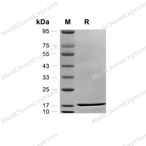
Purity - SDS-PAGE


Technical Parameters
-
Species Rhesus Macaque
-
Source E. coli
-
Tag Tag Free
-
Accession
P48089 (S113-A271)
-
Molecular Construction
-
N-term
-
IL-1α (S113-A271)
Accession # P48089 -
C-term
-
-
Protein Length
Full Length of Mature Protein
-
Synonyms
IL1A; IL-1A; Prev. IL1; Preinterleukin 1 Alpha; IL1F1; Interleukin-1 Alpha; Hematopoietin-1; IL-1 Alpha; IL1-ALPHA; Interleukin 1 Alpha; Pro-Interleukin-1-Alpha
-
AA Sequence
SAPFSFLSNMTYHFIRIIKHEFILNDTLNQTIIRANDQHLTAAAIHNLDEAVKFDMGAYTSSKDDTKVPVILRISKTQLYVSAQDEDQPVLLKEMPEINKTITGSETNFLFFWETHGTKNYFISVAHPNLFIATKHDNWVCLAKGLPSITDFQILENQA
-
Predicted Molecular Mass
18.1 kDa
-
Molecular Weight
Approximately 18-20 kDa, based on SDS-PAGE under reducing conditions.
-
Purity
≥ 95%, as determined by reducing SDS-PAGE.


Product Properties
Lyophilized powder.
Lyophilized from a 0.22 μm filtered solution of PBS, pH 7.4.
<1 EU/μg, determined by LAL method.
It is not recommended to reconstitute to a concentration less than 100 μg/mL in ddH2O. For long term storage it is recommended to add a carrier protein (0.1% BSA, 5% HSA, 10% FBS or 5% Trehalose).
Stored at -20°C for 2 years from date of receipt. After reconstitution, it is stable at 4°C for 1 week or -20°C for longer (with carrier protein). It is recommended to freeze aliquots at -20°C or -80°C for extended storage.
Room temperature in continental US; may vary elsewhere.


Documentation
-
Data Sheet (236 KB)
-
SDS (251 KB)
- English - EN (251 KB)
- Français - FR (251 KB)
- Deutsch - DE (251 KB)
- Norwegian - NO (251 KB)
- Español - ES (251 KB)
- Swedish - SV (251 KB)
- Italian - IT (251 KB)
- Korean - KR (251 KB)
- Portuguese - PT (251 KB)
-
Handling Instructions (2659 KB)

Calculators
Concentration (start) × Volume (start) = Concentration (final) × Volume (final)


 Powered by Bioz

