


GDC-0152
Based on 14 publication(s) in Google Scholar
GDC-0152 is a potent IAPs inhibitor, and binds to the BIR3 domains of XIAP, cIAP1, cIAP2 and the BIR domain of ML-IAP with Ki values of 28 nM, 17 nM, 43 nM and 14 nM, respectively.
For research use only. We do not sell to patients.
- Purity: 99.70%
- CAS No.: 873652-48-3
- Formula: C25H34N6O3S
- Molecular Weight:498.64
-
Storage:Powder -20°C, 3 years , 4°C, 2 years ; In solvent -80°C, 2 years , -20°C, 1 year

To place orders, for customer services and technical support, please contact: MedChemExpress USA
Tel: 609-228-6898 E-mail: [email protected] [email protected]


-
 Biological Activity
-
Chemical Information
-
Solvent & Solubility
- Protocol
- Purity & Documentation
- References
-
 Help & FAQs
-


Apoptosis Compound Library
HY-L003
-
Anti-Cancer Compound Library
HY-L025
-
Clinical Compound Library
HY-L026
-
Anti-Breast Cancer Compound Library
HY-L074
-
Protein-protein Interaction Inhibitor Library
HY-L109
-
Anti-Prostate Cancer Compound Library
HY-L124
-
Anti-Ovarian Cancer Compound Library
HY-L173
-
Bioactive Compound Library Max
HY-L181
-
MCE Bioactive Compound Library
HY-L001V
-
Clinical Compound Library Plus
HY-L026P
-
Bioactive Compound Library
HY-L001
-
High-Throughput Bioactive Compound Library
HY-L205


Publications Citing Use of MedChemExpress (MCE) GDC-0152
More- Sci Immunol. 2018 Aug 24;3(26):eaat2738. [Abstract]
- Cell Death Differ. 2020 May;27(5):1569-1587. [Abstract]
- J Exp Clin Cancer Res. 2024 Nov 28;43(1):311. [Abstract]
- Cell Rep. 2026 Mar 20;45(4):117130. [Abstract]
- J Med Chem. 2019 Jun 13;62(11):5616-5627. [Abstract]
- Sci Signal. 2018 Jul 17;11(539). pii: eaao3964. [Abstract]
- J Taiwan Inst Chem Eng. June 2022, 104394.
- Biomater Adv. 2023 Nov:154:213639. [Abstract]
- Biochem Pharmacol. 2026 Apr:246:117727. [Abstract]
- Sci Rep. 2018 Feb 1;8(1):2189. [Abstract]
- J Biol Chem. 2017 Jun 9;292(23):9666-9679. [Abstract]
- ACS Chem Biol. 2017 Dec 15;12(12):2981-2989. [Abstract]
- bioRxiv. 2024 Nov 28:2024.11.25.625306. [Abstract]
- bioRxiv. May 2, 2018.
 Customer Validation & Images

-
WB
-
WB


Biological Activity
Ki: 28 nM (XIAP BIR3), 14 nM (MLIAP-BIR3), 17 nM (cIAP1-BIR3), 43 nM (cIAP2-BIR3)
|
Cell Line
|
Type | Value | Description | References |
|---|---|---|---|---|
| SK-OV-3 | EC50 |
2.5 nM
Compound: GDC-0152
|
Induction of apoptosis in human SKOV3 cells assessed caspase-3 activation after 48 hrs by IncuCyte S3 live-cell analysis
Induction of apoptosis in human SKOV3 cells assessed caspase-3 activation after 48 hrs by IncuCyte S3 live-cell analysis
|
[PMID: 31095386] |
| SK-OV-3 | EC50 |
3 nM
Compound: GDC-0152
|
Induction of apoptosis in human SKOV3 cells assessed caspase-3 activation after 24 hrs by IncuCyte S3 live-cell analysis
Induction of apoptosis in human SKOV3 cells assessed caspase-3 activation after 24 hrs by IncuCyte S3 live-cell analysis
|
[PMID: 31095386] |
GDC-0152 can block protein?protein interactions that involve IAP proteins and pro-apoptotic molecules. Using transiently transfected HEK293T cells, GDC-0152 is shown to disrupt XIAP binding to partially processed caspase-9 and to disrupt the association of ML-IAP, cIAP1, and cIAP2 with Smac. In melanoma SK-MEL28 cells, the endogenous association of ML-IAP and Smac is also effectively abolished by GDC-0152. GDC-0152 leads to a decrease in cell viability in the MDA-MB-231 breast cancer cell line, while having no effect on normal human mammary epithelial cells (HMEC). GDC-0152 is found to activate caspases 3 and 7 in a dose- and time-dependent manner. GDC-0152 is shown to induce rapid degradation of cIAP1 in A2058 melanoma cells. It effectively induces degradation of cIAP1 at concentrations as low as 10 nM, consistent with its affinity for cIAP1[1].
MedChemExpress (MCE) has not independently confirmed the accuracy of these methods. They are for reference only.
MedChemExpress (MCE) has not independently confirmed the accuracy of these methods. They are for reference only.
| NCT Number | Sponsor | Condition | Start Date |
Phase
|
|---|---|---|---|---|
| NCT01329991 | Plexxikon| | 2011-05 | PHASE1 |


Chemical Information
-
CAS No. 873652-48-3
-
Appearance Solid
-
Molecular Weight 498.64
-
Formula C25H34N6O3S
-
Color White to off-white
-
SMILES
O=C(N1[C@H](C(NC2=C(C3=CC=CC=C3)N=NS2)=O)CCC1)[C@H](C4CCCCC4)NC([C@H](C)NC)=O
-
Shipping
Room temperature in continental US; may vary elsewhere.
-
Storage
Powder -20°C 3 years 4°C 2 years In solvent -80°C 2 years -20°C 1 year


Publications (14)
-
Journal Impact Factor
-
Most Recent
-
Sci Immunol
Chemical disruption of the pyroptotic pore-forming protein gasdermin D inhibits inflammatory cell death and sepsis. [Abstract]2018 Aug 24;3(26):eaat2738. PMID: 30143556 -
Cell Death Differ
Membrane-bound TNF mediates microtubule-targeting chemotherapeutics-induced cancer cytolysis via juxtacrine inter-cancer-cell death signaling. [Abstract]2020 May;27(5):1569-1587. PMID: 31645676 -
J Exp Clin Cancer Res
Tailoring glioblastoma treatment based on longitudinal analysis of post-surgical tumor microenvironment. [Abstract]2024 Nov 28;43(1):311. PMID: 39605004 -
Cell Rep
Autoinhibitory control of MLKL governs pseudokinase domain phosphorylation and oligomerization during necroptosis. [Abstract]2026 Mar 20;45(4):117130. PMID: 41863805 -
J Med Chem
Covalent Inhibitors of Protein-Protein Interactions Targeting Lysine, Tyrosine, or Histidine Residues. [Abstract]2019 Jun 13;62(11):5616-5627. PMID: 31095386 -
Sci Signal
Unique BIR domain sets determine inhibitor of apoptosis protein-driven cell death and NOD2 complex signal specificity. [Abstract]2018 Jul 17;11(539). pii: eaao3964. PMID: 30018081
GDC-0152 purchased from MedChemExpress. Usage Cited in: Sci Signal. 2018 Jul 17;11(539). pii: eaao3964. [Abstract]
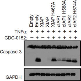
iBMDM XIAP-knockout cells reconstituted with the indicated IAP protein are treated with TNF (10 ng/mL) alone or in combination with GDC-0152 (200 nM) as indicated for 8 hours, and cellular lysates are assayed by Western blot for caspase-3 cleavage.
-
-
Biomater Adv
Optimized lipopolymers with curcumin to enhance AZD5582 and GDC0152 activity and downregulate inhibitors of apoptosis proteins in glioblastoma multiforme. [Abstract]2023 Nov:154:213639. PMID: 37793310 -
Biochem Pharmacol
Structural and molecular characterization of a small-molecule TNF-α-TNFR1 inhibitor modulating cell death signaling. [Abstract]2026 Apr:246:117727. PMID: 41571205 -
Sci Rep
Kaempferol mitigates Endoplasmic Reticulum Stress Induced Cell Death by targeting caspase 3/7. [Abstract]2018 Feb 1;8(1):2189. PMID: 29391535 -
J Biol Chem
Nucleotide-binding oligomerization domain (NOD) signaling defects and cell death susceptibility cannot be uncoupled in X-linked inhibitor of apoptosis (XIAP)-driven inflammatory disease. [Abstract]2017 Jun 9;292(23):9666-9679. PMID: 28404814
GDC-0152 purchased from MedChemExpress. Usage Cited in: J Biol Chem. 2017 Jun 9;292(23):9666-9679. [Abstract]
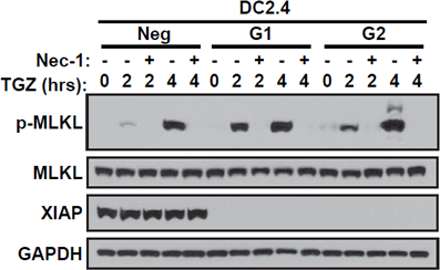
Western blot of lysates from DC2.4 Neg, XIAP-G1, and XIAP-G2 cell lines stimulated with TNF (10 ng/uL), GDC-0152 (2 μM), and zVAD (20 μM, TGZ) for 0, 2, or 4 hours in the presence (+) or absence (-) of nec-1 (20 μM). Representative blots from three independent replicates.
-
ACS Chem Biol
Enthalpy-Based Screening of Focused Combinatorial Libraries for the Identification of Potent and Selective Ligands. [Abstract]2017 Dec 15;12(12):2981-2989. PMID: 29094589 -
bioRxiv
2024 Nov 28:2024.11.25.625306. PMID: 39651304 -


Solvent & Solubility
Ethanol : 50 mg/mL (100.27 mM; Need ultrasonic)
DMSO : 50 mg/mL (100.27 mM; Need ultrasonic; Hygroscopic DMSO has a significant impact on the solubility of product, please use newly opened DMSO)
Please refer to the solubility information to select the appropriate solvent. Once prepared, please aliquot and store the solution to prevent product inactivation from repeated freeze-thaw cycles.
Storage method and period of stock solution: -80°C, 2 years; -20°C, 1 year. When stored at -80°C, please use it within 2 years. When stored at -20°C, please use it within 1 year.
Concentration (start) × Volume (start) = Concentration (final) × Volume (final)
Select the appropriate dissolution method based on your experimental animal and administration route.
- For the following dissolution methods, please ensure to first prepare a clear stock solution using an In Vitro approach and then sequentially add co-solvents:
- To ensure reliable experimental results, the clarified stock solution can be appropriately stored based on storage conditions. As for the working solution for In Vivo experiments, it is recommended to prepare freshly and use it on the same day.
- The percentages shown for the solvents indicate their volumetric ratio in the final prepared solution. If precipitation or phase separation occurs during preparation, heat and/or sonication can be used to aid dissolution.
Add each solvent one by one: 10% DMSO 40% PEG300 5% Tween-80 45% Saline
Solubility: ≥ 2.5 mg/mL (5.01 mM); Clear solution
This protocol yields a clear solution of ≥ 2.5 mg/mL (saturation unknown).
Taking 1 mL working solution as an example, add 100 μL DMSO stock solution (25.0 mg/mL) to 400 μL PEG300, and mix evenly; then add 50 μL Tween-80 and mix evenly; then add 450 μL Saline to adjust the volume to 1 mL.
Preparation of Saline: Dissolve 0.9 g sodium chloride in ddH₂O and dilute to 100 mL to obtain a clear Saline solution.
Add each solvent one by one: 10% DMSO 90% (20% SBE-β-CD in Saline)
Solubility: ≥ 2.5 mg/mL (5.01 mM); Clear solution
This protocol yields a clear solution of ≥ 2.5 mg/mL (saturation unknown).
Taking 1 mL working solution as an example, add 100 μL DMSO stock solution (25.0 mg/mL) to 900 μL 20% SBE-β-CD in Saline, and mix evenly.
Preparation of 20% SBE-β-CD in Saline (4°C, storage for one week): 2 g SBE-β-CD powder is dissolved in 10 mL Saline, completely dissolve until clear.
Please enter the basic information of animal experiments:
-
-
-
-
Recommended: Prepare an additional quantity of animals to account for potential losses during experiments.
Please enter your animal formula composition:
-
%DMSO +
Recommended: Keep the proportion of DMSO in working solution below 2% if your animal is weak.
-
%+
-
+%Tween-80 + +
-
%Saline +
The co-solvents required include: DMSO, . All of co-solvents are available by MedChemExpress (MCE). , Tween 80. All of co-solvents are available by MedChemExpress (MCE).
Working solution concentration: 0.22 mg/mL
Method for preparing stock solution: mg drug dissolved in μL DMSO. Stock solution concentration: mg/mL.
1. Take μL DMSO stock solution;
2. Add μL .
μL , mix evenly;
3. Then add μL Tween 80, mix evenly;
4. Then add μL
Please ensure that the stock solution in the first step is dissolved to a clear state, and add co-solvents in sequence. You can use ultrasonic heating (ultrasonic cleaner, recommended frequency 20-40 kHz), vortexing, etc. to assist dissolution.

Protocol
Inhibition constants (Ki) for the antagonists are determined by addition of the IAP protein constructs to wells containing serial dilutions of the antagonists or the peptide AVPW, and the Hid-FAM probe or AVP-diPhe-FAM probe, as appropriate, in the polarization buffer. Samples are read after a 30-minute incubation. Fluorescence polarization values are plotted as a function of the antagonist concentration, and the IC50 values are obtained by fitting the data to a 4-parameter equation using software. Ki values for the antagonists are determined from the IC50 valued.
MedChemExpress (MCE) has not independently confirmed the accuracy of these methods. They are for reference only.
Detached cells are washed with phosphate-buffered saline (PBS) and are resuspended in assay media (MDA-MB-231 cells: RPMI1640 supplemented with 10% fetal bovine serum and 2 mM L-glutamine [GlutaMAX-1]) or culture media (HMECs: MEBM® with MEGM SingleQuots®). Cells are placed in tissue culture-treated, white-wall or black-wall, clear-bottom, 96-well plates at 1×10⁴ cells/well in a volume of 50 μL. The plates are incubated at 37°C and 5% CO2 overnight, the media is removed, and GDC-0152 or it's enantiomer are added in assay media. Cells cultured in white-wall, clear-bottom plates are incubated at 37°C and 5% CO2 for 3 days before cell viability is measured using the CellTiter-Glo® luminescent cell viability assay kit.
MedChemExpress (MCE) has not independently confirmed the accuracy of these methods. They are for reference only.
Cells are resuspended in PBS and the cell suspension is mixed 1:1 with Matrigel. The cells (1.5×10⁷) are then implanted subcutaneously into the right flank of 130 female nude mice aged 6-8 weeks. Tumor volumes are calculated. Ten mice with the appropriate mean tumor volume are assigned randomLy to each of six groups. The mean tumor volume±the standard error of the mean (SEM) for all six groups is 168±3 mm3 at the initiation of treatment (Day 0). Mice are dosed 1 or vehicle (PBS) by oral gavage with a dose volume of 4.0 mL/kg. The mice are observed on each day of the study, and tumor volumes and body weights are measured twice each week. Percent tumor growth inhibition is calculated using the formula %TGI=100× (1- Tumor Volumedose/Tumor Volumevehicle).
MedChemExpress (MCE) has not independently confirmed the accuracy of these methods. They are for reference only.


Purity & Documentation
-
Data Sheet (283 KB)
-
SDS (393 KB)
- English - EN (393 KB)
- Français - FR (393 KB)
- Deutsch - DE (393 KB)
- Norwegian - NO (393 KB)
- Español - ES (393 KB)
- Swedish - SV (393 KB)
- Italian - IT (393 KB)
- Korean - KR (393 KB)
- Portuguese - PT (393 KB)
-
Handling Instructions (2659 KB)
References


Complete Stock Solution Preparation Table
Please refer to the solubility information to select the appropriate solvent. Once prepared, please aliquot and store the solution to prevent product inactivation from repeated freeze-thaw cycles.
Storage method and period of stock solution: -80°C, 2 years; -20°C, 1 year. When stored at -80°C, please use it within 2 years. When stored at -20°C, please use it within 1 year.
| Optional Solvent | Concentration Solvent Mass | 1 mg | 5 mg | 10 mg | 25 mg |
|---|---|---|---|---|---|
| Ethanol / DMSO | 1 mM | 2.0055 mL | 10.0273 mL | 20.0545 mL | 50.1364 mL |
| 5 mM | 0.4011 mL | 2.0055 mL | 4.0109 mL | 10.0273 mL | |
| 10 mM | 0.2005 mL | 1.0027 mL | 2.0055 mL | 5.0136 mL | |
| 15 mM | 0.1337 mL | 0.6685 mL | 1.3370 mL | 3.3424 mL | |
| 20 mM | 0.1003 mL | 0.5014 mL | 1.0027 mL | 2.5068 mL | |
| 25 mM | 0.0802 mL | 0.4011 mL | 0.8022 mL | 2.0055 mL | |
| 30 mM | 0.0668 mL | 0.3342 mL | 0.6685 mL | 1.6712 mL | |
| 40 mM | 0.0501 mL | 0.2507 mL | 0.5014 mL | 1.2534 mL | |
| 50 mM | 0.0401 mL | 0.2005 mL | 0.4011 mL | 1.0027 mL | |
| 60 mM | 0.0334 mL | 0.1671 mL | 0.3342 mL | 0.8356 mL | |
| 80 mM | 0.0251 mL | 0.1253 mL | 0.2507 mL | 0.6267 mL | |
| 100 mM | 0.0201 mL | 0.1003 mL | 0.2005 mL | 0.5014 mL |


 Powered by Bioz