



Hydroxysafflor yellow A
Based on 15 publication(s) in Google Scholar
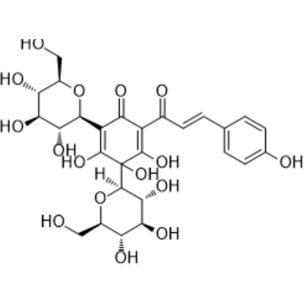
Hydroxysafflor yellow A (Safflomin A) is a natural product of flavonoids isolated from safflower. Hydroxysafflor yellow A can inhibit cell proliferation and promote apoptosis through the autophagy pathway. Hydroxysafflor yellow A has anti-inflammatory, antioxidant and antitumor effects. Hydroxysafflor yellow A can be used in the study of cardiovascular disease.
For research use only. We do not sell to patients.
- Purity : 99.46%
- CAS No.: 78281-02-4
- Formula: C27H32O16
- Molecular Weight:612.53
-
Storage:
4°C, protect from light
* In solvent : -80°C, 6 months; -20°C, 1 month (protect from light)

To place orders, for customer services and technical support, please contact: MedChemExpress USA
Tel: 609-228-6898 E-mail: [email protected] [email protected]


-
 Biological Activity
-
Chemical Information
-
Solvent & Solubility
- Protocols
- Purity & Documentation
- References
-
 Help & FAQs


-
HY-N0060
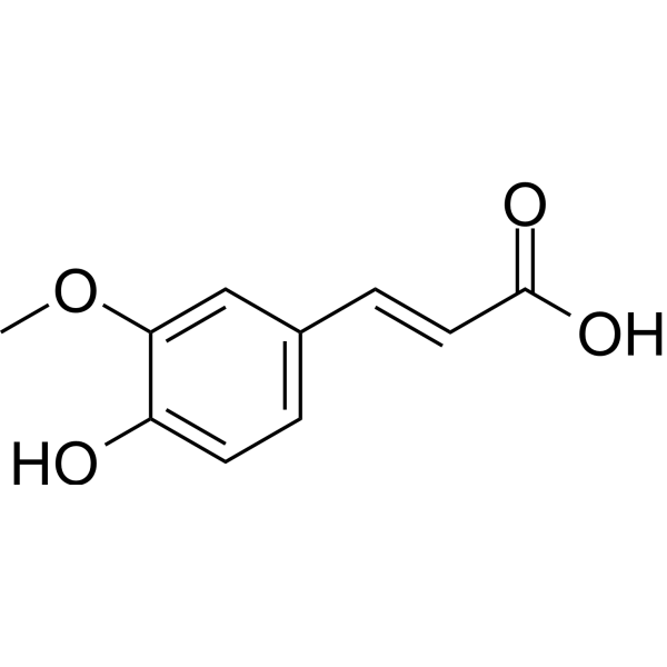
Ferulic acidFGFR1 Inhibitor -
HY-N0293
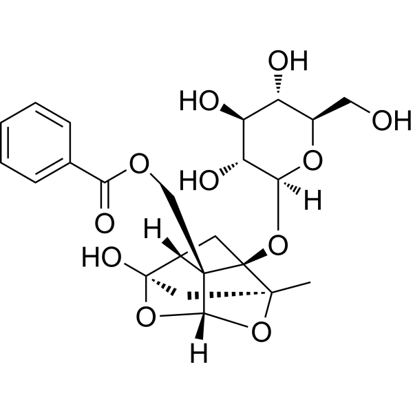
PaeoniflorinHSP Activator -
HY-14590
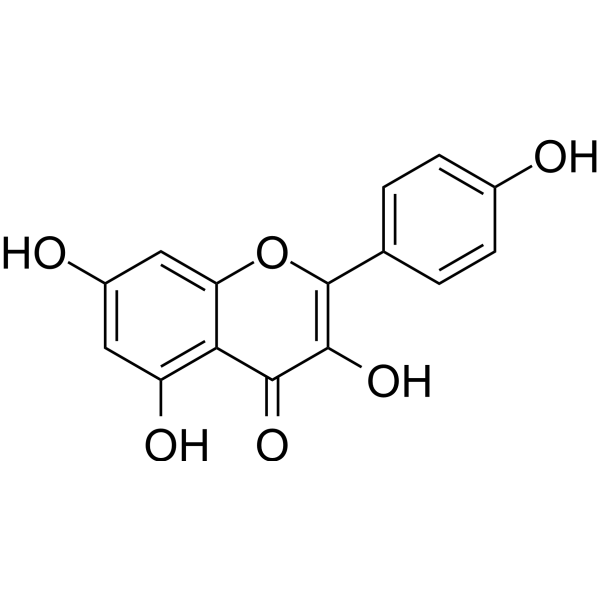
KaempferolEstrogen Receptor α Inhibitor -
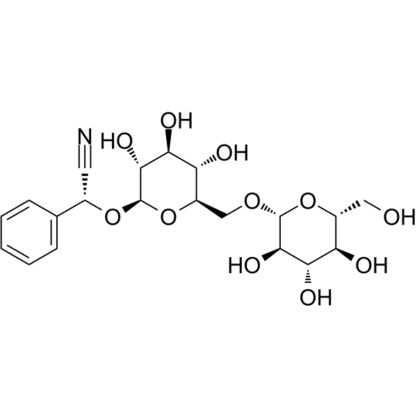
HY-N0190
AmygdalinGlucoside -
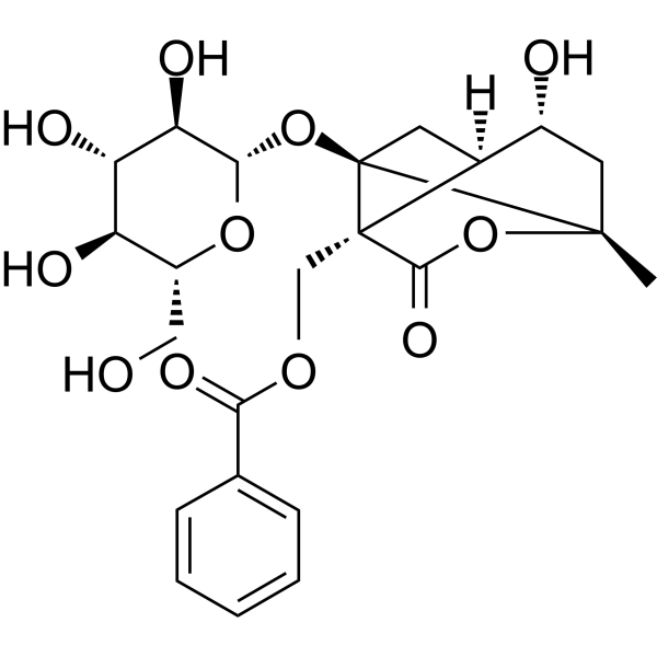
HY-N0037
AlbiflorinNeuroprotective Agent -
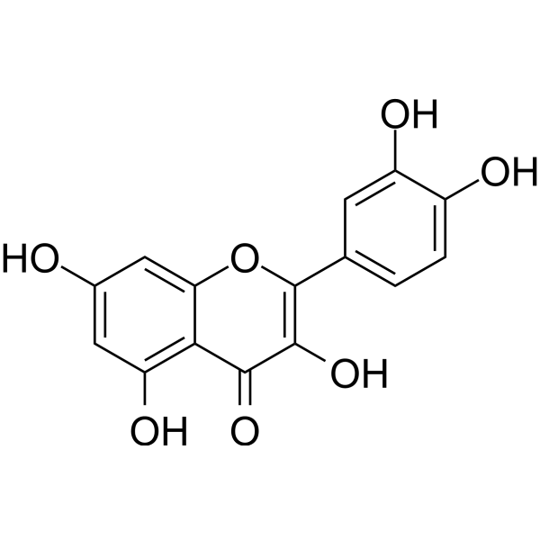
HY-18085
QuercetinFlavonoid Antioxidant -
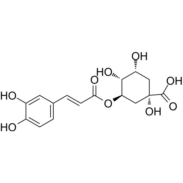
HY-N0055
Chlorogenic acidAntioxidant -
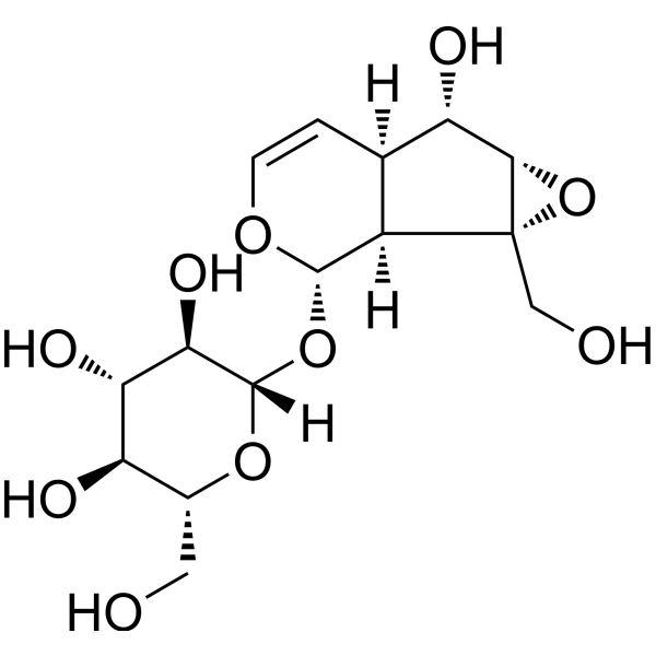
HY-N0820
CatalpolHBV Inhibitor -
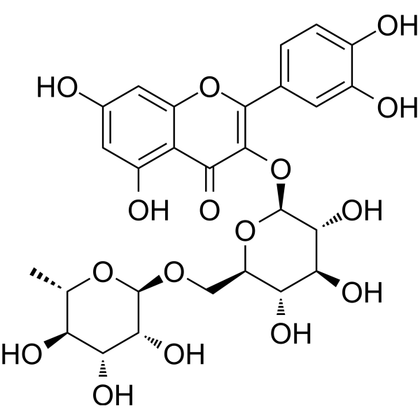
HY-N0148
RutinCBR1 Inhibitor -
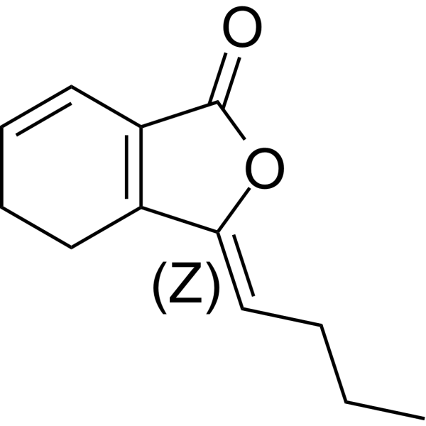
HY-N0401A
(Z)-LigustilideBacterial Inhibitor -
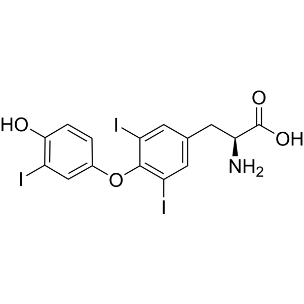
HY-A0070A
LiothyronineTIGIT/PVR Blocker -
HY-N0264
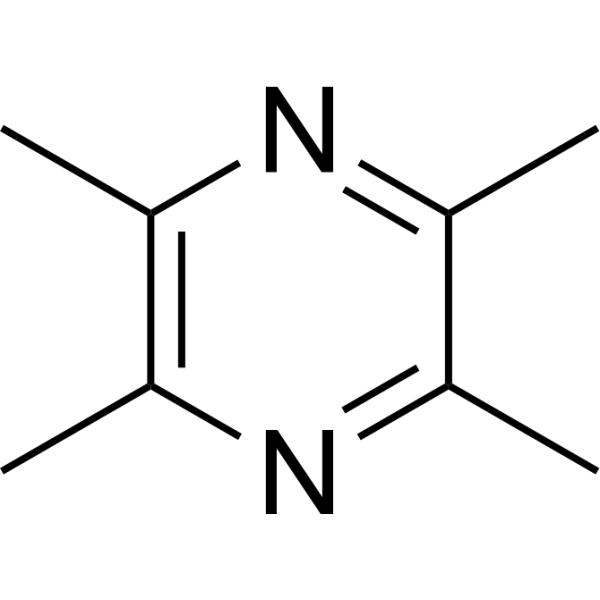
LigustrazineNeuroprotective Agent/Lipid Lowering Agent
Publications Citing Use of MedChemExpress (MCE) Hydroxysafflor yellow A
More- Acta Pharm Sin B. 2021 Jan;11(1):143-155. [Abstract]
- Pharmacol Res. 2020 May:155:104751. [Abstract]
- Food Chem. 2025 Dec 30:497:146992. [Abstract]
- Food Chem. 2025 Oct 15:489:144992. [Abstract]
- CNS Neurosci Ther. 2023 Jun;29 Suppl 1(Suppl 1):161-184. [Abstract]
- Drug Des Devel Ther. 2023 Jun 17:17:1819-1829. [Abstract]
- Pharmaceuticals (Basel). 2022 Jan 31;15(2):179. [Abstract]
- Int Immunopharmacol. 2026 Jun 8:185:116972. [Abstract]
- Int Immunopharmacol. 2026 May 14:182:116841. [Abstract]
- Int Immunopharmacol. 2024 Nov 5;143(Pt 3):113534. [Abstract]
- Cell Biochem Biophys. 2020 Dec;78(4):511-520. [Abstract]
- Exp Ther Med. 2022 Nov 3;24(6):741. [Abstract]
- Am J Transl Res. 2020 Aug 15;12(8):4781-4794. [Abstract]
- Acta Cir Bras. 2022 Jun 15;37(3):e370305. [Abstract]
- Evid Based Complement Alternat Med. 2022 Jul 4:2022:6326677. [Abstract]
 Customer Validation & Images

-
Cell Proliferation/Viability Assay
-
Histological Imaging/Staining
-
Cell Migration/Invasion Assay
-
Apoptosis Analysis
-
WB


Biological Activity
Description
In Vitro
Hydroxysafflor yellow A (10 μM, 24 h) Inhibition of LPS-induced proliferation and migration of vascular smooth muscle cells by toll-like receptor-4 pathway[1].
Hydroxysafflor yellow A (0.01-10 mg/L, 4-96 h) can promote the expression of fat-soluble specific enzyme HSL by increasing the activity of HSL promoter and inhibit the proliferation and adipogenesis of 3T3-L1 preadipocytes[2].
Hydroxysafflor yellow A (1-100 μM), 12-24 h) decreases LPS-induced EC inflammatory damage by inhibiting p38 MAPK phosphorylation, NF-κB activation, high expression of inflammatory factors and leukocyte adhesion to EC[3].
Hydroxysafflor yellow A (20-160 μM, 24-48 h) inhibits cell proliferation and promotes apoptosis by blocking the autophagy flux in liver cancer cells[6].
MedChemExpress (MCE) has not independently confirmed the accuracy of these methods. They are for reference only.
-
Cell Line:VSMCs
-
Concentration:0.1, 1, 10,100 μM
-
Incubation Time:24 h
-
Result:Inhibited LPS-induced VSMCs proliferation and migration with the concentration of 10 μM.
-
Cell Line:VSMCs
-
Concentration:10 μM
-
Incubation Time:24 h
-
Result:Inhibited LPS-induced upregulation of pro-inflammatory factors (TNF-α, IL-6, and IL-8). Inhibited LPS-induced upregulation of TLR-4 and activation of Rac1/Akt pathway.
-
Cell Line:3T3-L1
-
Concentration:0.01, 0.1, 1, 10 mg/L
-
Incubation Time:4, 8, 24, 48, 72, 96 h
-
Result:Inhibit the cell viability in a dose and time-dependent manner.
-
Cell Line:HepG2, Huh7
-
Concentration:20, 40, 80, 160 μM
-
Incubation Time:24 h
-
Result:Suppressed the growth and induced apoptotic cell death in both concentration and time-dependent manners. Augmented the induction of autophagosomes.
In Vivo
Hydroxysafflor yellow A (30, 48, 76.8 mg/kg, intraperitoneal injection) has a protective effect on inflammatory damage in rats with chronic obstructive pulmonary disease by reducing NF-κB and p38MAPK signal transduction[5].
Hydroxysafflor yellow A (1.135, 2.25 mg/kg, intraperitoneal injection) can inhibit tumor growth and block autophagy of liver cancer cells in tumor models of nude mice[6].
MedChemExpress (MCE) has not independently confirmed the accuracy of these methods. They are for reference only.
-
Animal Model:Liver injury model induced by alcohol[4]
-
Dosage:2.5 or 10 mg/kg
-
Administration:i.p. daily for 6 weeks
-
Result:Decreased the level of ALT and AST. Decreased the levels of HA, LN, and III-C. Increased the activities of SOD and GPx and decreased the level of TGF-β1 expression.
-
Animal Model:Chronic obstructive pulmonary disease (COPD)[5]
-
Dosage:30, 48, 76.8 mg/kg
-
Administration:i.p.
-
Result:Inhibited the elevated IL-6, IL-1β, TNF-α, ICAM-1 and VCAM-1 mRNA Levels. Inhibited the increased phosphorylation and NF-κB p65 levels.
- Annexin V plus membrane-impermeant dye apoptosis staining
- BrdU Incorporation Assay
- Somatic Cell Culture
- CFSE Dye Dilution Proliferation Assay
- Dye-dilution cell tracking and proliferation staining
- CCK-8/WST-8 Cell Proliferation Assay
- Protocol for Cell Cycle
- Research Protocol for Cardiovascular Diseases
- Cell Counting-Based Growth Curve Assay
- Research Protocol for Inflammation-related Diseases
- Colony Formation (Clonogenic) Assay
- Cell Viability Determination by MTT Colorimetric Assay
- EdU Incorporation Assay (Click Chemistry-Based DNA Synthesis Measurement)
- Ki-67 Immunostaining Proliferation Assay
- Detection of Apoptosis
- TUNEL staining for apoptotic DNA fragmentation
- PCNA Immunodetection Proliferation Assay
- Protocol for Cell Counting and Cell Density Analysis
- Autophagy
- Apoptosis Solutions
- Lysosome and acidic-vesicle live-cell staining
- MTT Cell Proliferation Assay
- Macroautophagy Solutions


Chemical Information
-
CAS No. 78281-02-4
-
Appearance Solid
-
Molecular Weight 612.53
-
Formula C27H32O16
-
Color Yellow to orange
-
SMILES
OC(C(O)=C1C(/C=C/C2=CC=C(O)C=C2)=O)([C@]([C@@H]([C@@H](O)[C@@H]3O)O)([H])O[C@@H]3CO)C(O)=C([C@@H]([C@@H]([C@@H](O)[C@@H]4O)O)O[C@@H]4CO)C1=O
-
Synonyms
Safflomin A; HSYA
-
Structure Classification
-
Shipping
Room temperature in continental US; may vary elsewhere.
-
Storage
4°C, protect from light
* In solvent : -80°C, 6 months; -20°C, 1 month (protect from light)


Publications (15)
-
Journal Impact Factor
-
Most Recent
-
Acta Pharm Sin B
Chrysin serves as a novel inhibitor of DGK α/FAK interaction to suppress the malignancy of esophageal squamous cell carcinoma (ESCC). [Abstract]2021 Jan;11(1):143-155. PMID: 33532186 -
Pharmacol Res
Cardamonin retards progression of autosomal dominant polycystic kidney disease via inhibiting renal cyst growth and interstitial fibrosis. [Abstract]2020 May:155:104751. PMID: 32151678 -
Food Chem
Effects of sun drying combined with baking processes on the flavor quality of Chongqing Tuocha raw tea. [Abstract]2025 Dec 30:497:146992. PMID: 41285060 -
Food Chem
Flavonoid-mediated metabolic underpinning quality variation in red bud-sport pear mutants. [Abstract]2025 Oct 15:489:144992. PMID: 40466530 -
CNS Neurosci Ther
Tongqiao Huoxue Decoction ameliorates traumatic brain injury-induced gastrointestinal dysfunction by regulating CD36/15-LO/NR4A1 signaling, which fails when CD36 and CX3CR1 are deficient. [Abstract]2023 Jun;29 Suppl 1(Suppl 1):161-184. PMID: 37157929 -
Drug Des Devel Ther
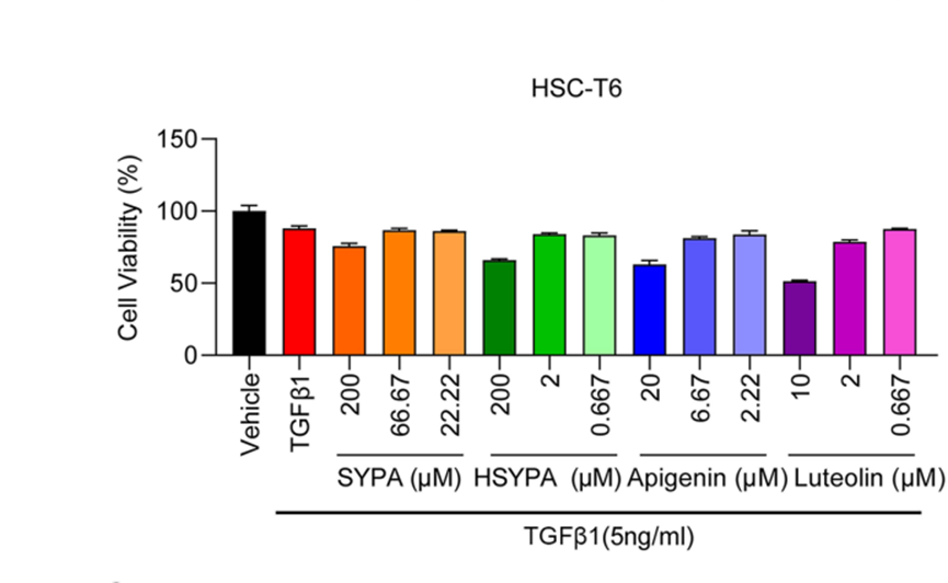
Luteolin Alleviates Liver Fibrosis in Rat Hepatic Stellate Cell HSC-T6: A Proteomic Analysis. [Abstract]2023 Jun 17:17:1819-1829. PMID: 37360572
Hydroxysafflor yellow A purchased from MedChemExpress. Usage Cited in: Drug Des Devel Ther. 2023 Jun 17:17:1819-1829. [Abstract]
Cell viabilities in the Hydroxysafflor yellow A (HSYPA) (200 µM), Apigenin (20 µM), and Luteolin (10 µM) groups were all significantly lower in the absence of TGFβ1, while no differences were observed in the SYPA group.
-
Pharmaceuticals (Basel)
Bruceine D Identified as a Drug Candidate against Breast Cancer by a Novel Drug Selection Pipeline and Cell Viability Assay. [Abstract]2022 Jan 31;15(2):179. PMID: 35215292 -
Int Immunopharmacol
Hydroxysafflor yellow A mitigates lipopolysaccharide-induced acute lung injury by enhancing pulmonary microvascular endothelial barrier function via Calpain-1/HIF-1α inhibition. [Abstract]2026 Jun 8:185:116972. PMID: 42259237 -
Int Immunopharmacol
Albiflorin contributes to Xuebijing-mediated protection against sepsis-associated acute kidney injury by modulating the succinate-PFKFB3 immunometabolic axis. [Abstract]2026 May 14:182:116841. PMID: 42134292 -
Int Immunopharmacol
HSYA ameliorates venous thromboembolism by depleting the formation of TLR4/NF-κB pathway-dependent neutrophil extracellular traps. [Abstract]2024 Nov 5;143(Pt 3):113534. PMID: 39504860 -
Cell Biochem Biophys
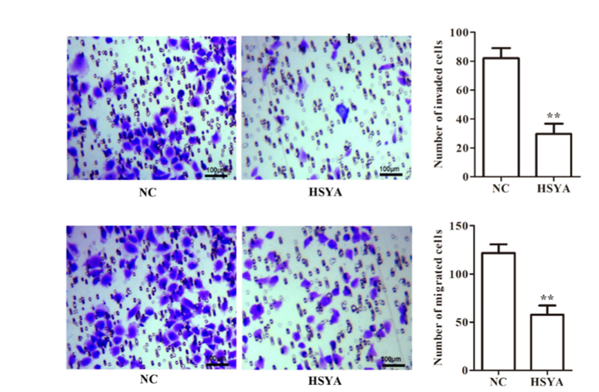
Hydroxysafflor Yellow A of Carthamus Tinctorius L., Represses the Malignant Development of Esophageal Cancer Cells via Regulating NF-κB Signaling Pathway. [Abstract]2020 Dec;78(4):511-520. PMID: 32705535
Hydroxysafflor yellow A purchased from MedChemExpress. Usage Cited in: Cell Biochem Biophys. 2020 Dec;78(4):511-520. [Abstract]
The invasive and migratory ability of KYSE-30 cells after being treated with Hydroxysafflor yellow A (HSYA) (20 μM, 24 h).
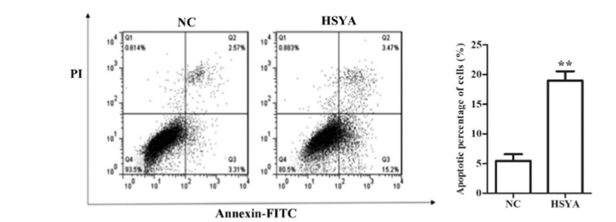
Hydroxysafflor yellow A purchased from MedChemExpress. Usage Cited in: Cell Biochem Biophys. 2020 Dec;78(4):511-520. [Abstract]
Induction of apoptosis in KYSE-30 cells after Hydroxysafflor yellow A (HSYA) (20 μM, 24 h) treatment.
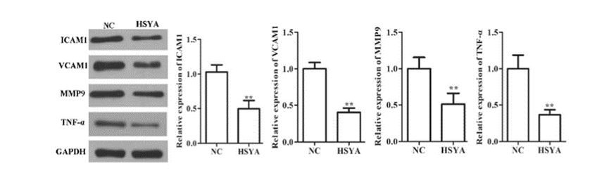
Hydroxysafflor yellow A purchased from MedChemExpress. Usage Cited in: Cell Biochem Biophys. 2020 Dec;78(4):511-520. [Abstract]
After KYSE-30 cells were treated with 20 µM Hydroxysafflor yellow A (HSYA) for 24 h, the protein expression levels of ICAM1, MMP9, TNF-α, and VCAM1 were all upregulated when compared with that in NC group via western blot analysis.
-
Exp Ther Med
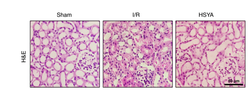
Protective effect of hydroxysafflor yellow A on renal ischemia‑-reperfusion injury by targeting the Akt‑Nrf2 axis in mice. [Abstract]2022 Nov 3;24(6):741. PMID: 36478883
Hydroxysafflor yellow A purchased from MedChemExpress. Usage Cited in: Exp Ther Med. 2022 Nov 3;24(6):741. [Abstract]
Representative photomicrographs of H&E stained kidney sections, pretreatment with Hydroxysafflor yellow A (HSYA) (100 mg/kg, i.p.) reduced the severity of the tubular injury.
-
Am J Transl Res
Hydroxysafflor yellow A promotes multiterritory perforating flap survival: an experimental study. [Abstract]2020 Aug 15;12(8):4781-4794. PMID: 32913550 -
Acta Cir Bras
Protective effect of hydroxysafflor yellow A on cyclosporin A-induced renal oxidative stress in vitro and in vivo. [Abstract]2022 Jun 15;37(3):e370305. PMID: 35730865 -
Evid Based Complement Alternat Med
Hydroxysafflor Yellow A (HSYA) Protects Endplate Chondrocytes Against IL-1 β-Induced Injury Through Promoting Autophagy. [Abstract]2022 Jul 4:2022:6326677. PMID: 35832517


Solvent & Solubility
In Vitro:
H2O : ≥ 100 mg/mL (163.26 mM)
DMSO : 87.5 mg/mL (142.85 mM; ultrasonic and warming and heat to 60°C; Hygroscopic DMSO has a significant impact on the solubility of product, please use newly opened DMSO)
* "≥" means soluble, but saturation unknown.
Please refer to the solubility information to select the appropriate solvent. Once prepared, please aliquot and store the solution to prevent product inactivation from repeated freeze-thaw cycles.
Storage method and period of stock solution: -80°C, 6 months; -20°C, 1 month (protect from light). When stored at -80°C, please use it within 6 months. When stored at -20°C, please use it within 1 month.
* Note: If you choose water as the stock solution, please dilute it to the working solution, then filter and sterilize it with a 0.22 μm filter before use.
Please refer to the solubility information to select the appropriate solvent. Once prepared, please aliquot and store the solution to prevent product inactivation from repeated freeze-thaw cycles.
Storage method and period of stock solution: -80°C, 6 months; -20°C, 1 month (protect from light). When stored at -80°C, please use it within 6 months. When stored at -20°C, please use it within 1 month.
* Note: If you choose water as the stock solution, please dilute it to the working solution, then filter and sterilize it with a 0.22 μm filter before use.
Concentration (start) × Volume (start) = Concentration (final) × Volume (final)
In Vivo:
Select the appropriate dissolution method based on your experimental animal and administration route.
- For the following dissolution methods, please ensure to first prepare a clear stock solution using an In Vitro approach and then sequentially add co-solvents:
- To ensure reliable experimental results, the clarified stock solution can be appropriately stored based on storage conditions. As for the working solution for In Vivo experiments, it is recommended to prepare freshly and use it on the same day.
- The percentages shown for the solvents indicate their volumetric ratio in the final prepared solution. If precipitation or phase separation occurs during preparation, heat and/or sonication can be used to aid dissolution.
Add each solvent one by one: 10% DMSO 40% PEG300 5% Tween-80 45% Saline
Solubility: ≥ 2.08 mg/mL (3.40 mM); Clear solution
This protocol yields a clear solution of ≥ 2.08 mg/mL (saturation unknown).
Taking 1 mL working solution as an example, add 100 μL DMSO stock solution (20.8 mg/mL) to 400 μL PEG300, and mix evenly; then add 50 μL Tween-80 and mix evenly; then add 450 μL Saline to adjust the volume to 1 mL.
Preparation of Saline: Dissolve 0.9 g sodium chloride in ddH₂O and dilute to 100 mL to obtain a clear Saline solution.
Add each solvent one by one: 10% DMSO 90% (20% SBE-β-CD in Saline)
Solubility: ≥ 2.08 mg/mL (3.40 mM); Clear solution
This protocol yields a clear solution of ≥ 2.08 mg/mL (saturation unknown).
Taking 1 mL working solution as an example, add 100 μL DMSO stock solution (20.8 mg/mL) to 900 μL 20% SBE-β-CD in Saline, and mix evenly.
Preparation of 20% SBE-β-CD in Saline (4°C, storage for one week): 2 g SBE-β-CD powder is dissolved in 10 mL Saline, completely dissolve until clear.
In Vivo Dissolution Calculator
Please enter the basic information of animal experiments:
-
-
-
-
Recommended: Prepare an additional quantity of animals to account for potential losses during experiments.
Working solution concentration: 0.22 mg/mL
This product has good water solubility, please refer to the measured solubility data in water/PBS/Saline for details.


Purity & Documentation
-
Data Sheet (286 KB)
-
SDS (393 KB)
- English - EN (393 KB)
- Français - FR (393 KB)
- Deutsch - DE (393 KB)
- Norwegian - NO (393 KB)
- Español - ES (393 KB)
- Swedish - SV (393 KB)
- Italian - IT (393 KB)
- Korean - KR (393 KB)
- Portuguese - PT (393 KB)
-
Handling Instructions (2659 KB)
References
[1]. Yang G, et al. Hydroxysafflor yellow A inhibits lipopolysaccharide-induced proliferation and migration of vascular smooth muscle cells via Toll-like receptor-4 pathway. Int J Clin Exp Med. 2015 Apr 15;8(4):5295-302. [Content Brief]
[2]. Zhu HJ, et al. Hydroxysafflor yellow A (HYSA) inhibited the proliferation and differentiation of 3T3-L1 preadipocytes. Cytotechnology. 2015 Oct;67(5):885-92. [Content Brief]
[3]. Jin M, et al. Hydroxysafflor yellow A attenuate lipopolysaccharide-induced endothelium inflammatory injury. Chin J Integr Med. 2016 Jan;22(1):36-41. [Content Brief]
[4]. He Y, et al. Protective effects of hydroxysafflor yellow A (HSYA) on alcohol-induced liver injury in rats. J Physiol Biochem. 2015 Mar;71(1):69-78. [Content Brief]
[5]. Jin M, Xue et al. Protective Effect of Hydroxysafflor Yellow A on Inflammatory Injury in Chronic Obstructive Pulmonary Disease Rats. Chin J Integr Med. 2019 Oct;25(10):750-756. [Content Brief]
[6]. Wu N, et al. Hydroxysafflor yellow A promotes apoptosis via blocking autophagic flux in liver cancer. Biomed Pharmacother. 2021 Apr;136:111227. [Content Brief]


Complete Stock Solution Preparation Table
Please refer to the solubility information to select the appropriate solvent. Once prepared, please aliquot and store the solution to prevent product inactivation from repeated freeze-thaw cycles.
Storage method and period of stock solution: -80°C, 6 months; -20°C, 1 month (protect from light). When stored at -80°C, please use it within 6 months. When stored at -20°C, please use it within 1 month.
| Optional Solvent | Concentration Solvent Mass | 1 mg | 5 mg | 10 mg | 25 mg |
|---|---|---|---|---|---|
| DMSO / H2O | 1 mM | 1.6326 mL | 8.1629 mL | 16.3257 mL | 40.8143 mL |
| 5 mM | 0.3265 mL | 1.6326 mL | 3.2651 mL | 8.1629 mL | |
| 10 mM | 0.1633 mL | 0.8163 mL | 1.6326 mL | 4.0814 mL | |
| 15 mM | 0.1088 mL | 0.5442 mL | 1.0884 mL | 2.7210 mL | |
| 20 mM | 0.0816 mL | 0.4081 mL | 0.8163 mL | 2.0407 mL | |
| 25 mM | 0.0653 mL | 0.3265 mL | 0.6530 mL | 1.6326 mL | |
| 30 mM | 0.0544 mL | 0.2721 mL | 0.5442 mL | 1.3605 mL | |
| 40 mM | 0.0408 mL | 0.2041 mL | 0.4081 mL | 1.0204 mL | |
| 50 mM | 0.0327 mL | 0.1633 mL | 0.3265 mL | 0.8163 mL | |
| 60 mM | 0.0272 mL | 0.1360 mL | 0.2721 mL | 0.6802 mL | |
| 80 mM | 0.0204 mL | 0.1020 mL | 0.2041 mL | 0.5102 mL | |
| 100 mM | 0.0163 mL | 0.0816 mL | 0.1633 mL | 0.4081 mL |
* Note: If you choose water as the stock solution, please dilute it to the working solution, then filter and sterilize it with a 0.22 μm filter before use.


Hydroxysafflor yellow A Related Classifications
HY-N0567 Related Classifications
 Powered by Bioz