



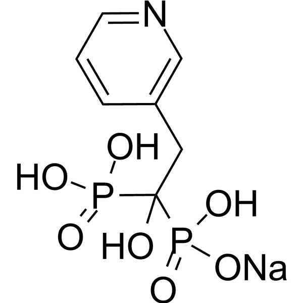
Risedronic acid sodium
Based on 2 publication(s) in Google Scholar
Risedronic acid (Risedronate) sodium, a bisphosphonate, is a potent anti-resorption agent that inhibits osteoclast-mediated bone resorption and changes the bone metabolism. Risedronic acid sodium suppresses osteoblast differentiation and induced caspase- and isoprenoid depletion-dependent apoptosis. Risedronic acid sodium inhibits blood stages of Plasmodium falciparum (IC50 of 20.3 μM). Risedronic acid sodium inhibits the transfer of the farnesyl pyrophosphate group to parasite proteins.
For research use only. We do not sell to patients.
- Purity : 98.0%
- CAS No.: 115436-72-1
- Formula: C7H10NNaO7P2
- Molecular Weight:305.09
-
Storage:
4°C, sealed storage, away from moisture
* In solvent : -80°C, 6 months; -20°C, 1 month (sealed storage, away from moisture)

To place orders, for customer services and technical support, please contact: MedChemExpress USA
Tel: 609-228-6898 E-mail: [email protected] [email protected]


-
 Biological Activity
-
Chemical Information
-
Solvent & Solubility
- Protocols
- Purity & Documentation
- References
-
 Help & FAQs


-
HY-B0515
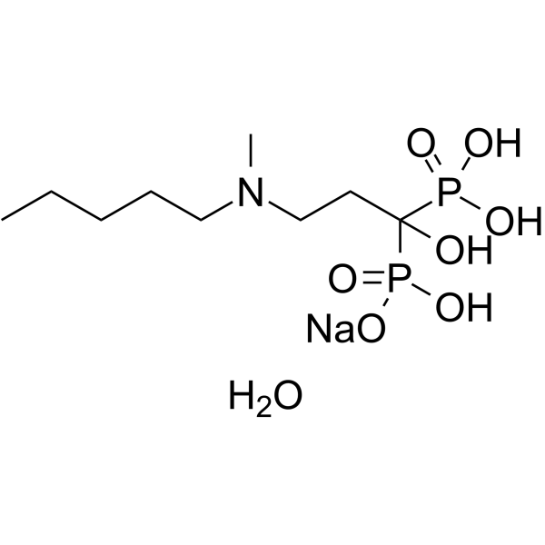
Ibandronate Sodium MonohydrateFPP synthase Inhibitor -
HY-11101
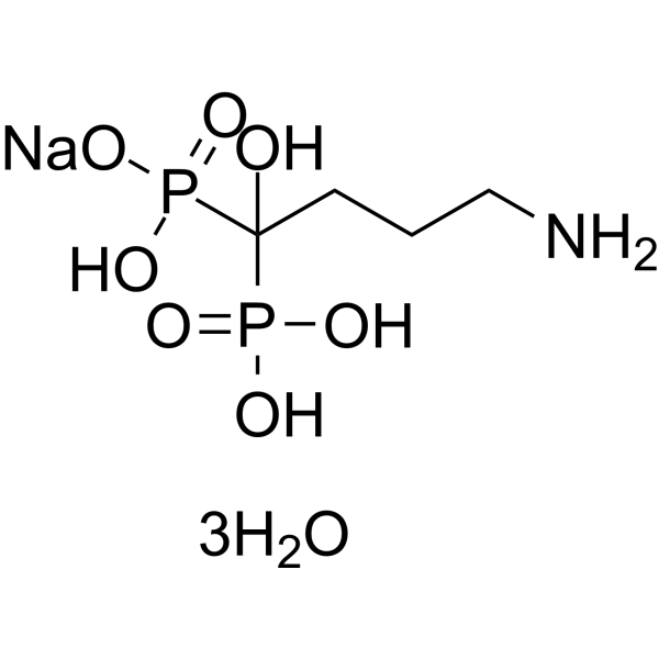
Alendronate sodium hydrateNitrogen-Containing Bisphosphonate -
HY-13777A
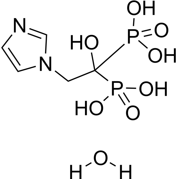
Zoledronic acid monohydrateAnti-resorptive Agant -
HY-B0012A
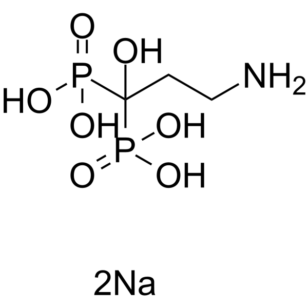
Pamidronate disodiumBone Loss Inhibitors -

HY-B0215
AcetylcysteineMucolytic Agent -
HY-17471A
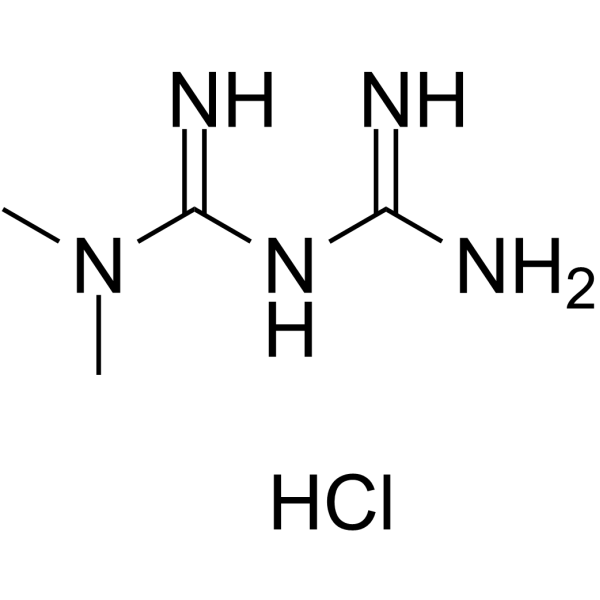
Metformin hydrochlorideAntihyperglycemic Agent -
HY-B0515B
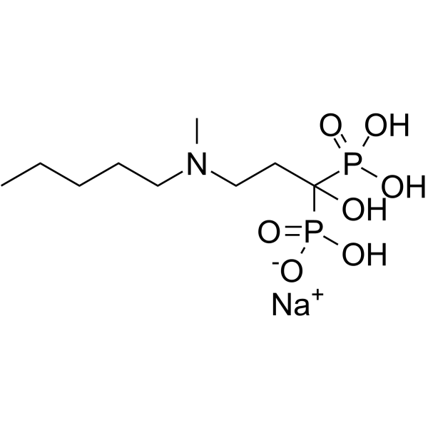
Ibandronate SodiumFPP synthase Inhibitor -
HY-B0657A
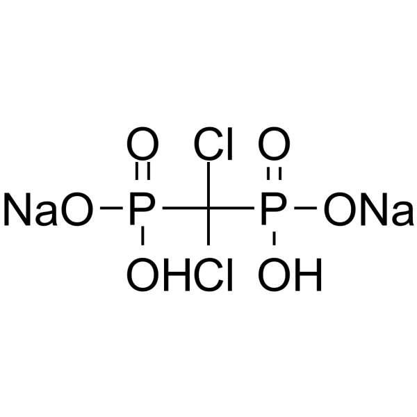
Clodronic acid disodium saltBisphosphonate -
HY-B0302A
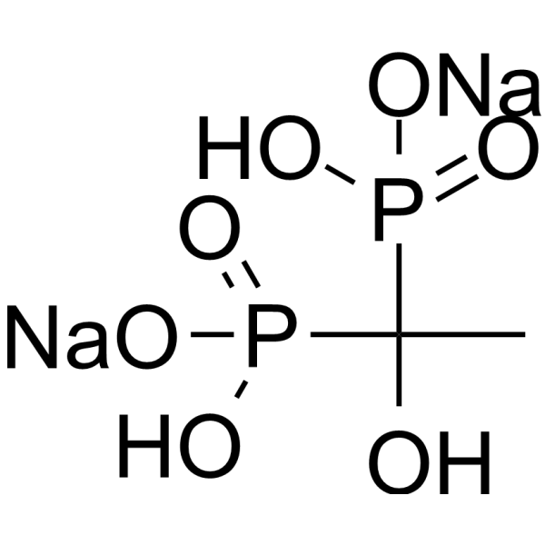
Etidronic acid disodiumApoptosis Inducer
Publications Citing Use of MedChemExpress (MCE) Risedronic acid sodium
MoreAll Parasite Isoforms
More

Biological Activity
Description
In Vitro
Risedronic acid (0-10 μM; 14 days) sodium suppresses the formation of mineralized nodules in human mesenchymal stem cells (hMSC12) and rat thymus-derived mesenchymal stem cells (ST1BIIb)1.
Risedronic acid (0-10 μM; 3 days) sodium suppresses the mRNA expression of osteoblast marker gene osteocalcin (OC) in human mesenchymal stem cells (hMSC12)1.
Risedronic acid (100-1000 μM; 48 h) sodium induces chromatin condensation and activates caspase-3 in human mesenchymal stem cells (hMSC12)1.
Risedronic acid (Risedronate) (15 μM; 36 h) sodium inhibits isoprenoid biosynthesis in Plasmodium falciparum, as shown by the decreased intensities of bands corresponding to farnesol (FOH) and geranylgeraniol (GGOH)3.
Risedronic acid (15 μM; 36 h) sodium interferes with protein isoprenylation in Plasmodium falciparum, reducing the intensities of farnesylated protein bands and increasing those of geranylgeranylated protein bands3.
MedChemExpress (MCE) has not independently confirmed the accuracy of these methods. They are for reference only.
-
Cell Line:Human mesenchymal stem cells (hMSC12)
-
Concentration:100 μM, 300 μM, 500 μM, 1000 μM
-
Incubation Time:48 h
-
Result:Induced chromatin condensation and activated caspase-3.
In Vivo
Risedronic acid (10-25 mg/kg; intraperitoneal injection; once a day; 7 days) sodium inhibits parasitemia in BALB/c mice infected with Plasmodium berghei strain ANKA, but does not prolong the survival time of mice3.
MedChemExpress (MCE) has not independently confirmed the accuracy of these methods. They are for reference only.
-
Animal Model:Female C57BL/6 mice (20-week-old, ~23 g) irradiated with 2 Gy X-rays whole-body2
-
Dosage:30 μg/kg
-
Administration:Subcutaneous injection, once every other day, for 3 weeks
-
Result:Prevented the radiation-induced increase in osteoclast number, surface, and TRAP5b.
-
Animal Model:Male BALB/c mice (3-4 weeks old) infected with Plasmodium berghei strain ANKA by intraperitoneal injection of 1×10⁶ blood-stage parasites3
-
Dosage:10 mg/kg, 15 mg/kg, 20 mg/kg, 25 mg/kg
-
Administration:Intraperitoneal injection, once a day, for 7 days
-
Result:Led to an 88.9% inhibition of the rodent parasite Plasmodium berghei in mice on the seventh day of treatment.


Chemical Information
-
CAS No. 115436-72-1
-
Appearance Solid
-
Molecular Weight 305.09
-
Formula C7H10NNaO7P2
-
Color White to off-white
-
SMILES
OC(P(O)(O[Na])=O)(P(O)(O)=O)CC1=CN=CC=C1
-
Synonyms
Risedronate sodium
-
Shipping
Room temperature in continental US; may vary elsewhere.
-
Storage
4°C, sealed storage, away from moisture
* In solvent : -80°C, 6 months; -20°C, 1 month (sealed storage, away from moisture)


Publications (2)
-
Journal Impact Factor
-
Most Recent
-
Respir Res
Nebulization of risedronate alleviates airway obstruction and inflammation of chronic obstructive pulmonary diseases via suppressing prenylation-dependent RAS/ERK/NF-κB and RhoA/ROCK1/MLCP signaling. [Abstract]2022 Dec 28;23(1):380. PMID: 36575527 -
Int J Parasitol Drugs Drug Resist
Promising efficacy of nitrogen-containing bisphosphonates against the infection of Cryptosporidium spp. [Abstract]2025 Aug 6:29:100607. PMID: 40782657


Solvent & Solubility
In Vitro:
H2O : 8.33 mg/mL (27.30 mM; Need ultrasonic)
DMSO : < 1 mg/mL (insoluble or slightly soluble)
Please refer to the solubility information to select the appropriate solvent. Once prepared, please aliquot and store the solution to prevent product inactivation from repeated freeze-thaw cycles.
Storage method and period of stock solution: -80°C, 6 months; -20°C, 1 month (sealed storage, away from moisture). When stored at -80°C, please use it within 6 months. When stored at -20°C, please use it within 1 month.
* Note: If you choose water as the stock solution, please dilute it to the working solution, then filter and sterilize it with a 0.22 μm filter before use.
Please refer to the solubility information to select the appropriate solvent. Once prepared, please aliquot and store the solution to prevent product inactivation from repeated freeze-thaw cycles.
Storage method and period of stock solution: -80°C, 6 months; -20°C, 1 month (sealed storage, away from moisture). When stored at -80°C, please use it within 6 months. When stored at -20°C, please use it within 1 month.
* Note: If you choose water as the stock solution, please dilute it to the working solution, then filter and sterilize it with a 0.22 μm filter before use.
Concentration (start) × Volume (start) = Concentration (final) × Volume (final)
In Vivo:
For the following dissolution methods, please prepare the working solution directly:
It is recommended to prepare fresh solutions and use them promptly within a short period of time.
The percentages shown for the solvents indicate their volumetric ratio in the final prepared solution. If precipitation or phase separation occurs during preparation, heat and/or sonication can be used to aid dissolution.
Add each solvent one by one: PBS
Solubility: 16.67 mg/mL (54.64 mM); Clear solution; Need ultrasonic


Purity & Documentation
-
Data Sheet (287 KB)
-
SDS (393 KB)
- English - EN (393 KB)
- Français - FR (393 KB)
- Deutsch - DE (393 KB)
- Norwegian - NO (393 KB)
- Español - ES (393 KB)
- Swedish - SV (393 KB)
- Italian - IT (393 KB)
- Korean - KR (393 KB)
- Portuguese - PT (393 KB)
-
Handling Instructions (2659 KB)
References
[1]. Fujita H, et al. Effect of risedronate on osteoblast differentiation, expression of receptor activator of NF-κB ligand and apoptosis in mesenchymal stem cells. Basic Clin Pharmacol Toxicol. 2011 Aug;109(2):78-84. [Content Brief]
[2]. Willey JS, et al. Risedronate prevents early radiation-induced osteoporosis in mice at multiple skeletal locations. Bone. 2010 Jan;46(1):101-11. [Content Brief]
[3]. Jordao FM, et al. In vitro and in vivo antiplasmodial activities of risedronate and its interference with protein prenylation in Plasmodium falciparum. Antimicrob Agents Chemother. 2011 May;55(5):2026-31. [Content Brief]


Complete Stock Solution Preparation Table
Please refer to the solubility information to select the appropriate solvent. Once prepared, please aliquot and store the solution to prevent product inactivation from repeated freeze-thaw cycles.
Storage method and period of stock solution: -80°C, 6 months; -20°C, 1 month (sealed storage, away from moisture). When stored at -80°C, please use it within 6 months. When stored at -20°C, please use it within 1 month.
| Optional Solvent | Concentration Solvent Mass | 1 mg | 5 mg | 10 mg | 25 mg |
|---|---|---|---|---|---|
| H2O | 1 mM | 3.2777 mL | 16.3886 mL | 32.7772 mL | 81.9430 mL |
| 5 mM | 0.6555 mL | 3.2777 mL | 6.5554 mL | 16.3886 mL | |
| 10 mM | 0.3278 mL | 1.6389 mL | 3.2777 mL | 8.1943 mL | |
| 15 mM | 0.2185 mL | 1.0926 mL | 2.1851 mL | 5.4629 mL | |
| 20 mM | 0.1639 mL | 0.8194 mL | 1.6389 mL | 4.0972 mL | |
| 25 mM | 0.1311 mL | 0.6555 mL | 1.3111 mL | 3.2777 mL |
* Note: If you choose water as the stock solution, please dilute it to the working solution, then filter and sterilize it with a 0.22 μm filter before use.


Risedronic acid sodium Related Classifications
HY-B0119 Related Classifications
 Powered by Bioz