



ATP5C1 Antibody (YA7819)
(Synonyms: ATP5C; ATP5CL1)
Based on 1 Customer Validation
ATP5C1 Antibody (YA7819) is a Mouse-derived and non-conjugated IgG1 monoclonal antibody, targeting to ATP5C1.
-
Host:
Mouse
-
Isotype:
IgG1
-
Application:
WB, IHC-P
-
Reactivity :
Human
-
Formulation:
Spplied in PBS (pH 7.3) containing 1% BSA, 50% glycerol and 0.02% sodium azide.
-
Conjugation:
Non-conjugated


To place orders, for customer services and technical support, please contact: MedChemExpress USA
Tel: 609-228-6898 E-mail: [email protected] [email protected]





Applications
| Application |
WB

WB: Western Blot
|
IHC-P
IHC-P: Immunohistochemistry-Paraffin
|
|---|---|---|
| Dilution Ratio | 1:500-2000 | 1:150-500 |


Product Details
ATP5C1 Antibody (YA7819) is a Mouse-derived and non-conjugated IgG1 monoclonal antibody, targeting to ATP5C1.
-
Host Mouse

-
Clonality Monoclonal

-
Species ReactivityHuman
-
Calculated Molecular Weight Predicted band size: 30.1 kDa

Full length human recombinant protein of human ATP5C1 produced in E.coli.
Endogenous
Affinity purified
Non-conjugated
Unmodified
IgG1


Product Properties
-
Appearance
Liquid
-
Formulation
Spplied in PBS (pH 7.3) containing 1% BSA, 50% glycerol and 0.02% sodium azide.
-
Concentration
Batch-dependent, Please check the COA for the concentration of each lot. Check Lot Concentration
-
Storage & Stability
Stored at -20°C for 1 year. Avoid repeated freeze / thaw cycles.
-
Shipping
Shipping with blue ice.


Verification Images
-
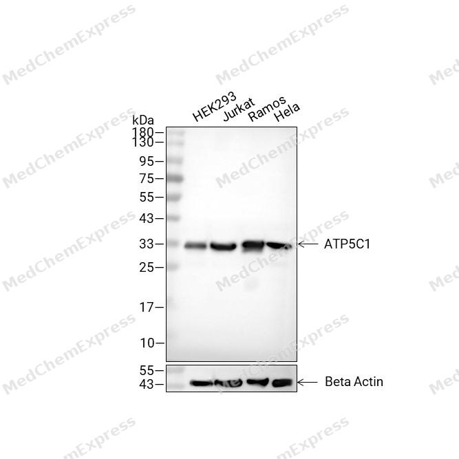 Western blot analysis of extracts from HEK293 (lane2(20μg), Jurkat (lane3(20μg), Ramos (lane4(20μg) and Hela (lane5(20μg) using ATP5C1 Antibody (HY-P88135). Proteins were transferred to a PVDF membrane and blocked with 5% non-fat milk in TBST for 2 hour at room temperature. The primary antibody (1/1000) and Loading control antibody (Beta Actin, HY-P80993, 1/10,000) was used in 5% non-fat milk in TBST at 4°C overnight. Goat Anti-Mouse IgG-HRP Secondary Antibody (HY-P8004 ,1/10,000) was used for 1 hour at room temperature.
 -
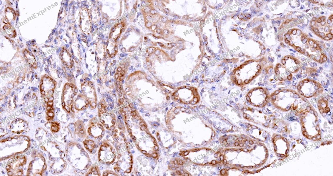 Immunohistochemical analysis of paraffin-embedded human kidney tissue using ATP5C1 Antibody (HY-P88135, 1/500). The section was pretreated using heat mediated antigen retrieval with sodium citrate buffer (pH 6.0) for 8 minutes. The tissues were blocked with quick block buffer for 0.5 hours at room temperature, washed with PBS and PBST, and then incubated with the primary antibody overnight at 4℃. The detection was performed using an HRP conjugated compact polymer system. DAB was used as the chromogen. Tissues were counterstained with hematoxylin and mounted with DPX.
 -
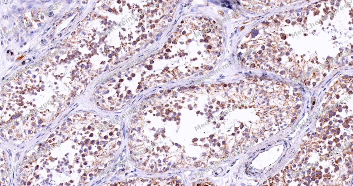 Immunohistochemical analysis of paraffin-embedded human testis tissue using ATP5C1 Antibody (HY-P88135, 1/500). The section was pretreated using heat mediated antigen retrieval with sodium citrate buffer (pH 6.0) for 8 minutes. The tissues were blocked with quick block buffer for 0.5 hours at room temperature, washed with PBS and PBST, and then incubated with the primary antibody overnight at 4℃. The detection was performed using an HRP conjugated compact polymer system. DAB was used as the chromogen. Tissues were counterstained with hematoxylin and mounted with DPX.
 -
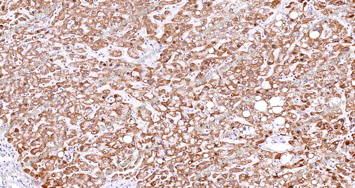 Immunohistochemical analysis of paraffin-embedded human liver tissue using ATP5C1 Antibody (HY-P88135, 1/500). The section was pretreated using heat mediated antigen retrieval with sodium citrate buffer (pH 6.0) for 8 minutes. The tissues were blocked with quick block buffer for 0.5 hours at room temperature, washed with PBS and PBST, and then incubated with the primary antibody overnight at 4℃. The detection was performed using an HRP conjugated compact polymer system. DAB was used as the chromogen. Tissues were counterstained with hematoxylin and mounted with DPX.
 -
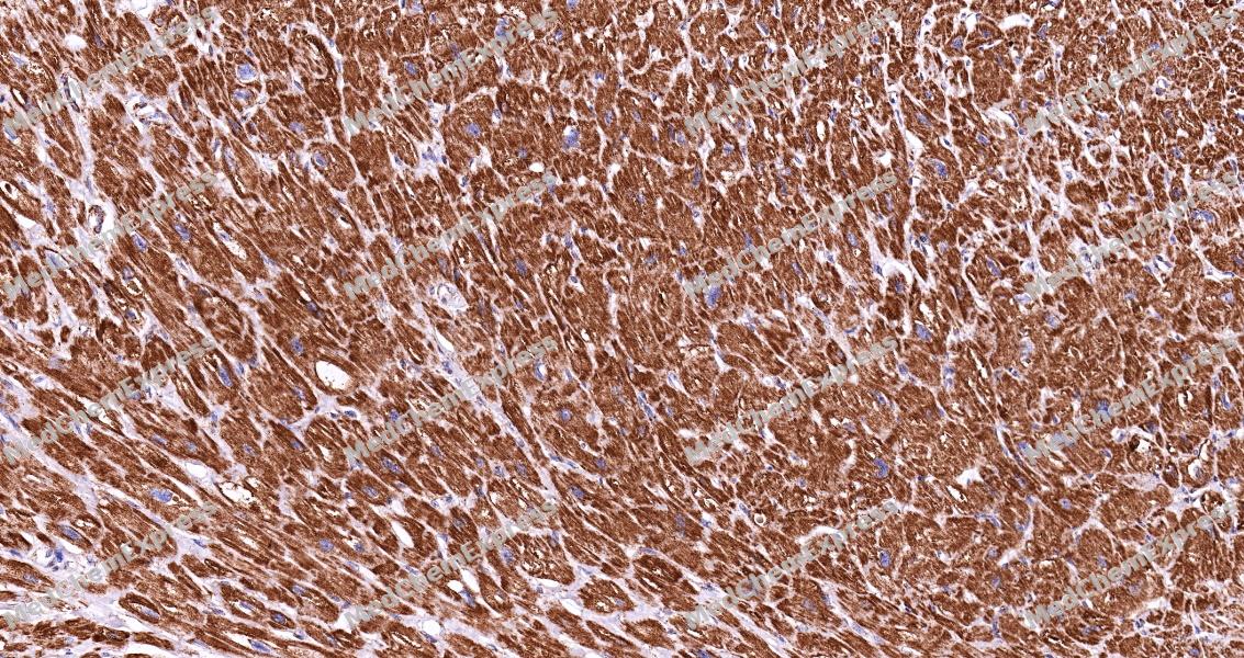 Immunohistochemical analysis of paraffin-embedded human heart tissue using ATP5C1 Antibody (HY-P88135, 1/500). The section was pretreated using heat mediated antigen retrieval with sodium citrate buffer (pH 6.0) for 8 minutes. The tissues were blocked with quick block buffer for 0.5 hours at room temperature, washed with PBS and PBST, and then incubated with the primary antibody overnight at 4℃. The detection was performed using an HRP conjugated compact polymer system. DAB was used as the chromogen. Tissues were counterstained with hematoxylin and mounted with DPX.
 -
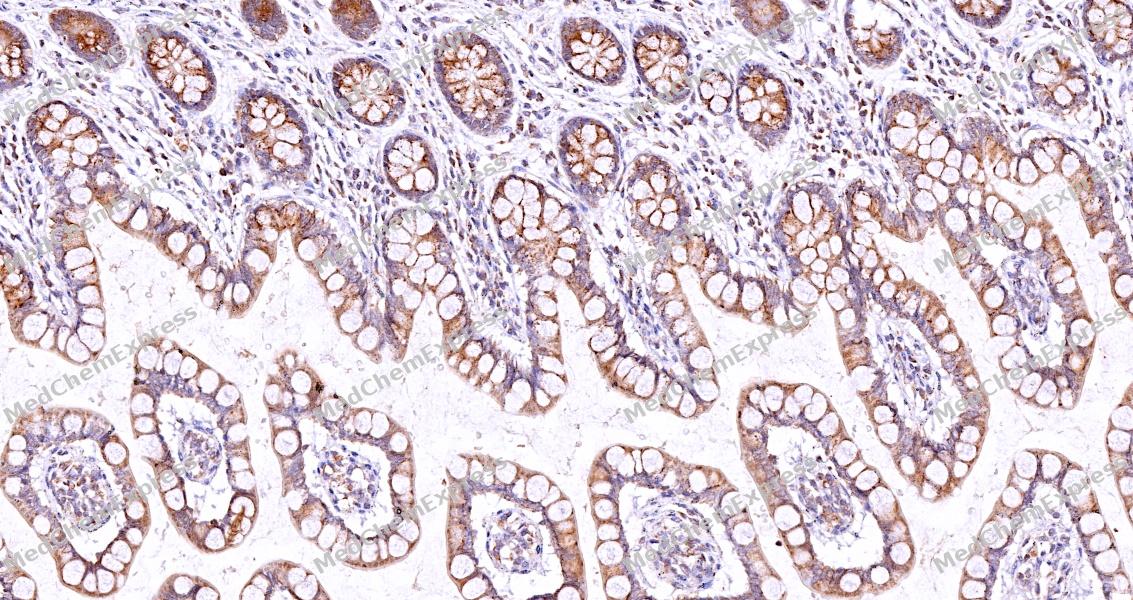 Immunohistochemical analysis of paraffin-embedded human small intestine tissue using ATP5C1 Antibody (HY-P88135, 1/500). The section was pretreated using heat mediated antigen retrieval with sodium citrate buffer (pH 6.0) for 8 minutes. The tissues were blocked with quick block buffer for 0.5 hours at room temperature, washed with PBS and PBST, and then incubated with the primary antibody overnight at 4℃. The detection was performed using an HRP conjugated compact polymer system. DAB was used as the chromogen. Tissues were counterstained with hematoxylin and mounted with DPX.
 -
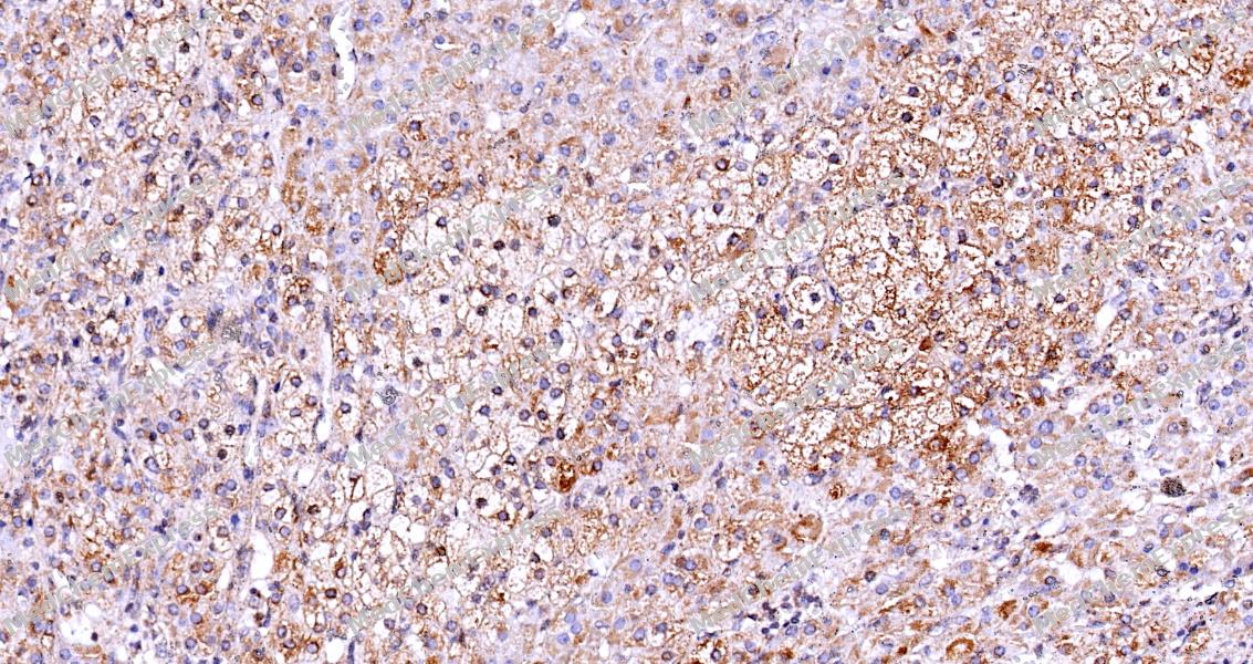 Immunohistochemical analysis of paraffin-embedded human adrenal gland tissue using ATP5C1 Antibody (HY-P88135, 1/500). The section was pretreated using heat mediated antigen retrieval with sodium citrate buffer (pH 6.0) for 8 minutes. The tissues were blocked with quick block buffer for 0.5 hours at room temperature, washed with PBS and PBST, and then incubated with the primary antibody overnight at 4℃. The detection was performed using an HRP conjugated compact polymer system. DAB was used as the chromogen. Tissues were counterstained with hematoxylin and mounted with DPX.


Background
-
Function
ATP5C1 is a Subunit gamma, of the mitochondrial membrane ATP synthase complex (F(1)F(0) ATP synthase or Complex V) that produces ATP from ADP in the presence of a proton gradient across the membrane which is generated by electron transport complexes of the respiratory chain. ATP synthase complex consist of a soluble F(1) head domain - the catalytic core - and a membrane F(1) domain - the membrane proton channel. These two domains are linked by a central stalk rotating inside the F(1) region and a stationary peripheral stalk. During catalysis, ATP synthesis in the catalytic domain of F(1) is coupled via a rotary mechanism of the central stalk subunits to proton translocation (Probable). In vivo, can only synthesize ATP although its ATP hydrolase activity can be activated artificially in vitro. With the central stalk subunit delta, is essential for the biogenesis of F(1) catalytic part of the ATP synthase complex namely in the formation of F1 assembly intermediate[1][2].
-
Subcellular Localization
Mitochondrion inner membrane
-
Expression
Tissue_Specificity: Isoform Heart is expressed specifically in the heart and skeletal muscle, which require rapid energy supply. Isoform Liver is expressed in the brain, liver and kidney. Isoform Heart and Isoform Liver are expressed in the skin, intestine, stomach and aorta -
Isoforms & Post-Translational Modification
P36542 has two isomers: P36542-1: 32996 Da (predicted); P36542-2: 32881 Da (predicted).
-
Subunit
Component of the ATP synthase complex composed at least of ATP5F1A/subunit alpha, ATP5F1B/subunit beta, ATP5MC1/subunit c (homooctomer), MT-ATP6/subunit a, MT-ATP8/subunit 8, ATP5ME/subunit e, ATP5MF/subunit f, ATP5MG/subunit g, ATP5MK/subunit k, ATP5MJ/subunit j, ATP5F1C/subunit gamma, ATP5F1D/subunit delta, ATP5F1E/subunit epsilon, ATP5PF/subunit F6, ATP5PB/subunit b, ATP5PD/subunit d, ATP5PO/subunit OSCP (PubMed:37244256)
-
SwissProt ID
-
Synonyms
ATP5C; ATP5CL1


Documentation

 Powered by Bioz
