



Calreticulin Antibody (YA3926)
(Synonyms: RO; CRT; SSA; cC1qR; HEL-S-99n)Based on 2 publication(s) in Google Scholar
Calreticulin Antibody (YA3926) is a Mouse-derived and non-conjugated IgG1 monoclonal antibody, targeting to Calreticulin.
-
Host:
Mouse
-
Isotype:
IgG1
-
Application:
WB, IHC-P, FC, ELISA
-
Reactivity :
Human, Mouse, Rat
-
Formulation:
Supplied in PBS with 0.05% sodium azide
-
Conjugation:
Non-conjugated


To place orders, for customer services and technical support, please contact: MedChemExpress USA
Tel: 609-228-6898 E-mail: [email protected] [email protected]



Publications Citing Use of MedChemExpress (MCE) Calreticulin Antibody (YA3926)
More

Applications
| Application |
WB

WB: Western Blot
|
IHC-P
IHC-P: Immunohistochemistry-Paraffin
|
FC
FC: Flow Cytometry
|
ELISA
ELISA: Enzyme Linked Immunosorbent Assay
|
|---|---|---|---|---|
| Dilution Ratio | 1:500-1:2000 | 1:200-1:1000 | 1:200-1:400 | 1:10000 |


Product Details
Calreticulin Antibody (YA3926) is a Mouse-derived and non-conjugated IgG1 monoclonal antibody, targeting to Calreticulin.
-
Host Mouse

-
Clonality Monoclonal

-
Species ReactivityHuman, Mouse, Rat
-
Observed Molecular WeightObserved band size: 60 kDa
Note: Due to possible protein modifications or aggregation, the molecular weight should be confirmed by actual measurement, and the predicted value is for reference only.

-
Calculated Molecular Weight Predicted band size: 48 kDa

Purified recombinant fragment of human CALR (AA: 18-417) expressed in E. Coli.
affinity purified.
Non-conjugated
Unmodified
IgG1


Product Properties
-
Appearance
Liquid
-
Formulation
Supplied in PBS with 0.05% sodium azide
-
Concentration
Batch-dependent, Please check the COA for the concentration of each lot. Check Lot Concentration
-
Storage & Stability
Stored at -20°C for 1 year. Avoid repeated freeze / thaw cycles.
-
Shipping
Shipping with blue ice.


Publications (2)
-
Journal Impact Factor
-
Most Recent
-
ACS Nano
Magnetic Microcarrier-Based Whole Tumor Cell Vaccines for Tumor Prevention and Immunotherapy. [Abstract]2025 Jun 17;19(23):21468-21483. PMID: 40476671 -
Biochem Biophys Res Commun
Non-fused ring A-D-A-type dyes for near-infrared II fluorescence imaging guided phototherapy and immune activation of tumour. [Abstract]2026 Jun 16:829:154158. PMID: 42361740


Verification Images
-
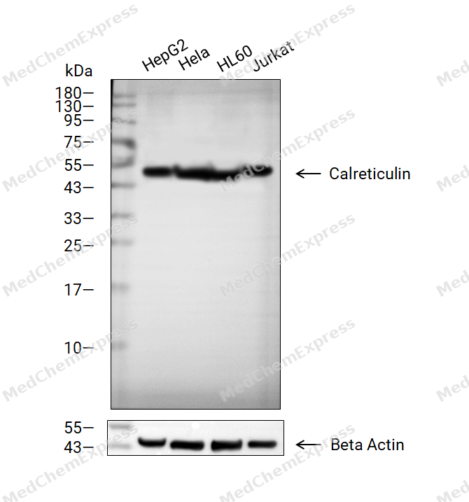 Western blot analysis of extracts from HepG2 (lane 2(20μg), Hela (lane 3(20μg), HL60 (lane 4(20μg) and Jurkat (lane 5(20μg) using Calreticulin Antibody (HY-P84229). Proteins were transferred to a PVDF membrane and blocked with 5% non-fat milk in TBST for 2 hour at room temperature. The primary antibody (1/1000) and Loading control antibody (Beta Actin, HY-P80993, 1/10000) was used in 5% non-fat milk in TBST at 4°C overnight. Goat Anti-Mouse IgG-HRP Secondary Antibody (1/10000) was used for 1 hour at room temperature.
 -
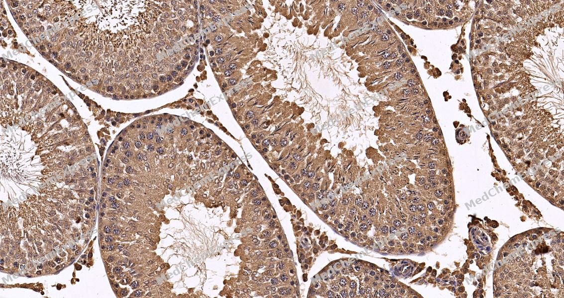 Immunohistochemical analysis of paraffin-embedded human testis tissue using Calreticulin antibody. The section was pre-treated using heat mediated antigen retrieval with sodium citrate buffer (pH 6.0) for 2 minutes. The tissues were blocked with QuickBlock at room temperature for 30 minutes, washed with PBSand PBST, and then incubated with the primary antibody (HY-P84229, 1/500) overnight at 4℃. The detection was performed using an HRP conjugated compact polymer system. DAB was used as the chromogen. Tissues were counterstained with hematoxylin and mounted with DPX.
 -
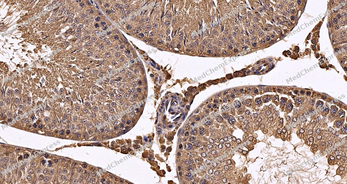 Immunohistochemical analysis of paraffin-embedded rat testis tissue using Calreticulin antibody. The section was pre-treated using heat mediated antigen retrieval with sodium citrate buffer (pH 6.0) for 2 minutes. The tissues were blocked with QuickBlock at room temperature for 30 minutes, washed with PBSand PBST, and then incubated with the primary antibody (HY-P84229, 1/500) overnight at 4℃. The detection was performed using an HRP conjugated compact polymer system. DAB was used as the chromogen. Tissues were counterstained with hematoxylin and mounted with DPX.
 -
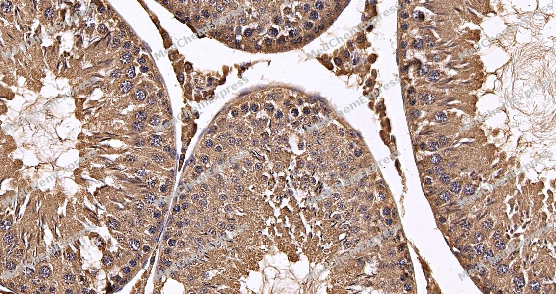 Immunohistochemical analysis of paraffin-embedded mouse testis tissue using Calreticulin antibody. The section was pre-treated using heat mediated antigen retrieval with sodium citrate buffer (pH 6.0) for 2 minutes. The tissues were blocked with QuickBlock at room temperature for 30 minutes, washed with PBSand PBST, and then incubated with the primary antibody (HY-P84229, 1/500) overnight at 4℃. The detection was performed using an HRP conjugated compact polymer system. DAB was used as the chromogen. Tissues were counterstained with hematoxylin and mounted with DPX.
 -
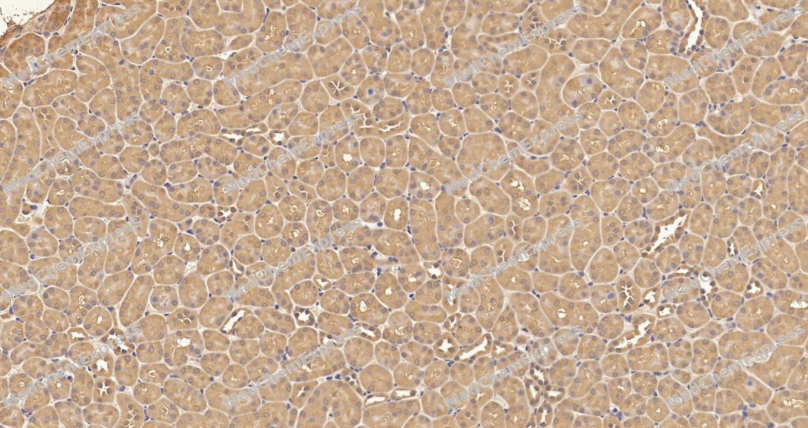 Immunohistochemical analysis of paraffin-embedded human kidney tissue using Calreticulin antibody. The section was pre-treated using heat mediated antigen retrieval with sodium citrate buffer (pH 6.0) for 2 minutes. The tissues were blocked with QuickBlock at room temperature for 30 minutes, washed with PBSand PBST, and then incubated with the primary antibody (HY-P84229, 1/500) overnight at 4℃. The detection was performed using an HRP conjugated compact polymer system. DAB was used as the chromogen. Tissues were counterstained with hematoxylin and mounted with DPX.
 -
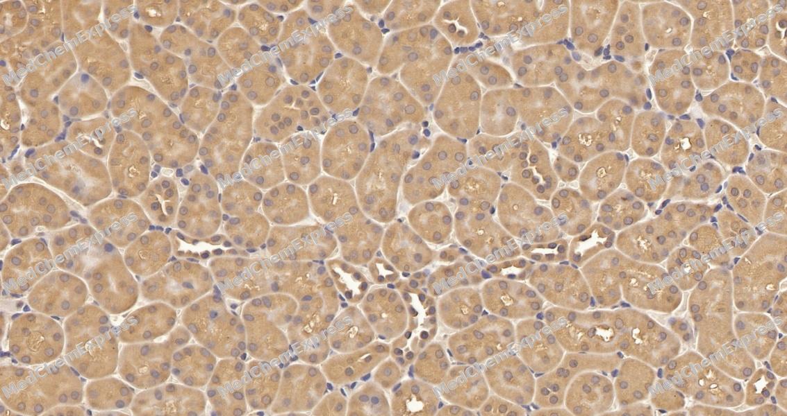 Immunohistochemical analysis of paraffin-embedded rat kidney tissue using Calreticulin antibody. The section was pre-treated using heat mediated antigen retrieval with sodium citrate buffer (pH 6.0) for 2 minutes. The tissues were blocked with QuickBlock at room temperature for 30 minutes, washed with PBSand PBST, and then incubated with the primary antibody (HY-P84229, 1/500) overnight at 4℃. The detection was performed using an HRP conjugated compact polymer system. DAB was used as the chromogen. Tissues were counterstained with hematoxylin and mounted with DPX.
 -
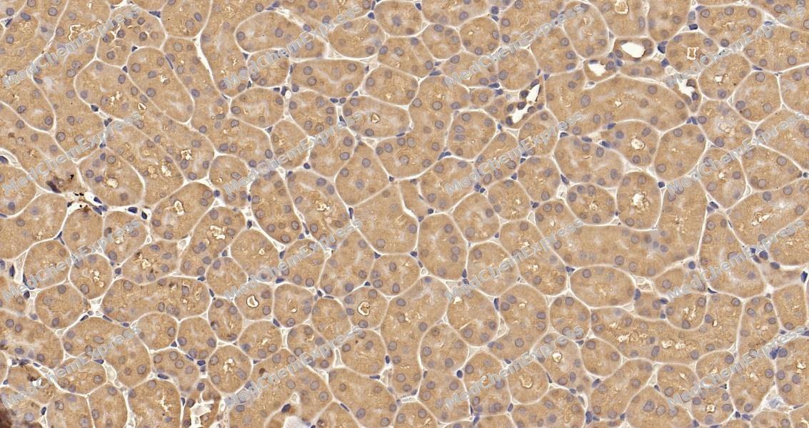 Immunohistochemical analysis of paraffin-embedded mouse kidney tissue using Calreticulin antibody. The section was pre-treated using heat mediated antigen retrieval with sodium citrate buffer (pH 6.0) for 2 minutes. The tissues were blocked with QuickBlock at room temperature for 30 minutes, washed with PBSand PBST, and then incubated with the primary antibody (HY-P84229, 1/500) overnight at 4℃. The detection was performed using an HRP conjugated compact polymer system. DAB was used as the chromogen. Tissues were counterstained with hematoxylin and mounted with DPX.


Background
-
Function
Calreticulin is a Calcium-binding chaperone that promotes folding, oligomeric assembly and quality control in the endoplasmic reticulum (ER) via the calreticulin/calnexin cycle. This lectin interacts transiently with almost all of the monoglucosylated glycoproteins that are synthesized in the ER. Interacts with the DNA-binding domain of NR3C1 and mediates its nuclear export. Involved in maternal gene expression regulation. May participate in oocyte maturation via the regulation of calcium homeostasis. Present in the cortical granules of non-activated oocytes, is exocytosed during the cortical reaction in response to oocyte activation and might participate in the block to polyspermy[1][2].
-
Subcellular Localization
Endoplasmic reticulum lumen; Cytoplasm, cytosol; Secreted, extracellular space, extracellular matrix; Cell surface; Sarcoplasmic reticulum lumen; Cytoplasmic vesicle, secretory vesicle, Cortical granule; Cytolytic granule
-
Subunit
Monomer. Component of an EIF2 complex at least composed of CELF1/CUGBP1, CALR, CALR3, EIF2S1, EIF2S2, HSP90B1 and HSPA5. Interacts with PDIA3/ERp57 and SPACA9 (By similarity). Interacts with TRIM21 (PubMed:8666824). Interacts with NR3C1 (PubMed:11149926). Interacts with PPIB (PubMed:20801878). Interacts (via P-domain) with PDIA5 (PubMed:23614004). Interacts with GABARAP (PubMed:19154346). Interacts with HLA-E-B2M and HLA-G-B2M complexes (PubMed:9427624, PubMed:9640257). Interacts with HLA-F (PubMed:10605026). Interacts with CLCC1 (PubMed:30157172)
-
SwissProt ID
-
Synonyms
RO; CRT; SSA; cC1qR; HEL-S-99n
Documentation
-
Data Sheet (234 KB)
-
SDS (251 KB)
- English - EN (251 KB)
- Français - FR (251 KB)
- Deutsch - DE (251 KB)
- Norwegian - NO (251 KB)
- Español - ES (251 KB)
- Swedish - SV (251 KB)
- Italian - IT (251 KB)
- Korean - KR (251 KB)
- Portuguese - PT (251 KB)
-
User Guide for Antibodies (1077 KB)

 Powered by Bioz
