



CD130 Antibody (YA3990)
(Synonyms: IL6ST; GP130; CDW130; IL-6RB)Based on 1 Customer Validation
CD130 Antibody (YA3990) is a Mouse-derived and non-conjugated IgG1 monoclonal antibody, targeting to CD130.
-
Host:
Mouse
-
Isotype:
IgG
-
Application:
WB, IHC-P, ICC/IF, FC, ELISA
-
Reactivity :
Human, Monkey
-
Formulation:
Supplied in PBS with 0.05% sodium azide
-
Conjugation:
Non-conjugated


To place orders, for customer services and technical support, please contact: MedChemExpress USA
Tel: 609-228-6898 E-mail: [email protected] [email protected]





Applications
| Application |
WB

WB: Western Blot
|
IHC-P
IHC-P: Immunohistochemistry-Paraffin
|
ICC/IF
ICC/IF: Immunocytochemistry/
Immunofluorescence |
FC
FC: Flow Cytometry
|
ELISA
ELISA: Enzyme Linked Immunosorbent Assay
|
|---|---|---|---|---|---|
| Dilution Ratio | 1:500-1:2000 | 1:200-1:1000 | 1:200-1:1000 | 1:200-1:400 | 1:10000 |


Product Details
CD130 Antibody (YA3990) is a Mouse-derived and non-conjugated IgG1 monoclonal antibody, targeting to CD130.
-
Host Mouse

-
Clonality Monoclonal

-
Species ReactivityHuman, Monkey
-
Observed Molecular WeightObserved band size: 130-140 kDa
Note: Due to possible protein modifications or aggregation, the molecular weight should be confirmed by actual measurement, and the predicted value is for reference only.

-
Calculated Molecular Weight Predicted band size: 104 kDa

Purified recombinant fragment of human CD130 (AA: extra 73-231) expressed in E. Coli.
affinity purified.
Non-conjugated
Unmodified
IgG


Product Properties
-
Appearance
Solution
-
Formulation
Supplied in PBS with 0.05% sodium azide
-
Concentration
Batch-dependent, Please check the COA for the concentration of each lot. Check Lot Concentration
-
Storage & Stability
Stored at -20°C for 1 year. Avoid repeated freeze / thaw cycles.
-
Shipping
Shipping with blue ice.


Verification Images
-
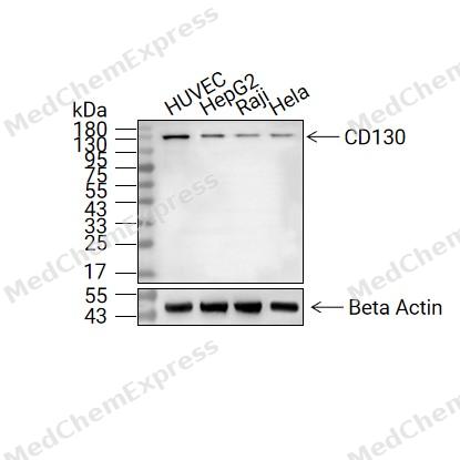 Western blot analysis of extracts from PC-12(lane 1(40μg) )
 -
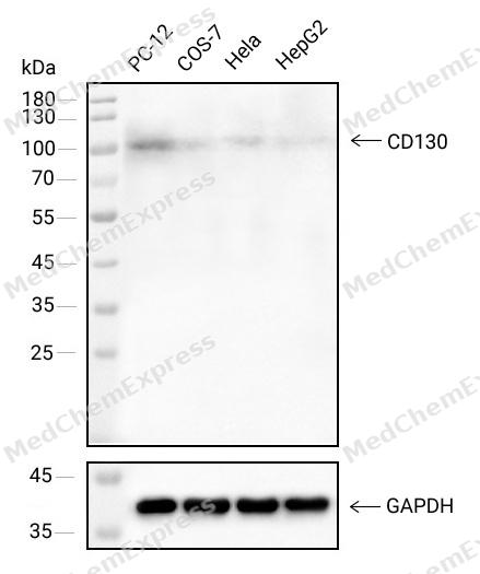 Western blot analysis of extracts from PC-12(lane 1(40μg) ),COS-7(lane 2(40μg) ),Hela(lane 3(40μg) )and HepG2(lane 4(40μg) ) using CD130 antibody(31685P). Proteins were transferred to a NC membrane and blocked with 5% Skim milk in TBST for 2 hour at room temperature. The primary antibody ( 1/1000) and Loading control antibody (GAPDH,20035, 1/1000) was used in 5% Skim milk in TBST at 4°C overnight. Goat Anti-Mouse IgG-HRP Secondary Antibody (1/10,000) was used for 1 hour at room temperature.
 -
 Immunohistochemical analysis of paraffin-embedded human lung cancer tissue using CD130 Antibody (HY-P84293, 1/800). The section was pretreated using heat mediated antigen retrieval with sodium citrate buffer (pH 6.0) for 8 minutes. The tissues were blocked with quick block buffer for 0.5 hours at room temperature, washed with PBS and PBST, and then incubated with the primary antibody overnight at 4℃. The detection was performed using an HRP conjugated compact polymer system. DAB was used as the chromogen. Tissues were counterstained with hematoxylin and mounted with DPX.
 -
 Immunohistochemical analysis of paraffin-embedded human testis cancer tissue using CD130 Antibody (HY-P84293, 1/800). The section was pretreated using heat mediated antigen retrieval with sodium citrate buffer (pH 6.0) for 8 minutes. The tissues were blocked with quick block buffer for 0.5 hours at room temperature, washed with PBS and PBST, and then incubated with the primary antibody overnight at 4℃. The detection was performed using an HRP conjugated compact polymer system. DAB was used as the chromogen. Tissues were counterstained with hematoxylin and mounted with DPX.
 -
 Immunohistochemical analysis of paraffin-embedded human liver cancer tissue using CD130 Antibody (HY-P84293, 1/800). The section was pretreated using heat mediated antigen retrieval with sodium citrate buffer (pH 6.0) for 8 minutes. The tissues were blocked with quick block buffer for 0.5 hours at room temperature, washed with PBS and PBST, and then incubated with the primary antibody overnight at 4℃. The detection was performed using an HRP conjugated compact polymer system. DAB was used as the chromogen. Tissues were counterstained with hematoxylin and mounted with DPX.
 -
 Immunohistochemical analysis of paraffin-embedded human breast cancer tissue using CD130 Antibody (HY-P84293, 1/800). The section was pretreated using heat mediated antigen retrieval with sodium citrate buffer (pH 6.0) for 8 minutes. The tissues were blocked with quick block buffer for 0.5 hours at room temperature, washed with PBS and PBST, and then incubated with the primary antibody overnight at 4℃. The detection was performed using an HRP conjugated compact polymer system. DAB was used as the chromogen. Tissues were counterstained with hematoxylin and mounted with DPX.
 -
 Immunohistochemical analysis of paraffin-embedded human renal cancer tissue using CD130 Antibody (HY-P84293, 1/800). The section was pretreated using heat mediated antigen retrieval with sodium citrate buffer (pH 6.0) for 8 minutes. The tissues were blocked with quick block buffer for 0.5 hours at room temperature, washed with PBS and PBST, and then incubated with the primary antibody overnight at 4℃. The detection was performed using an HRP conjugated compact polymer system. DAB was used as the chromogen. Tissues were counterstained with hematoxylin and mounted with DPX.
 -
 Immunohistochemical analysis of paraffin-embedded human thyroid cancer tissue using CD130 Antibody (HY-P84293, 1/800). The section was pretreated using heat mediated antigen retrieval with sodium citrate buffer (pH 6.0) for 8 minutes. The tissues were blocked with quick block buffer for 0.5 hours at room temperature, washed with PBS and PBST, and then incubated with the primary antibody overnight at 4℃. The detection was performed using an HRP conjugated compact polymer system. DAB was used as the chromogen. Tissues were counterstained with hematoxylin and mounted with DPX.
 -
 Immunohistochemical analysis of paraffin-embedded human glioma tissue using CD130 Antibody (HY-P84293, 1/800). The section was pretreated using heat mediated antigen retrieval with sodium citrate buffer (pH 6.0) for 8 minutes. The tissues were blocked with quick block buffer for 0.5 hours at room temperature, washed with PBS and PBST, and then incubated with the primary antibody overnight at 4℃. The detection was performed using an HRP conjugated compact polymer system. DAB was used as the chromogen. Tissues were counterstained with hematoxylin and mounted with DPX.
 -
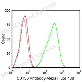 Flow cytometric analysis of 1X10^6 HL-60 cells labeling CD130 Antibody(HY-P84293
 -
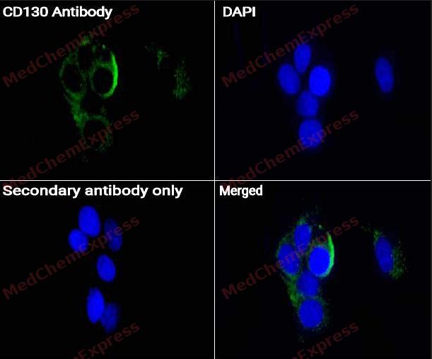 Immunofluorescence analysis of Hela cells labeling CD130 antibody (HY-P84293) at 1/50 dilution. Cells were fixed in 4% paraformaldehyde for 15 minutes at room temperature
 -
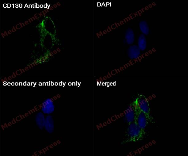 Immunocytochemistry analysis of HUVEC cells labeling CD130 with CD130 antibody (HY-P84293) at 1/100 dilution. Cells were fixed in 4% paraformaldehyde for 15 minutes at room temperature, permeabilized with 0.1% Triton X-100 for 10 minutes at room temperature, then blocked with BSA for Immunol Staining for 10 min at room temperature. Cells were then incubated with CD130 antibody (HY-P84293) at 1/100 dilution in BSA for Immunol Staining at 4℃, Stay overnight. AF488-conjugated AffiniPure Goat Anti-Mouse IgG H&L(HY-P8005, Green) was used as the secondary antibody at 1/1,000 dilution. PBS instead of the primary antibody was used as the secondary antibody only control. The Nuclear counterstain was DAPI (Blue).
 -
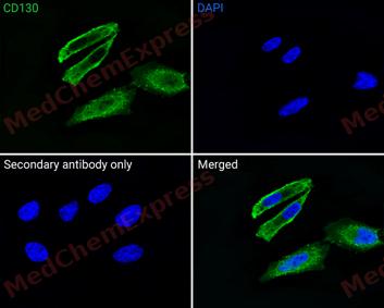 Immunofluorescence analysis of Hela cells labeling CD130 antibody (31685P) at 1/50 dilution. Cells were fixed in 4% paraformaldehyde for 15 minutes at room temperature, permeabilized with 0.1% Triton X-100 in PBS for 15 minutes at room temperature, then blocked with 1% BSA in 10% negative goat serum for 1 hour at room temperature.Cells were then incubated with CD130 antibody (31685P) at 1/50 dilution in 1% BSA in PBST overnight at 4 ℃. Alexa Fluor® 488 Goat Anti-mouse IgG H&L (invitrogen A11001, green) was used as the secondary antibody at 1/500 dilution. PBS instead of the primary antibody was used as the secondary antibody only control. The Nuclear counterstain was DAPI (Blue)


Background
-
Function
CD130 is a Signal-transducing molecule. The receptor systems for IL6, LIF, OSM, CNTF, IL11, CTF1 and BSF3 can utilize IL6ST for initiating signal transmission. Binding of IL6 to IL6R induces IL6ST homodimerization and formation of a high-affinity receptor complex, which activates the intracellular JAK-MAPK and JAK-STAT3 signaling pathways. That causes phosphorylation of IL6ST tyrosine residues which in turn activates STAT3. In parallel, the IL6 signaling pathway induces the expression of two cytokine receptor signaling inhibitors, SOCS1 and SOCS3, which inhibit JAK and terminate the activity of the IL6 signaling pathway as a negative feedback loop. Also activates the yes-associated protein 1 (YAP) and NOTCH pathways to control inflammation-induced epithelial regeneration, independently of STAT3. Acts as a receptor for the neuroprotective peptide humanin as part of a complex with IL27RA/WSX1 and CNTFR. Mediates signals which regulate immune response, hematopoiesis, pain control and bone metabolism. Has a role in embryonic development. Essential for survival of motor and sensory neurons and for differentiation of astrocytes. Required for expression of TRPA1 in nociceptive neurons. Required for the maintenance of PTH1R expression in the osteoblast lineage and for the stimulation of PTH-induced osteoblast differentiation. Required for normal trabecular bone mass and cortical bone composition; Binds to the soluble IL6:sIL6R complex (hyper-IL6), thereby blocking IL6 trans-signaling. Inhibits sIL6R-dependent acute phase response. Also blocks IL11 cluster signaling through IL11R[1][2][3][4][5][6][7][8].
-
Subcellular Localization
Cell membrane; Single-pass type I membrane protein; Secreted
-
Expression
Tissue_specificity:Found in all the tissues and cell lines examined (PubMed:2261637) . Expression not restricted to IL6 responsive cells (PubMed:2261637) ; Expressed in blood serum (at protein level) (PubMed:24629561)
Induction:LIF and OSM activate the type I OSM receptor while only OSM can activate the type II OSM receptor -
Isoforms & Post-Translational Modification
P40189 has 3 isomers: P40189-1: 103537 Da (predicted); P40189-2: 37499 Da (predicted); P40189-3: 96276 Da (predicted).
Phosphorylation of Ser-782 down-regulates cell surface expression;Heavily N-glycosylated (PubMed:11098061, PubMed:11251120, PubMed:16335952, PubMed:19139490, PubMed:19159218). Glycosylation is required for protein stability and localization in plasma membrane but not for ligand binding (PubMed:19915009) -
Subunit
Component of a hexamer of two molecules each of IL6, IL6R and IL6ST; associates with the complex IL6:IL6R but does not interact with IL6 (PubMed:12829785, PubMed:2261637). Forms heterodimers composed of LIFR and IL6ST (type I OSM receptor) which are activated by LIF and OSM (PubMed:8999038). Also forms heterodimers composed of OSMR and IL6ST (type II receptor) which are activated by OSM but not by LIF (PubMed:8999038). Component of a receptor complex composed of IL6ST/GP130, IL27RA/WSX1 and CNTFR which interacts with the neuroprotective peptide humanin (PubMed:19386761). Interacts with HCK (PubMed:9406996). Interacts with INPP5D/SHIP1 (By similarity). Interacts with SRC and YES (PubMed:25731159). Interacts with ARMH4; this interaction prevents IL6ST protein homodimerization and bridges ARMH4 with IL6R and STAT3 and therefore inhibits phosphorylation of STAT3 at 'Tyr-705' (PubMed:26927669)
-
SwissProt ID
-
Synonyms
IL6ST; GP130; CDW130; IL-6RB


Documentation
-
Data Sheet (235 KB)
-
SDS (251 KB)
- English - EN (251 KB)
- Français - FR (251 KB)
- Deutsch - DE (251 KB)
- Norwegian - NO (251 KB)
- Español - ES (251 KB)
- Swedish - SV (251 KB)
- Italian - IT (251 KB)
- Korean - KR (251 KB)
- Portuguese - PT (251 KB)
-
User Guide for Antibodies (1077 KB)

 Powered by Bioz
