



CSNK1E Antibody (YA7795)
(Synonyms: CKIepsilon; HCKIE)
Based on 1 Customer Validation
CSNK1E Antibody (YA7795) is a Mouse-derived and non-conjugated IgG1 monoclonal antibody, targeting to CSNK1E.
-
Host:
Mouse
-
Isotype:
IgG1
-
Application:
WB, IHC-P
-
Reactivity :
Human, Mouse, Rat
-
Formulation:
Spplied in PBS (pH 7.3) containing 1% BSA, 50% glycerol and 0.02% sodium azide.
-
Conjugation:
Non-conjugated


To place orders, for customer services and technical support, please contact: MedChemExpress USA
Tel: 609-228-6898 E-mail: [email protected] [email protected]





Applications
| Application |
WB

WB: Western Blot
|
IHC-P
IHC-P: Immunohistochemistry-Paraffin
|
|---|---|---|
| Dilution Ratio | 1:500-2000 | 1:150-500 |


Product Details
CSNK1E Antibody (YA7795) is a Mouse-derived and non-conjugated IgG1 monoclonal antibody, targeting to CSNK1E.
-
Host Mouse

-
Clonality Monoclonal

-
Species ReactivityHuman, Mouse, Rat
-
Calculated Molecular Weight Predicted band size: 47.1 kDa

Human recombinant protein fragment corresponding to amino acids 153-416 of human CSNK1E produced in E.coli.
Endogenous
Affinity purified
Non-conjugated
Unmodified
IgG1


Product Properties
-
Appearance
Liquid
-
Formulation
Spplied in PBS (pH 7.3) containing 1% BSA, 50% glycerol and 0.02% sodium azide.
-
Concentration
Batch-dependent, Please check the COA for the concentration of each lot. Check Lot Concentration
-
Storage & Stability
Stored at -20°C for 1 year. Avoid repeated freeze / thaw cycles.
-
Shipping
Shipping with blue ice.


Verification Images
-
 Western blot analysis was performed by loading 0.1 μg (Lane 2), 0.5 μg (Lane 3), and 1 μg (Lane 4) of CSNK1E Protein, Human (Inactive, His) using CSNK1E antibody. Proteins were transferred onto a 0.45 μm PVDF membrane using the Trans-Blot® Turbo™ system for 13 min. The membrane was then blocked with 5% nonfat milk in TBST (HY-K1025) for 1 h at room temperature. The primary antibody (1:1000) was diluted in 5% nonfat milk in TBST and incubated with the membrane overnight at 4°C. After washing, the membrane of primary antibody was incubated with HRP-conjugated goat anti-rabbit/mouse IgG secondary antibody (HY-P8001/HY-P8004) (1:5000) diluted in 5% nonfat milk in TBST for 1 h at room temperature. Protein bands were visualized using an Ultra High Sensitivity ECL detection kit (HY-K1005).
 -
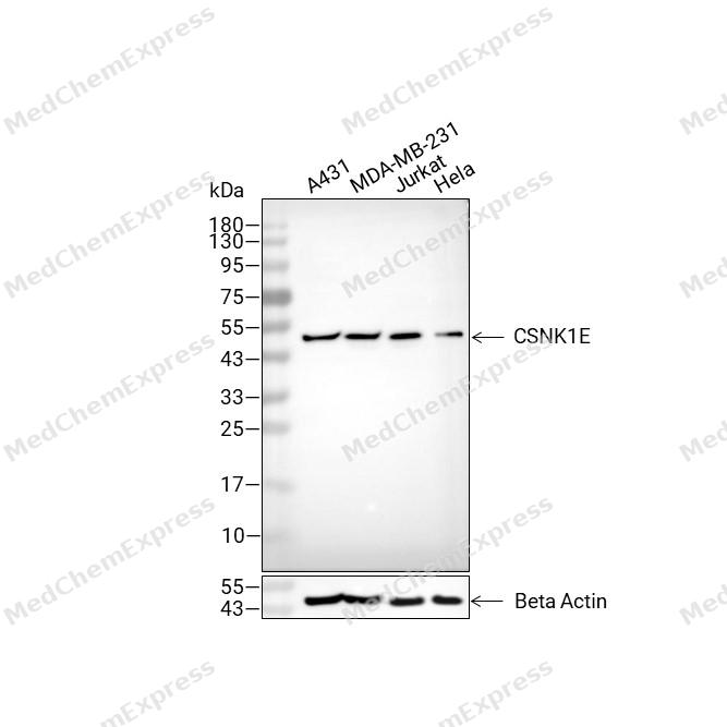 Western blot analysis of extracts from A431 (lane2(20μg), MDA-MB-231 (lane3(20μg), Jurkat (lane4(20μg) and Hela (lane5(20μg) using CSNK1E Antibody (HY-P88111). Proteins were transferred to a PVDF membrane and blocked with 5% non-fat milk in TBST for 2 hour at room temperature. The primary antibody (1/1000) and Loading control antibody (Beta Actin, HY-P80993, 1/10,000) was used in 5% non-fat milk in TBST at 4°C overnight. Goat Anti-Mouse IgG-HRP Secondary Antibody (HY-P8004 ,1/10,000) was used for 1 hour at room temperature.
 -
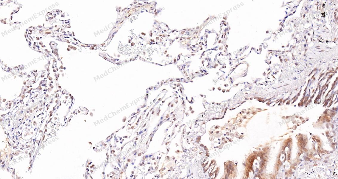 Immunohistochemical analysis of paraffin-embedded human lung tissue using CSNK1E Antibody (HY-P88111, 1/500). The section was pretreated using heat mediated antigen retrieval with sodium citrate buffer (pH 6.0) for 8 minutes. The tissues were blocked with quick block buffer for 0.5 hours at room temperature, washed with PBS and PBST, and then incubated with the primary antibody overnight at 4℃. The detection was performed using an HRP conjugated compact polymer system. DAB was used as the chromogen. Tissues were counterstained with hematoxylin and mounted with DPX.
 -
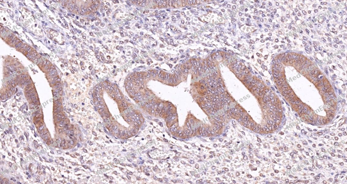 Immunohistochemical analysis of paraffin-embedded human endometrium tissue using CSNK1E Antibody (HY-P88111, 1/500). The section was pretreated using heat mediated antigen retrieval with sodium citrate buffer (pH 6.0) for 8 minutes. The tissues were blocked with quick block buffer for 0.5 hours at room temperature, washed with PBS and PBST, and then incubated with the primary antibody overnight at 4℃. The detection was performed using an HRP conjugated compact polymer system. DAB was used as the chromogen. Tissues were counterstained with hematoxylin and mounted with DPX.
 -
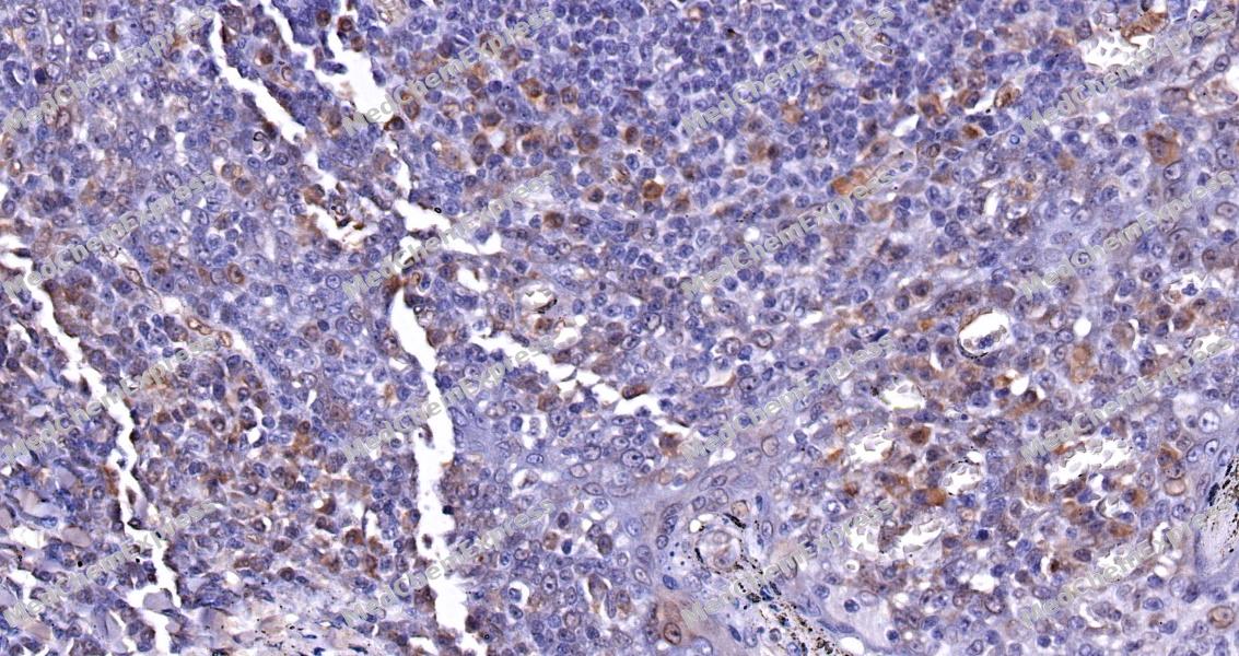 Immunohistochemical analysis of paraffin-embedded human tonsil tissue using CSNK1E Antibody (HY-P88111, 1/500). The section was pretreated using heat mediated antigen retrieval with sodium citrate buffer (pH 6.0) for 8 minutes. The tissues were blocked with quick block buffer for 0.5 hours at room temperature, washed with PBS and PBST, and then incubated with the primary antibody overnight at 4℃. The detection was performed using an HRP conjugated compact polymer system. DAB was used as the chromogen. Tissues were counterstained with hematoxylin and mounted with DPX.
 -
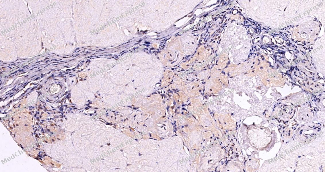 Immunohistochemical analysis of paraffin-embedded human ovary tissue using CSNK1E Antibody (HY-P88111, 1/500). The section was pretreated using heat mediated antigen retrieval with sodium citrate buffer (pH 6.0) for 8 minutes. The tissues were blocked with quick block buffer for 0.5 hours at room temperature, washed with PBS and PBST, and then incubated with the primary antibody overnight at 4℃. The detection was performed using an HRP conjugated compact polymer system. DAB was used as the chromogen. Tissues were counterstained with hematoxylin and mounted with DPX.
 -
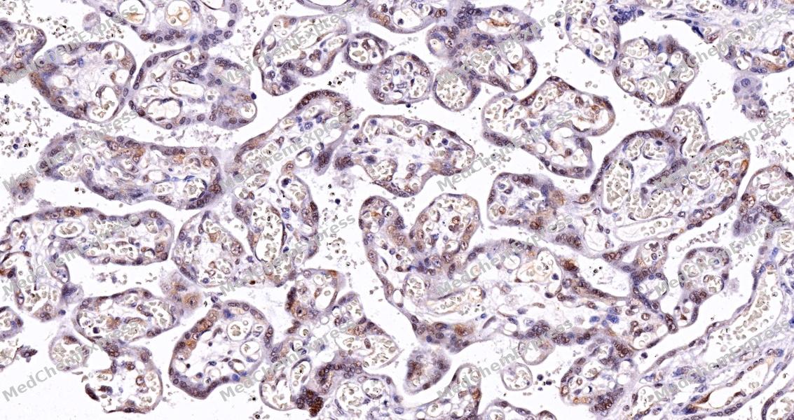 Immunohistochemical analysis of paraffin-embedded human placenta tissue using CSNK1E Antibody (HY-P88111, 1/500). The section was pretreated using heat mediated antigen retrieval with sodium citrate buffer (pH 6.0) for 8 minutes. The tissues were blocked with quick block buffer for 0.5 hours at room temperature, washed with PBS and PBST, and then incubated with the primary antibody overnight at 4℃. The detection was performed using an HRP conjugated compact polymer system. DAB was used as the chromogen. Tissues were counterstained with hematoxylin and mounted with DPX.
 -
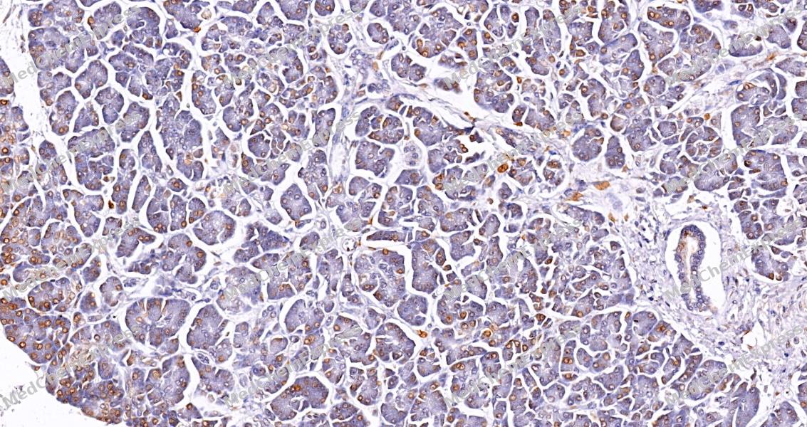 Immunohistochemical analysis of paraffin-embedded human pancreas tissue using CSNK1E Antibody (HY-P88111, 1/500). The section was pretreated using heat mediated antigen retrieval with sodium citrate buffer (pH 6.0) for 8 minutes. The tissues were blocked with quick block buffer for 0.5 hours at room temperature, washed with PBS and PBST, and then incubated with the primary antibody overnight at 4℃. The detection was performed using an HRP conjugated compact polymer system. DAB was used as the chromogen. Tissues were counterstained with hematoxylin and mounted with DPX.


Background
-
Function
CSNK1E is a Casein kinases are operationally defined by their preferential utilization of acidic proteins such as caseins as substrates (Probable). Participates in Wnt signaling. Phosphorylates DVL1. Phosphorylates DVL2. Phosphorylates NEDD9/HEF1. Central component of the circadian clock. In balance with PP1, determines the circadian period length, through the regulation of the speed and rhythmicity of PER1 and PER2 phosphorylation. Controls PER1 and PER2 nuclear transport and degradation. Inhibits cytokine-induced granuloytic differentiation[1][2][3][4][5].
-
Subcellular Localization
Cytoplasm,Nucleus
-
Expression
Tissue_Specificity: Expressed in all tissues examined, including brain, heart, lung, liver, pancreas, kidney, placenta and skeletal muscle. Expressed in monocytes and lymphocytes but not in granulocytes
Induction: Down-regulated during granulocytic differentiation -
Isoforms & Post-Translational Modification
P49674: 416 amino acids, molecular weight 47315 Da.
-
Subunit
Monomer (PubMed:23106386)
-
SwissProt ID
-
Synonyms
CKIepsilon; HCKIE


Documentation

 Powered by Bioz
