



EPHA7 Antibody (YA4835)
(Synonyms: EPHA7; EHK3; HEK11; Ephrin type-A receptor 7; EPH homology kinase 3; EHK-3; EPH-like kinase 11; EK11; hEK11)
Based on 1 Customer Validation
EPHA7 Antibody (YA4835) is a Mouse-derived and non-conjugated monoclonal antibody, targeting to EPHA7.
-
Host:
Mouse
-
Application:
WB, IHC-P, ICC/IF, ELISA
-
Reactivity :
Human
-
Formulation:
Supplied in PBS containing 50% glycerol, 0.5% BSA and 0.02% sodium azide.
-
Conjugation:
Non-conjugated


To place orders, for customer services and technical support, please contact: MedChemExpress USA
Tel: 609-228-6898 E-mail: [email protected] [email protected]





Applications
| Application |
WB

WB: Western Blot
|
IHC-P
IHC-P: Immunohistochemistry-Paraffin
|
ELISA
ELISA: Enzyme Linked Immunosorbent Assay
|
ICC/IF
ICC/IF: Immunocytochemistry/
Immunofluorescence |
|---|---|---|---|---|
| Dilution Ratio | 1:500-1:2000 | 1:200-1:1000 | 1:10000 | 1:50-200 |


Product Details
EPHA7 Antibody (YA4835) is a Mouse-derived and non-conjugated monoclonal antibody, targeting to EPHA7.
-
Host Mouse

-
Clonality Monoclonal

-
Species ReactivityHuman
-
Calculated Molecular Weight Predicted band size: 112 kDa

Purified recombinant fragment of EphA7 (aa27-210) expressed in E. Coli.
affinity purified.
Non-conjugated
Unmodified


Product Properties
-
Appearance
Liquid
-
Formulation
Supplied in PBS containing 50% glycerol, 0.5% BSA and 0.02% sodium azide.
-
Concentration
Batch-dependent, Please check the COA for the concentration of each lot. Check Lot Concentration
-
Storage & Stability
Stored at -20°C for 1 year. Avoid repeated freeze / thaw cycles.
-
Shipping
Shipping with blue ice.


Verification Images
-
 Western blot analysis of extracts from Hela (lane 2, 20ug) and Hela (lane 3, 40ug) using EPHA7 Antibody (HY-P85143). Proteins were transferred to a PVDF membrane and blocked with 5% BSA in TBST for 2 hour at room temperature. The primary antibody (1/1000) and Loading control antibody (Beta Actin, HY-P80993, 1/10,000) was used in 5% BSA in TBST at 4°C overnight. Goat Anti-Mouse IgG-HRP Secondary Antibody (HY-P8004,1/10,000) was used for 1 hour at room temperature.
 -
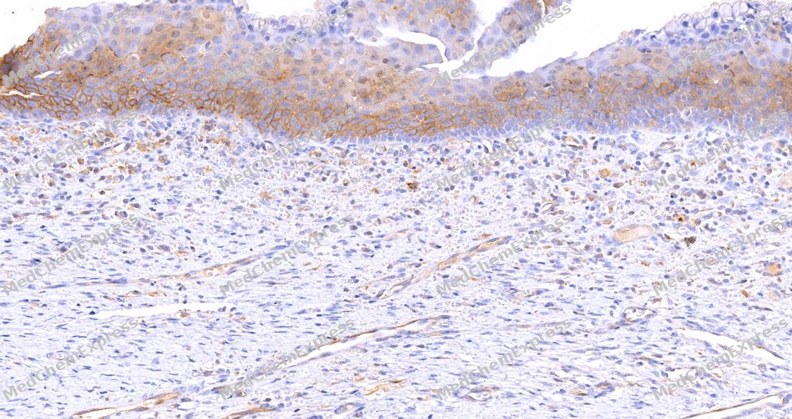 Immunohistochemical analysis of paraffin-embedded human cervix tissue using EPHA7 Antibody (HY-P85143, 1/800). The section was pretreated using heat mediated antigen retrieval with sodium citrate buffer (pH 6.0) for 8 minutes. The tissues were blocked with quick block buffer for 0.5 hours at room temperature, washed with PBS and PBST, and then incubated with the primary antibody overnight at 4℃. The detection was performed using an HRP conjugated compact polymer system. DAB was used as the chromogen. Tissues were counterstained with hematoxylin and mounted with DPX.
 -
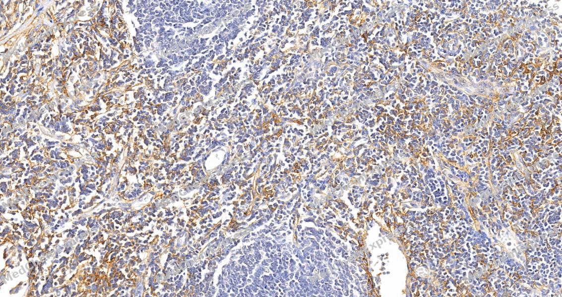 Immunohistochemical analysis of paraffin-embedded human lymph node tissue using EPHA7 Antibody (HY-P85143, 1/800). The section was pretreated using heat mediated antigen retrieval with sodium citrate buffer (pH 6.0) for 8 minutes. The tissues were blocked with quick block buffer for 0.5 hours at room temperature, washed with PBS and PBST, and then incubated with the primary antibody overnight at 4℃. The detection was performed using an HRP conjugated compact polymer system. DAB was used as the chromogen. Tissues were counterstained with hematoxylin and mounted with DPX.
 -
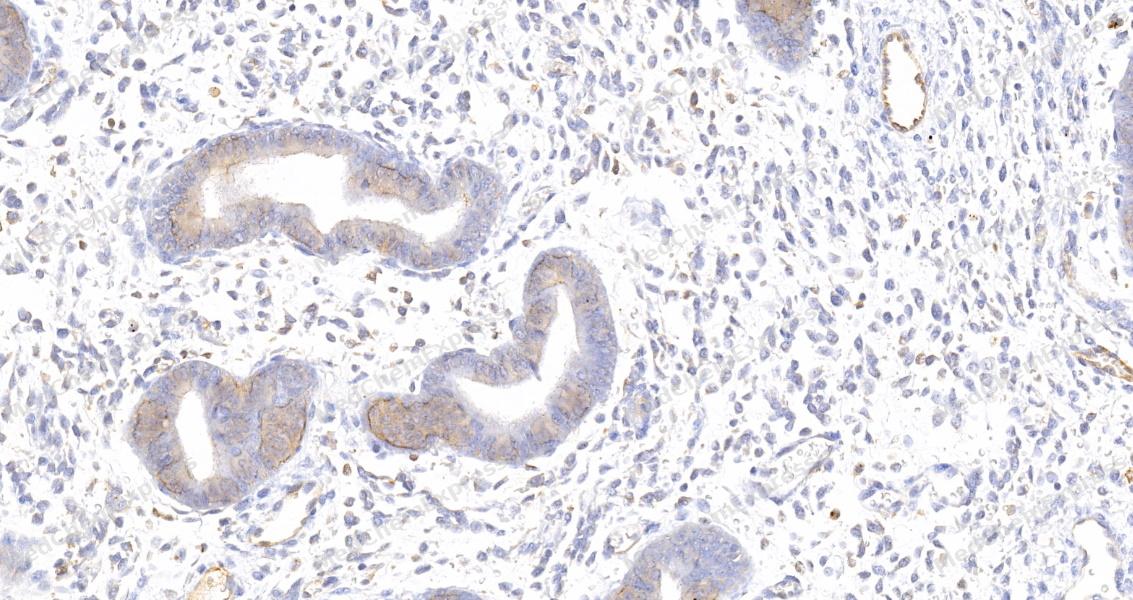 Immunohistochemical analysis of paraffin-embedded human endometrium tissue using EPHA7 Antibody (HY-P85143, 1/800). The section was pretreated using heat mediated antigen retrieval with sodium citrate buffer (pH 6.0) for 8 minutes. The tissues were blocked with quick block buffer for 0.5 hours at room temperature, washed with PBS and PBST, and then incubated with the primary antibody overnight at 4℃. The detection was performed using an HRP conjugated compact polymer system. DAB was used as the chromogen. Tissues were counterstained with hematoxylin and mounted with DPX.
 -
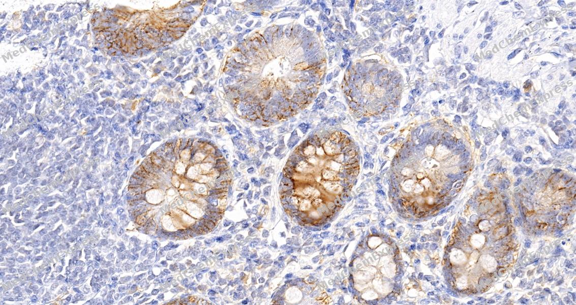 Immunohistochemical analysis of paraffin-embedded human small intestine tissue using EPHA7 Antibody (HY-P85143, 1/800). The section was pretreated using heat mediated antigen retrieval with sodium citrate buffer (pH 6.0) for 8 minutes. The tissues were blocked with quick block buffer for 0.5 hours at room temperature, washed with PBS and PBST, and then incubated with the primary antibody overnight at 4℃. The detection was performed using an HRP conjugated compact polymer system. DAB was used as the chromogen. Tissues were counterstained with hematoxylin and mounted with DPX.
 -
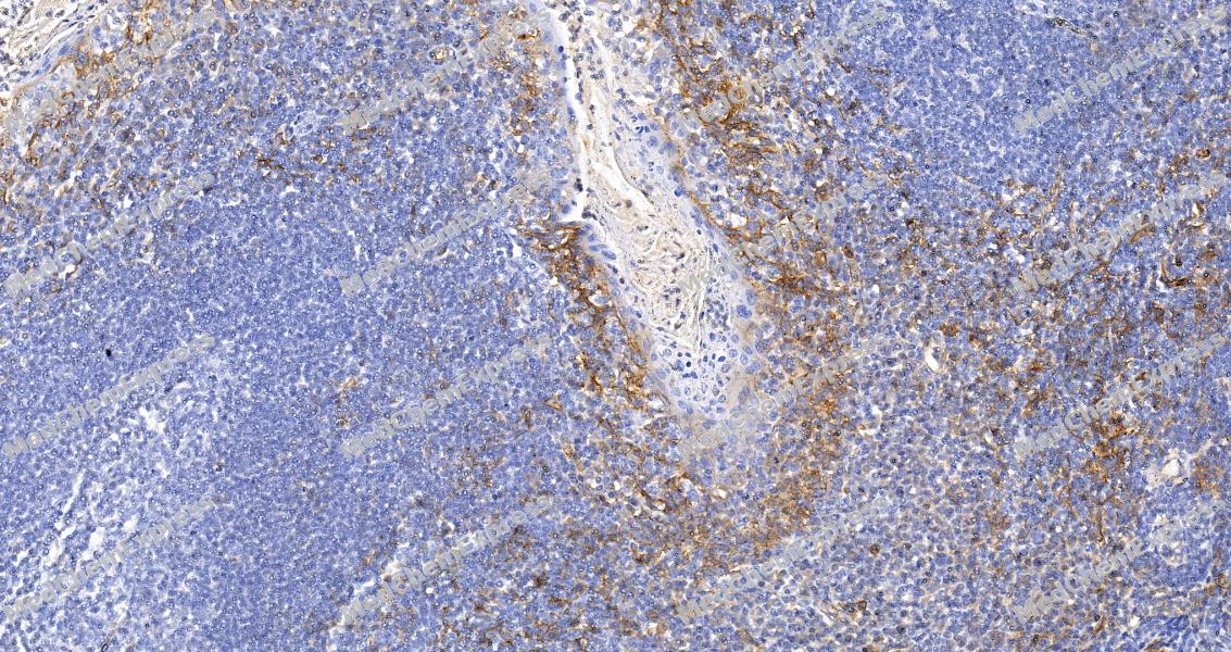 Immunohistochemical analysis of paraffin-embedded human tonsil tissue using EPHA7 Antibody (HY-P85143, 1/800). The section was pretreated using heat mediated antigen retrieval with sodium citrate buffer (pH 6.0) for 8 minutes. The tissues were blocked with quick block buffer for 0.5 hours at room temperature, washed with PBS and PBST, and then incubated with the primary antibody overnight at 4℃. The detection was performed using an HRP conjugated compact polymer system. DAB was used as the chromogen. Tissues were counterstained with hematoxylin and mounted with DPX.
 -
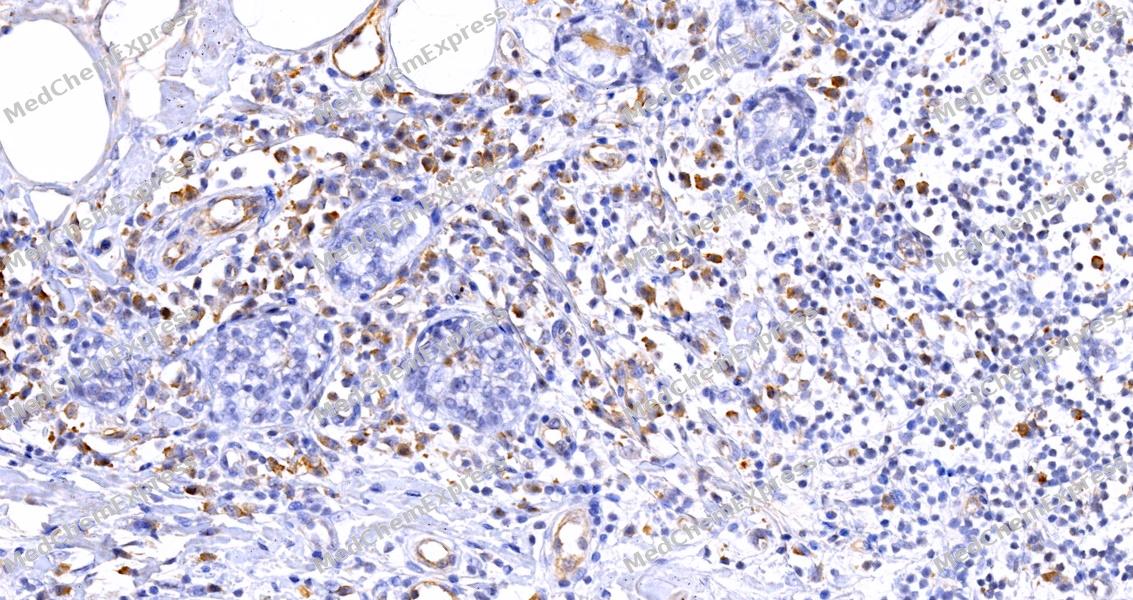 Immunohistochemical analysis of paraffin-embedded human breast tissue using EPHA7 Antibody (HY-P85143, 1/800). The section was pretreated using heat mediated antigen retrieval with sodium citrate buffer (pH 6.0) for 8 minutes. The tissues were blocked with quick block buffer for 0.5 hours at room temperature, washed with PBS and PBST, and then incubated with the primary antibody overnight at 4℃. The detection was performed using an HRP conjugated compact polymer system. DAB was used as the chromogen. Tissues were counterstained with hematoxylin and mounted with DPX.


Background
-
Function
EPHA7 receptor tyrosine kinase which binds promiscuously GPI-anchored ephrin-A family ligands residing on adjacent cells, leading to contact-dependent bidirectional signaling into neighboring cells. The signaling pathway downstream of the receptor is referred to as forward signaling while the signaling pathway downstream of the ephrin ligand is referred to as reverse signaling. Among GPI-anchored ephrin-A ligands, EFNA5 is a cognate/functional ligand for EPHA7 and their interaction regulates brain development modulating cell-cell adhesion and repulsion. Has a repellent activity on axons and is for instance involved in the guidance of corticothalamic axons and in the proper topographic mapping of retinal axons to the colliculus. May also regulate brain development through a caspase(CASP3)-dependent proapoptotic activity. Forward signaling may result in activation of components of the ERK signaling pathway including MAP2K1, MAP2K2, MAPK1 and MAPK3 which are phosphorylated upon activation of EPHA7
-
Subcellular Localization
Cell membrane; Single-pass type I membrane protein
-
Expression
Tissue_specificity:Widely expressed -
Isoforms & Post-Translational Modification
Q15375 has 5 isomers: Q15375-1: 112097 Da (predicted); Q15375-2: 111504 Da (predicted); Q15375-3: 31833 Da (predicted); Q15375-4: 111623 Da (predicted); Q15375-5: 50521 Da (predicted).
Phosphorylated -
Subunit
Heterotetramer upon binding of the ligand. The heterotetramer is composed of an ephrin dimer and a receptor dimer.
-
SwissProt ID
-
Synonyms
EPHA7; EHK3; HEK11; Ephrin type-A receptor 7; EPH homology kinase 3; EHK-3; EPH-like kinase 11; EK11; hEK11


Documentation
-
Data Sheet (234 KB)
-
SDS (252 KB)
- English - EN (252 KB)
- Français - FR (252 KB)
- Deutsch - DE (252 KB)
- Norwegian - NO (252 KB)
- Español - ES (252 KB)
- Swedish - SV (252 KB)
- Italian - IT (252 KB)
- Korean - KR (252 KB)
- Portuguese - PT (252 KB)
-
User Guide for Antibodies (1077 KB)

 Powered by Bioz
