



NF-κB1 p105/p50 Antibody (YA6065)
(Synonyms: NFKB1; Nuclear factor NF-kappa-B p105 subunit; DNA-binding factor KBF1; EBP-1; Nuclear factor of kappa light polypeptide gene enhancer in B-cells 1)
Based on 1 Customer Validation
NF-κB1 p105/p50 Antibody (YA6065) is a Rabbit-derived and non-conjugated IgG monoclonal antibody, targeting to NF-κB1 p105/p50.
-
Host:
Rabbit
-
Isotype:
IgG
-
Application:
WB, IHC-P, ICC/IF, IP, ELISA
-
Reactivity :
Human, Mouse, Rat
-
Formulation:
Supplied in PBS, 50% glycerol, 0.05% Proclin 300, 0.05%BSA
-
Conjugation:
Non-conjugated


To place orders, for customer services and technical support, please contact: MedChemExpress USA
Tel: 609-228-6898 E-mail: [email protected] [email protected]





Applications
| Application |
WB

WB: Western Blot
|
IHC-P
IHC-P: Immunohistochemistry-Paraffin
|
ICC/IF
ICC/IF: Immunocytochemistry/
Immunofluorescence |
ELISA
ELISA: Enzyme Linked Immunosorbent Assay
|
IP
IP: Immunoprecipitation
|
|---|---|---|---|---|---|
| Dilution Ratio | 1:2000-1:10000 | 1:200-1:1000 | 1:200-1:1000 | 1:5000-1:20000 | 1:50-1:200 |


Product Details
NF-κB1 p105/p50 Antibody (YA6065) is a Rabbit-derived and non-conjugated IgG monoclonal antibody, targeting to NF-κB1 p105/p50.
-
Host Rabbit

-
Clonality Monoclonal

-
Species ReactivityHuman, Mouse, Rat
-
Observed Molecular WeightObserved band size: 50 kDa,120 kDa
Note: Due to possible protein modifications or aggregation, the molecular weight should be confirmed by actual measurement, and the predicted value is for reference only.

-
Calculated Molecular Weight Predicted band size: 50 kDa,105 kDa

Protein A
Non-conjugated
Unmodified
IgG


Product Properties
-
Appearance
Solution
-
Formulation
Supplied in PBS, 50% glycerol, 0.05% Proclin 300, 0.05%BSA
-
Concentration
Batch-dependent, Please check the COA for the concentration of each lot. Check Lot Concentration
-
Storage & Stability
Stored at -20°C for 1 year. Avoid repeated freeze / thaw cycles.
-
Shipping
Shipping with blue ice.


Verification Images
-
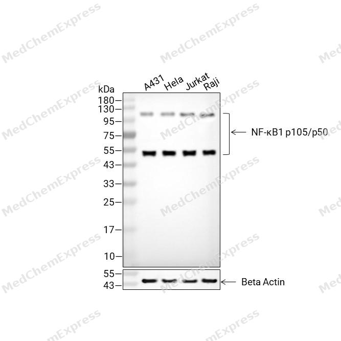 Western blot analysis of extracts from A431 (lane2(20μg), Hela (lane3(20μg), Jurkat (lane4(20μg) and Raji (lane5(20μg) using NF-κB1 p105/p50 Antibody (HY-P86373). Proteins were transferred to a PVDF membrane and blocked with 5% non-fat milk in TBST for 2 hour at room temperature. The primary antibody (1/5000) and Loading control antibody (Beta Actin, HY-P80993, 1/10,000) was used in 5% non-fat milk in TBST at 4°C overnight. Goat Anti-Rabbit IgG-HRP Secondary Antibody (HY-P8001 ,1/10,000) was used for 1 hour at room temperature.
 -
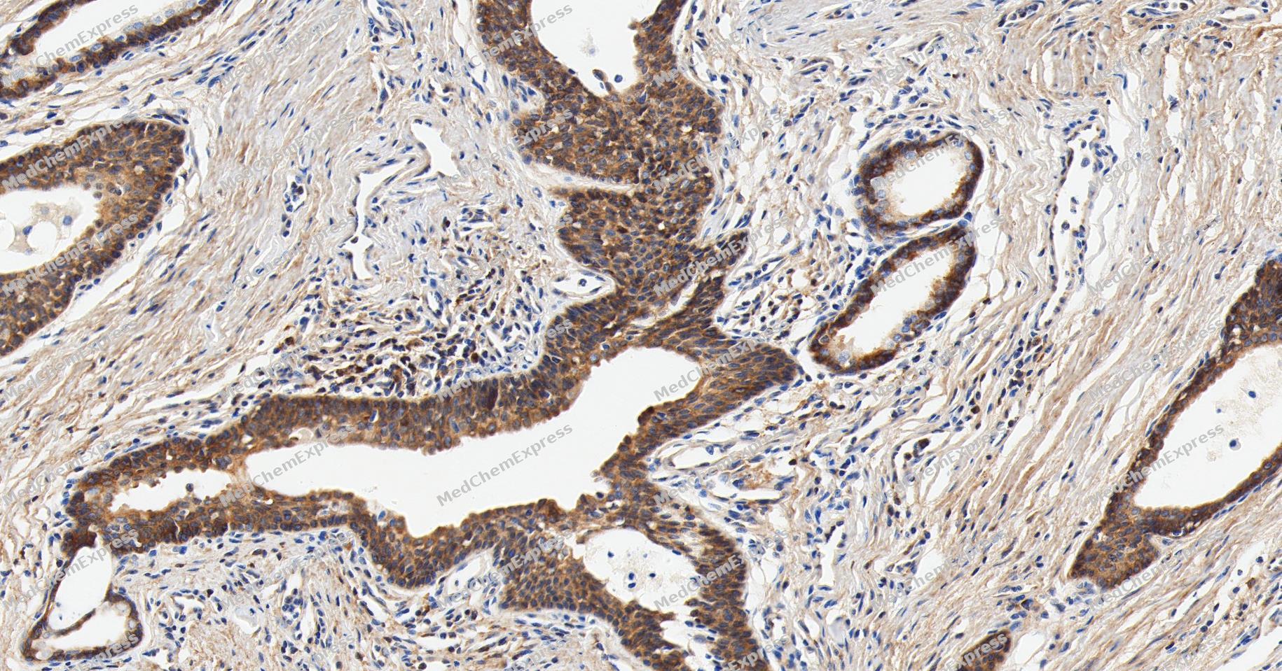 Immunohistochemical analysis of paraffin-embedded human prostate cancer tissue using NF-κB1 p105/p50 Antibody (YA6065). The section was pre-treated using heat mediated antigen retrieval with Tris/EDTA buffer (pH 9.0) for 20 minutes. The tissues were probed with the primary antibody (HY-P86373, 1/150) overnight at 4℃. The detection was performed using an HRP conjugated compact polymer system. DAB was used as the chromogen. Tissues were counterstained with hematoxylin and mounted with neutral balsam.
 -
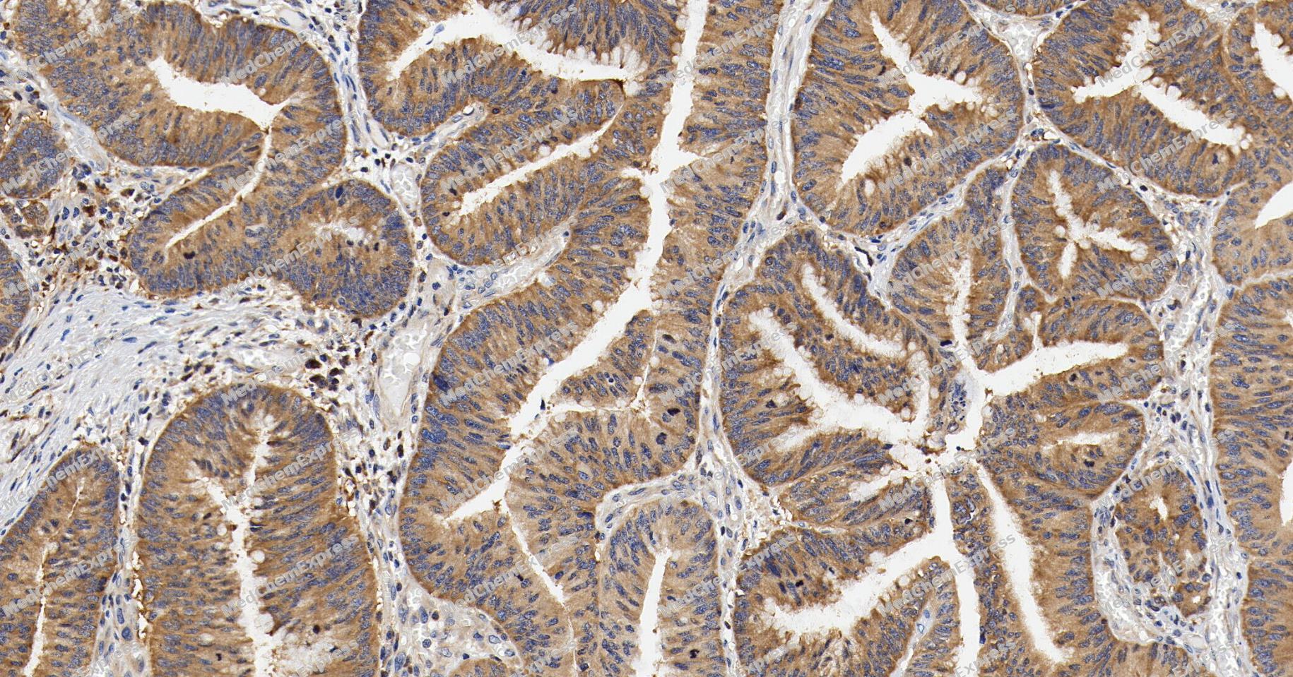 Immunohistochemical analysis of paraffin-embedded human colon cancer tissue using NF-κB1 p105/p50 Antibody (YA6065). The section was pre-treated using heat mediated antigen retrieval with Tris/EDTA buffer (pH 9.0) for 20 minutes. The tissues were probed with the primary antibody (HY-P86373, 1/150) overnight at 4℃. The detection was performed using an HRP conjugated compact polymer system. DAB was used as the chromogen. Tissues were counterstained with hematoxylin and mounted with neutral balsam.
 -
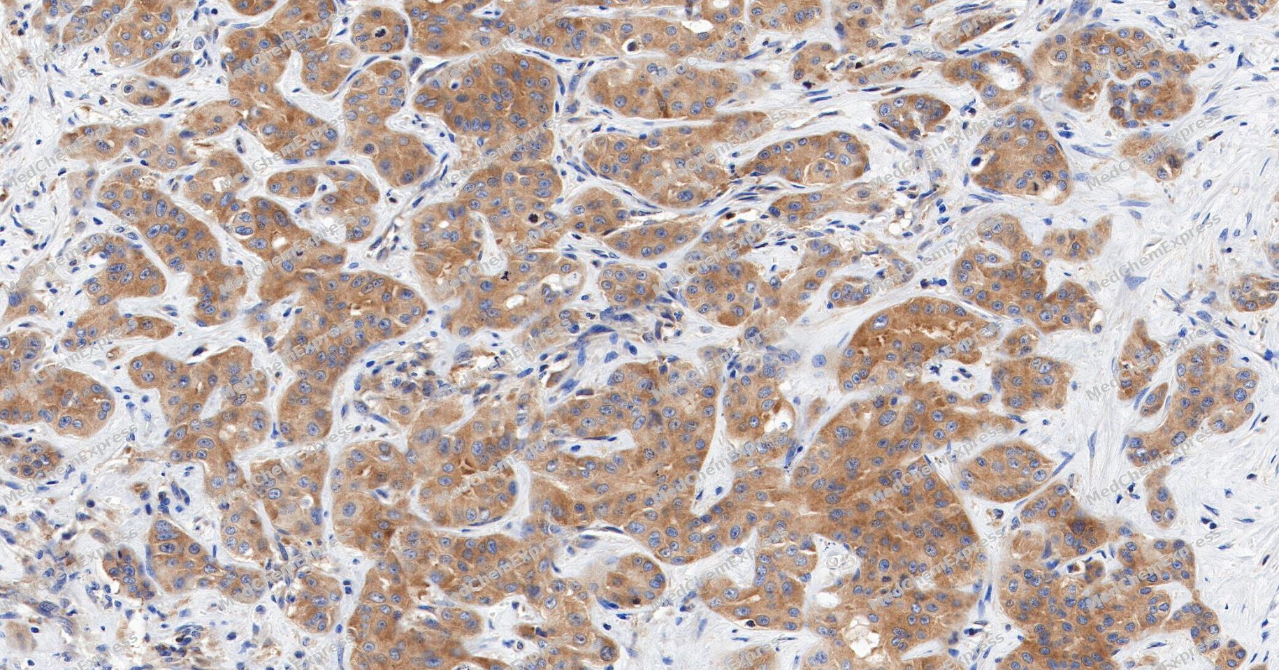 Immunohistochemical analysis of paraffin-embedded human liver cancer tissue using NF-κB1 p105/p50 Antibody (YA6065). The section was pre-treated using heat mediated antigen retrieval with Tris/EDTA buffer (pH 9.0) for 20 minutes. The tissues were probed with the primary antibody (HY-P86373, 1/150) overnight at 4℃. The detection was performed using an HRP conjugated compact polymer system. DAB was used as the chromogen. Tissues were counterstained with hematoxylin and mounted with neutral balsam.
 -
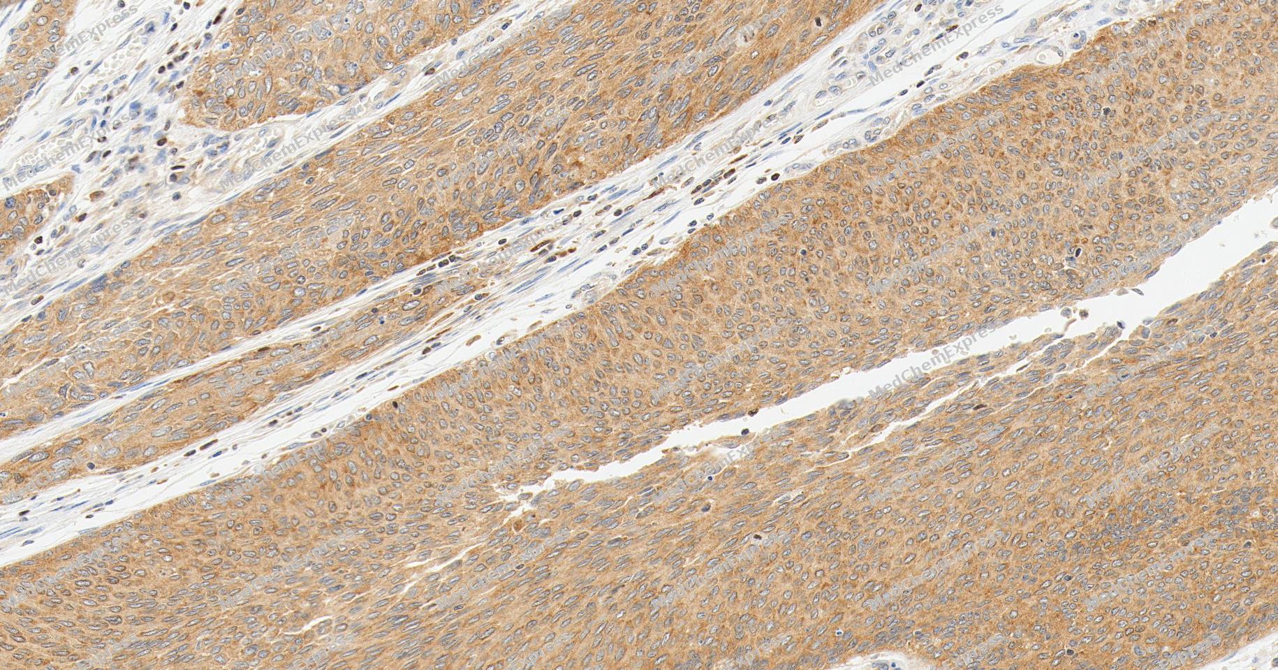 Immunohistochemical analysis of paraffin-embedded human bladder cancer tissue using NF-κB1 p105/p50 Antibody (YA6065). The section was pre-treated using heat mediated antigen retrieval with Tris/EDTA buffer (pH 9.0) for 20 minutes. The tissues were probed with the primary antibody (HY-P86373, 1/150) overnight at 4℃. The detection was performed using an HRP conjugated compact polymer system. DAB was used as the chromogen. Tissues were counterstained with hematoxylin and mounted with neutral balsam.
 -
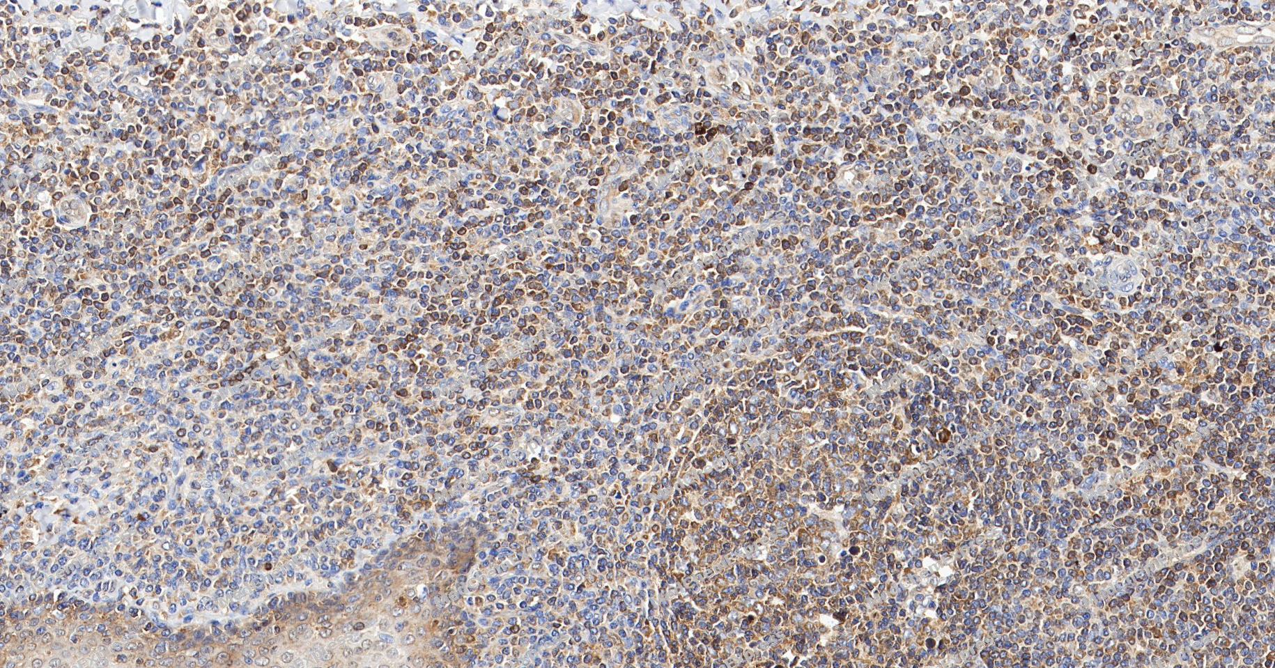 Immunohistochemical analysis of paraffin-embedded human tonsil tissue using NF-κB1 p105/p50 Antibody (YA6065). The section was pre-treated using heat mediated antigen retrieval with Tris/EDTA buffer (pH 9.0) for 20 minutes. The tissues were probed with the primary antibody (HY-P86373, 1/150) overnight at 4℃. The detection was performed using an HRP conjugated compact polymer system. DAB was used as the chromogen. Tissues were counterstained with hematoxylin and mounted with neutral balsam.
 -
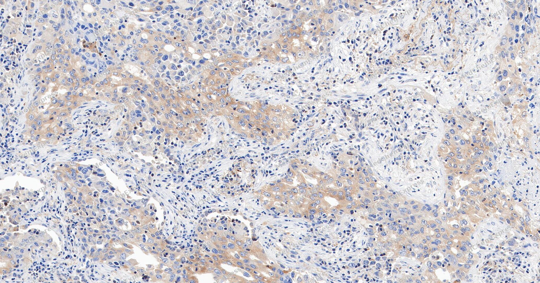 Immunohistochemical analysis of paraffin-embedded human lung cancer tissue using NF-κB1 p105/p50 Antibody (YA6065). The section was pre-treated using heat mediated antigen retrieval with Tris/EDTA buffer (pH 9.0) for 20 minutes. The tissues were probed with the primary antibody (HY-P86373, 1/150) overnight at 4℃. The detection was performed using an HRP conjugated compact polymer system. DAB was used as the chromogen. Tissues were counterstained with hematoxylin and mounted with neutral balsam.
 -
 Immunohistochemical analysis of paraffin-embedded human kidney tissue using NF-κB1 p105/p50 Antibody (HY-P86373, 1/1000). The section was pretreated using heat mediated antigen retrieval with sodium citrate buffer (pH 6.0) for 8 minutes. The tissues were blocked with quick block buffer for 0.5 hours at room temperature, washed with PBS and PBST, and then incubated with the primary antibody overnight at 4℃. The detection was performed using an HRP conjugated compact polymer system. DAB was used as the chromogen. Tissues were counterstained with hematoxylin and mounted with DPX.
 -
 Immunohistochemical analysis of paraffin-embedded human lung tissue using NF-κB1 p105/p50 Antibody (HY-P86373, 1/1000). The section was pretreated using heat mediated antigen retrieval with sodium citrate buffer (pH 6.0) for 8 minutes. The tissues were blocked with quick block buffer for 0.5 hours at room temperature, washed with PBS and PBST, and then incubated with the primary antibody overnight at 4℃. The detection was performed using an HRP conjugated compact polymer system. DAB was used as the chromogen. Tissues were counterstained with hematoxylin and mounted with DPX.
 -
 Immunohistochemical analysis of paraffin-embedded human testis tissue using NF-κB1 p105/p50 Antibody (HY-P86373, 1/1000). The section was pretreated using heat mediated antigen retrieval with sodium citrate buffer (pH 6.0) for 8 minutes. The tissues were blocked with quick block buffer for 0.5 hours at room temperature, washed with PBS and PBST, and then incubated with the primary antibody overnight at 4℃. The detection was performed using an HRP conjugated compact polymer system. DAB was used as the chromogen. Tissues were counterstained with hematoxylin and mounted with DPX.
 -
 Immunohistochemical analysis of paraffin-embedded human placenta tissue using NF-κB1 p105/p50 Antibody (HY-P86373, 1/1000). The section was pretreated using heat mediated antigen retrieval with sodium citrate buffer (pH 6.0) for 8 minutes. The tissues were blocked with quick block buffer for 0.5 hours at room temperature, washed with PBS and PBST, and then incubated with the primary antibody overnight at 4℃. The detection was performed using an HRP conjugated compact polymer system. DAB was used as the chromogen. Tissues were counterstained with hematoxylin and mounted with DPX.
 -
 Immunohistochemical analysis of paraffin-embedded human spleen tissue using NF-κB1 p105/p50 Antibody (HY-P86373, 1/1000). The section was pretreated using heat mediated antigen retrieval with sodium citrate buffer (pH 6.0) for 8 minutes. The tissues were blocked with quick block buffer for 0.5 hours at room temperature, washed with PBS and PBST, and then incubated with the primary antibody overnight at 4℃. The detection was performed using an HRP conjugated compact polymer system. DAB was used as the chromogen. Tissues were counterstained with hematoxylin and mounted with DPX.
 -
 Immunohistochemical analysis of paraffin-embedded human colon tissue using NF-κB1 p105/p50 Antibody (HY-P86373, 1/1000). The section was pretreated using heat mediated antigen retrieval with sodium citrate buffer (pH 6.0) for 8 minutes. The tissues were blocked with quick block buffer for 0.5 hours at room temperature, washed with PBS and PBST, and then incubated with the primary antibody overnight at 4℃. The detection was performed using an HRP conjugated compact polymer system. DAB was used as the chromogen. Tissues were counterstained with hematoxylin and mounted with DPX.
 -
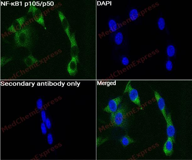 Immunocytochemistry analysis of NIH-3T3 cells labeling NF-κB1 p105/p50 with NF-κB1 p105/p50 Antibody (HY-P86373) at 1:200 dilution. Cells were fixed in 4% paraformaldehyde for 15 minutes at room temperature, permeabilized with 0.1% Triton X-100 in PBS for 15 minutes at room temperature, then blocked with quick block buffer for 10 minutes at room temperature. Cells were then incubated with NF-κB1 p105/p50 Antibody (HY-P86373) at 1:200 dilution in quick block buffer overnight at 4 ℃. AF488-conjugated Goat Anti-Rabbit IgG H&L(HY-P8002, Green) was used as the secondary antibody at 1/1,000 dilution. PBS instead of the primary antibody was used as the secondary antibody only control. The Nuclear counterstain was DAPI (Blue).
 -
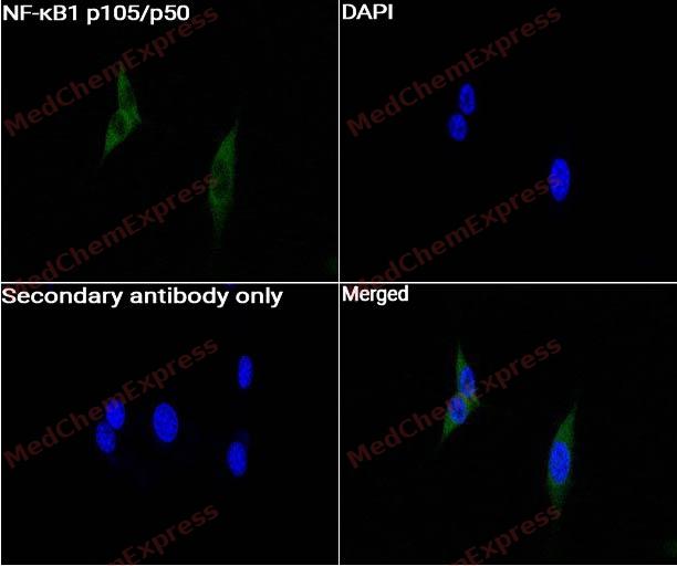 Immunocytochemistry analysis of NIH-3T3 cells labeling NF-κB1 p105/p50 with NF-κB1 p105/p50 Antibody (HY-P86373) at 1:400 dilution. Cells were fixed in 4% paraformaldehyde for 15 minutes at room temperature, permeabilized with 0.1% Triton X-100 in PBS for 15 minutes at room temperature, then blocked with quick block buffer for 10 minutes at room temperature. Cells were then incubated with NF-κB1 p105/p50 Antibody (HY-P86373) at 1:400 dilution in quick block buffer overnight at 4 ℃. AF488-conjugated Goat Anti-Rabbit IgG H&L(HY-P8002, Green) was used as the secondary antibody at 1/1,000 dilution. PBS instead of the primary antibody was used as the secondary antibody only control. The Nuclear counterstain was DAPI (Blue).


Background
-
Function
NF-κB1 p105/p50 is a NF-kappa-B is a pleiotropic transcription factor present in almost all cell types and is the endpoint of a series of signal transduction events that are initiated by a vast array of stimuli related to many biological processes such as inflammation, immunity, differentiation, cell growth, tumorigenesis and apoptosis. NF-kappa-B is a homo- or heterodimeric complex formed by the Rel-like domain-containing proteins RELA/p65, RELB, NFKB1/p105, NFKB1/p50, REL and NFKB2/p52 and the heterodimeric p65-p50 complex appears to be most abundant one. The dimers bind at kappa-B sites in the DNA of their target genes and the individual dimers have distinct preferences for different kappa-B sites that they can bind with distinguishable affinity and specificity. Different dimer combinations act as transcriptional activators or repressors, respectively. NF-kappa-B is controlled by various mechanisms of post-translational modification and subcellular compartmentalization as well as by interactions with other cofactors or corepressors. NF-kappa-B complexes are held in the cytoplasm in an inactive state complexed with members of the NF-kappa-B inhibitor (I-kappa-B) family. In a conventional activation pathway, I-kappa-B is phosphorylated by I-kappa-B kinases (IKKs) in response to different activators, subsequently degraded thus liberating the active NF-kappa-B complex which translocates to the nucleus. NF-kappa-B heterodimeric p65-p50 and RelB-p50 complexes are transcriptional activators. The NF-kappa-B p50-p50 homodimer is a transcriptional repressor, but can act as a transcriptional activator when associated with BCL3. NFKB1 appears to have dual functions such as cytoplasmic retention of attached NF-kappa-B proteins by p105 and generation of p50 by a cotranslational processing. The proteasome-mediated process ensures the production of both p50 and p105 and preserves their independent function, although processing of NFKB1/p105 also appears to occur post-translationally. p50 binds to the kappa-B consensus sequence 5'-GGRNNYYCC-3', located in the enhancer region of genes involved in immune response and acute phase reactions. Plays a role in the regulation of apoptosis. In a complex with MAP3K8, NFKB1/p105 represses MAP3K8-induced MAPK signaling; active MAP3K8 is released by proteasome-dependent degradation of NFKB1/p105; P105 is the precursor of the active p50 subunit (Nuclear factor NF-kappa-B p50 subunit) of the nuclear factor NF-kappa-B. Acts as a cytoplasmic retention of attached NF-kappa-B proteins by p105; Constitutes the active form, which associates with RELA/p65 to form the NF-kappa-B p65-p50 complex to form a transcription factor. Together with RELA/p65, binds to the kappa-B consensus sequence 5'-GGRNNYYCC-3', located in the enhancer region of genes involved in immune response and acute phase reactions; Isoform 3 (p98) (but not p84 or p105) acts as a transactivator of NF-kappa-B-regulated gene expression; Acts as an inhibitor of transactivation of p50 NF-kappa-B subunit, probably by sequestering it in the cytoplasm; Acts as an inhibitor of transactivation of p50 NF-kappa-B subunit, probably by sequestering it in the cytoplasm; Acts as an inhibitor of transactivation of p50 NF-kappa-B subunit, probably by sequestering it in the cytoplasm
-
Subcellular Localization
Cytoplasm; Nucleus; Cytoplasm; Cytoplasm; Nucleus; Cytoplasm; Nucleus
-
Expression
Induction:By phorbol ester and TNF-alpha -
Isoforms & Post-Translational Modification
P25799 has 7 isomers: P25799-1: 105615 Da (predicted); P25799-2: 84817 Da (predicted); P25799-3: 97413 Da (predicted); P25799-4: 39756 Da (predicted); P25799-5: 64782 Da (predicted); P25799-6: 56580 Da (predicted); P25799-7: 43983 Da (predicted).
Generation of the NF-kappa-B p50 (Nuclear factor NF-kappa-B p50 subunit) transcription factor takes place both cotranslationally and post-translationally via non-mutually exclusive mechanisms (By similarity). A cotranslational processing allows the production of both p50 and p105 (Nuclear factor NF-kappa-B p105 subunit) from a single NFKB1 mRNA (PubMed:9529257). While translation occurs, the particular unfolded structure after the GRR repeat region acts as a substrate for the proteasome, promoting degradation of the C-terminus (PubMed:9529257). The GRR acts as a proteasomal 'stop signal', protecting the region upstream of the GRR from degradation and promoting generation of p50 (PubMed:9529257). It is unclear if limited proteasome degradation during cotranslational processing depends on ubiquitination (PubMed:9529257). NF-kappa-B p50 is also generated post-translationally following ubiquitination by the KPC complex, leading to limited processing by the proteasome downstream of the GRR region, thereby generating p50 (By similarity);Phosphorylation at the C-terminus by IKBKB/IKKB acts as a signal for ubiquitination and promotes either complete degradation or processing to generate the NF-kappa-B p50 (Nuclear factor NF-kappa-B p50 subunit) (By similarity). Phosphorylation at Ser-910 primes p105 for proteolytic processing in response to TNF-alpha stimulation (By similarity). Phosphorylation at Ser-926, Ser-930 and Ser-935 are required for BTRC/BTRCP-mediated ubiquitination and proteolysis (By similarity). Phosphorylation at Ser-930 is also required for ubiquitination by the KPC complex and limited processing to generate NF-kappa-B p50 (Nuclear factor NF-kappa-B p50 subunit) (By similarity);Polyubiquitinated at multiple Lys residues in the C-terminus (By similarity). Polyubiquitinated by the SCF(FBXW11) and SCF(BTRC) complexes following phosphorylation at Ser-926, Ser-930 and Ser-935, leading to its complete degradation (By similarity). In contrast, polyubiquitination by the KPC complex following phosphorylation at Ser-930 leads to limited proteosomal processing and generation of the active NF-kappa-B p50 (Nuclear factor NF-kappa-B p50 subunit) (By similarity);S-nitrosylation of Cys-59 affects DNA binding;The covalent modification of cysteine by 15-deoxy-Delta12,14-prostaglandin-J2 is autocatalytic and reversible. It may occur as an alternative to other cysteine modifications, such as S-nitrosylation and S-palmitoylation -
Subunit
Component of the NF-kappa-B p65-p50 complex (By similarity). Homodimer; component of the NF-kappa-B p50-p50 complex (By similarity). Component of the NF-kappa-B p105-p50 complex (By similarity). Component of the NF-kappa-B p50-c-Rel complex (By similarity). Component of a complex consisting of the NF-kappa-B p50-p50 homodimer and BCL3 (By similarity). Also interacts with MAP3K8 (By similarity). NF-kappa-B p50 subunit interacts with NCOA3 coactivator, which may coactivate NF-kappa-B dependent expression via its histone acetyltransferase activity (By similarity). Interacts with TSC22D3; this interaction prevents nuclear translocation and DNA-binding (By similarity). Interacts with SPAG9 and UNC5CL (By similarity). NFKB1/p105 interacts with CFLAR; the interaction inhibits p105 processing into p50 (By similarity). NFKB1/p105 forms a ternary complex with MAP3K8 and TNIP2 (By similarity). Interacts with GSK3B; the interaction prevents processing of p105 to p50 (By similarity). NFKB1/p50 interacts with NFKBIE (By similarity). NFKB1/p50 interacts with NFKBIZ (PubMed:11356851, PubMed:15241416). Nuclear factor NF-kappa-B p50 subunit interacts with NFKBID (PubMed:11931770). Directly interacts with MEN1 (By similarity). Interacts with HIF1AN (By similarity). Interacts with FEM1AA; interaction is direct (PubMed:18270204)
-
SwissProt ID
-
Synonyms
NFKB1; Nuclear factor NF-kappa-B p105 subunit; DNA-binding factor KBF1; EBP-1; Nuclear factor of kappa light polypeptide gene enhancer in B-cells 1


Documentation
-
Data Sheet (237 KB)
-
SDS (252 KB)
- English - EN (252 KB)
- Français - FR (252 KB)
- Deutsch - DE (252 KB)
- Norwegian - NO (252 KB)
- Español - ES (252 KB)
- Swedish - SV (252 KB)
- Italian - IT (252 KB)
- Korean - KR (252 KB)
- Portuguese - PT (252 KB)
-
User Guide for Antibodies (1077 KB)

 Powered by Bioz
